Skip to content

Showing 1–10 of 46 results

Cell marker antibodies

Cell marker antibodies are highly specific antibodies that recognize and bind to proteins or other molecules that are uniquely expressed or enriched in specific cell types. These antibodies are crucial tools in cell biology and research as they allow scientists to identify, isolate, and study different cell populations in complex biological samples. By using cell marker antibodies in techniques like flow cytometry, immunohistochemistry, and immunofluorescence, researchers can distinguish between various cell types based on their unique surface markers or intracellular proteins. This information is vital for characterizing cell populations, understanding cellular heterogeneity, and studying cell differentiation and development. Additionally, cell marker antibodies are essential in biomedical research, aiding in the diagnosis and classification of diseases and identifying biomarkers associated with specific cell types or pathological conditions. As advances in antibody development continue, the use of cell marker antibodies is expanding, providing valuable insights into cellular biology and contributing to the development of personalized medicine and targeted therapies.


target type reactivity applications
1007 4-Hydroxynonenal (4-HNE) rabbit polyclonal other WB
1102 8-hydroxy-guanosine mouse monoclonal (15A3) other WB ICC IHC ELISA
9067 Alpha-synuclein mouse monoclonal human mouse rat WB ICC IHC
9481 AMPK α 1 rabbit polyclonal human rat cow WB IHC
4805 AMPK α 2 rabbit polyclonal human rat cow WB
1034 AMPK (AMP-activated protein kinase) rabbit polyclonal human mouse IHC ELISA
9012 BrdU chicken polyclonal other ELISA
9015 Calreticulin rabbit polyclonal human mouse rat WB ICC IHC
9059 Doublecortin rabbit polyclonal human mouse rat WB ICC IHC
1039 FOXO (Forkhead box O) Transcription Factor 3a rabbit polyclonal human mouse WB ELISA
1024 FOXO (Forkhead box O) Transcription Factors rabbit monoclonal (BLR140H) human mouse WB ELISA
9024 GFAP mouse monoclonal human mouse rat WB ICC IHC
1009 HIF-1α (Hypoxia-inducible factor 1-alpha) rabbit monoclonal (BL-124-3F7) human WB ICC IHC ELISA
1029 HIF-1α (Hypoxia-inducible factor 1-alpha) goat polyclonal human ELISA
1008 HO-1 (Heme oxygenase-1) rabbit polyclonal human WB ELISA
1028 HO-1 (Heme oxygenase-1) rabbit polyclonal human ELISA
1017 JNK (c-Jun N-terminal kinases) rabbit polyclonal human mouse WB IHC ELISA
1037 JNK (c-Jun N-terminal kinases) rabbit polyclonal human mouse WB IHC ELISA
9056 Ki-67 rabbit monoclonal human WB ICC IHC
9022 LAMP1 mouse monoclonal human WB ICC IHC
1006 Malondialdehyde (MDA) rabbit other WB
1027 Malondialdehyde (MDA) rabbit polyclonal other WB ELISA
9007 MAP2 mouse monoclonal (A4) human mouse rat WB ICC IHC
1015 MDA ApoB rabbit other WB ELISA
1014 MDA LDL rabbit other WB ELISA
9018 NeuN mouse monoclonal (2Q158) human mouse rat rabbit dog WB ICC IHC
1012 Nitrated LDL (NitratedLDL) rabbit other WB ELISA
1010 Nitrotyrosine rabbit polyclonal other WB ELISA
1030 Nitrotyrosine goat polyclonal other WB ELISA
1011 Oxidized LDL (oxLDL) rabbit other WB ELISA
1019 p38 MAPK rabbit polyclonal human mouse WB ELISA
1032 p38 MAPK rabbit polyclonal human mouse WB ELISA
1023 p53 rabbit monoclonal (BL-250-1-H11) human mouse WB ICC IHC ELISA
8319 p53 mouse monoclonal (D01) human WB ICC IHC ELISA
1020 PI3K/Akt Pathway (Phosphoinositide 3-kinase/Akt) rabbit monoclonal (BLR245L) human mouse WB ICC
1033 PI3K/Akt Pathway (Phosphoinositide 3-kinase/Akt) rabbit polyclonal human WB IHC ELISA
1035 PKC (Protein kinase C) alpha rabbit polyclonal human mouse WB ELISA
1022 PKC-eta (Protein kinase C) rabbit polyclonal human WB ELISA
1025 Sirtuins (SIRT1) rabbit polyclonal human WB ELISA
1038 Sirtuins (SIRT1) rabbit polyclonal human WB ELISA
1001 Superoxide Dismutase 1 (SOD) rabbit polyclonal human WB IHC
1026 Superoxide Dismutase 1 (SOD) rabbit polyclonal human WB IHC
1004 Thioredoxin (Trx) rabbit polyclonal human WB ELISA
9009 Tyrosine Hydroxylase rabbit monoclonal human mouse rat WB ICC
9044 Ubiquitin rabbit polyclonal (BLR100G) human mouse rat rabbit goat cat sheep hamster cow dog guinea pig pig h pylori other WB ICC IHC
9046 Vimentin rabbit monoclonal human mouse rat WB ICC IHC

ICC/IF-image-mouse-anti-GFP-monoclonal-antibody-3B11-3128
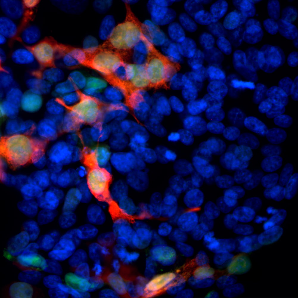
ICC/IF with antibody [3128] : Immunofluorescent analysis of transfected HEK293 cells with a GFP-construct in green stained with mouse mAb to GFP, 3128, dilution 1:1,000 in red. The blue is Hoechst staining of nuclear DNA. 3128 antibody reveals GFP protein expressed only in transfected cells, as a result cells are appeared in orange-golden color.
ICC/IF-image-rabbit-anti-GFP-polyclonal-antibody-1290
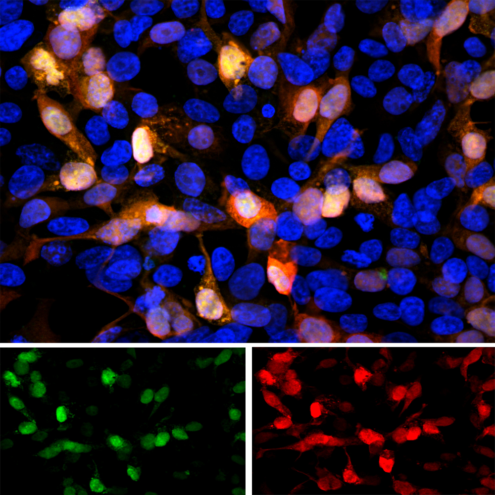
ICC/IF with antibody [1290] : Immunofluorescent analysis of transfected HEK293 cells with a GFP construct, in green, and stained with rabbit pAb to GFP, 1290, dilution 1:2,000 in red. The blue is Hoechst staining of nuclear DNA. The 1290 antibody reveals GFP protein expressed only in transfected cells, and as a result these cells appear orange-yellow in color.
IHC-image-goat-anti-GFP-polyclonal-antibody-1379
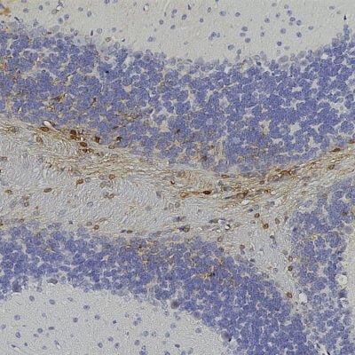
IHC with antibody [1379] : Chromogenic immunostaining of formalin fixed paraffin embedded GFP transduced mouse brain with goat pAb to GFP, 1379, dilution 1:5,000, detected with DAB (brown) using the Vector Elite 8718C-HRP detection and reagents with citra buffer retrieval. Hematoxylin (blue) was used as the counterstain. Antibody 1379 specifically detected GFP positive cells in the cerebellum as expected for this model.