| Weight | 1 lbs |
|---|---|
| Dimensions | 9 × 5 × 2 in |
| host | rabbit |
| isotype | IgG |
| clonality | polyclonal |
| concentration | 1 mg/mL |
| applications | ICC/IF, IHC, WB |
| available sizes | 100 µg |


rabbit anti-Ki67 polyclonal antibody 9056
$409.00
Antibody summary
- Rabbit polyclonal to Ki67
- Suitable for: WB, ICC/IF, IHC
- Reacts with: mouse, rat
- Isotype: IgG
- 100 µg
rabbit anti-Ki67 polyclonal antibody 9056
| antibody |
|---|
| Database link: P46013 |
| Tested applications WB,ICC/IF, Flow, IHC |
| Recommended dilutions WB: 1:1000-5000 IF 1:2000-5000 |
| Immunogen Recombinant construct containing the N-terminal 300 amino acids of the human sequence expressed in and purified from E. coli. |
| Size and concentration 100µg and 1 mg/mL |
| Form liquid |
| Storage Instructions 2-8°C for short term, for longer term at -20°C. Avoid freeze / thaw cycles. |
| Storage buffer PBS, 50% glycerol, 0.04% NaN3 |
| Purity affinity purified |
| Clonality monoclonal |
| Isotype IgG |
| Compatible secondaries goat anti-rabbit IgG, H&L chain specific, peroxidase conjugated, conjugated polyclonal antibody 9512 goat anti-rabbit IgG, H&L chain specific, biotin conjugated polyclonal antibody 2079 goat anti-rabbit IgG, H&L chain specific, FITC conjugated polyclonal antibody 7863 goat anti-rabbit IgG, H&L chain specific, Cross Absorbed polyclonal antibody 2371 goat anti-rabbit IgG, H&L chain specific, biotin conjugated polyclonal antibody, crossabsorbed 1715 goat anti-rabbit IgG, H&L chain specific, FITC conjugated polyclonal antibody, crossabsorbed 1720 |
| Isotype control Rabbit monocolonal IgG - Isotype Control |
| target relevance |
|---|
| Homo sapiens MKI67 Proliferation marker protein Ki-67 |
| Protein names Proliferation marker protein Ki-67 |
| Alternative names Antigen identified by monoclonal antibody Ki-67 |
| Gene names MKI67 |
| Function Protein that associates with the surface of mitotic chromosomes and acts both as a chromosome repellent during early mitosis and chromosome attractant during late mitosis (PubMed:27362226, PubMed:32879492, PubMed:35513709, PubMed:39153474). Required to maintain individual mitotic chromosomes dispersed in the cytoplasm following nuclear envelope disassembly (PubMed:27362226). During early mitosis, relocalizes from nucleoli to the chromosome surface where it forms extended brush structures that cover a substantial fraction of the chromosome surface (PubMed:27362226). The MKI67 brush structure prevents chromosomes from collapsing into a single chromatin mass by forming a steric and electrostatic charge barrier: the protein has a high net electrical charge and acts as a surfactant, dispersing chromosomes and enabling independent chromosome motility (PubMed:27362226). During mitotic anaphase, the MKI67 brush structure collapses and MKI67 switches from a chromosome repellent to a chromosome attractant to promote chromosome clustering and facilitate the exclusion of large cytoplasmic particles from the future nuclear space (PubMed:32879492, PubMed:39153474). Mechanistically, dephosphorylation during mitotic exit and simultaneous exposure of a conserved basic patch induce the RNA-dependent formation of a liquid-like condensed phase on the chromosome surface, promoting coalescence of neighboring chromosome surfaces and clustering of chromosomes (PubMed:39153474). Binds premature ribosomal RNAs during anaphase; promoting liquid-liquid phase separation (PubMed:28935370, PubMed:39153474). Binds DNA, with a preference for supercoiled DNA and AT-rich DNA (PubMed:10878551). Does not contribute to the internal structure of mitotic chromosomes (By similarity). May play a role in chromatin organization; it is however unclear whether it plays a direct role in chromatin organization or whether it is an indirect consequence of its function in mitotic chromosome (PubMed:24867636) |
| Subcellular location Chromosome, Nucleus, Nucleus, nucleolus |
| Structure Interacts with KIF15 (PubMed:10878014). Interacts (via the FHA domain) with NIFK (PubMed:11342549, PubMed:14659764, PubMed:16244663). Interacts with PPP1CC (PubMed:24867636, PubMed:25012651). Component of a complex at least composed of ZNF335, HCFC1, CCAR2, EMSY, MKI67, RBBP5, ASH2L and WDR5; the complex is formed as a result of interactions between components of a nuclear receptor-mediated transcription complex and a histone methylation complex (PubMed:19131338). Interacts with ZNF335 (PubMed:19131338, PubMed:23178126) |
| Post-translational modification Hyperphosphorylated by CDK1 in mitosis; hyperphosphorylatiom prevents undergoing liquid-liquid phase separation (PubMed:10502411, PubMed:10653604, PubMed:25012651, PubMed:39153474). Dephosphorylated by PPP1CC at the onset of anaphase (PubMed:25012651). Dephosphorylated by protein phosphatase 2A (PP2A) at the onset of anaphase (By similarity). Dephosphorylation by protein phosphatase 2A (PP2A) and simultaneous exposure of the positively charged patch (CP) during mitotic exit induce the RNA-dependent formation of a liquid-like condensed phase on the chromosome surface (PubMed:39153474) Ubiquitinated by the APC/C complex after neuronal progenitors exit mitosis during brain development, leading to clearance from constitutive heterochromatin |
| Keywords 3D-structure, Acetylation, Alternative splicing, ATP-binding, Cell cycle, Chromosome, DNA-binding, Isopeptide bond, Nucleotide-binding, Nucleus, Phosphoprotein, Proteomics identification, Reference proteome, Repeat, Ubl conjugation |
| Sequence MWPTRRLVTIKRSGVDGPHFPLSLSTCLFGRGIECDIRIQLPVVSKQHCKIEIHEQEAIL HNFSSTNPTQVNGSVIDEPVRLKHGDVITIIDRSFRYENESLQNGRKSTEFPRKIREQEP ARRVSRSSFSSDPDEKAQDSKAYSKITEGKVSGNPQVHIKNVKEDSTADDSKDSVAQGTT NVHSSEHAGRNGRNAADPISGDFKEISSVKLVSRYGELKSVPTTQCLDNSKKNESPFWKL YESVKKELDVKSQKENVLQYCRKSGLQTDYATEKESADGLQGETQLLVSRKSRPKSGGSG HAVAEPASPEQELDQNKGKGRDVESVQTPSKAVGASFPLYEPAKMKTPVQYSQQQNSPQK HKNKDLYTTGRRESVNLGKSEGFKAGDKTLTPRKLSTRNRTPAKVEDAADSATKPENLSS KTRGSIPTDVEVLPTETEIHNEPFLTLWLTQVERKIQKDSLSKPEKLGTTAGQMCSGLPG LSSVDINNFGDSINESEGIPLKRRRVSFGGHLRPELFDENLPPNTPLKRGEAPTKRKSLV MHTPPVLKKIIKEQPQPSGKQESGSEIHVEVKAQSLVISPPAPSPRKTPVASDQRRRSCK TAPASSSKSQTEVPKRGGRKSGNLPSKRVSISRSQHDILQMICSKRRSGASEANLIVAKS WADVVKLGAKQTQTKVIKHGPQRSMNKRQRRPATPKKPVGEVHSQFSTGHANSPCTIIIG KAHTEKVHVPARPYRVLNNFISNQKMDFKEDLSGIAEMFKTPVKEQPQLTSTCHIAISNS ENLLGKQFQGTDSGEEPLLPTSESFGGNVFFSAQNAAKQPSDKCSASPPLRRQCIRENGN VAKTPRNTYKMTSLETKTSDTETEPSKTVSTANRSGRSTEFRNIQKLPVESKSEETNTEI VECILKRGQKATLLQQRREGEMKEIERPFETYKENIELKENDEKMKAMKRSRTWGQKCAP MSDLTDLKSLPDTELMKDTARGQNLLQTQDHAKAPKSEKGKITKMPCQSLQPEPINTPTH TKQQLKASLGKVGVKEELLAVGKFTRTSGETTHTHREPAGDGKSIRTFKESPKQILDPAA RVTGMKKWPRTPKEEAQSLEDLAGFKELFQTPGPSEESMTDEKTTKIACKSPPPESVDTP TSTKQWPKRSLRKADVEEEFLALRKLTPSAGKAMLTPKPAGGDEKDIKAFMGTPVQKLDL AGTLPGSKRQLQTPKEKAQALEDLAGFKELFQTPGHTEELVAAGKTTKIPCDSPQSDPVD TPTSTKQRPKRSIRKADVEGELLACRNLMPSAGKAMHTPKPSVGEEKDIIIFVGTPVQKL DLTENLTGSKRRPQTPKEEAQALEDLTGFKELFQTPGHTEEAVAAGKTTKMPCESSPPES ADTPTSTRRQPKTPLEKRDVQKELSALKKLTQTSGETTHTDKVPGGEDKSINAFRETAKQ KLDPAASVTGSKRHPKTKEKAQPLEDLAGLKELFQTPVCTDKPTTHEKTTKIACRSQPDP VDTPTSSKPQSKRSLRKVDVEEEFFALRKRTPSAGKAMHTPKPAVSGEKNIYAFMGTPVQ KLDLTENLTGSKRRLQTPKEKAQALEDLAGFKELFQTRGHTEESMTNDKTAKVACKSSQP DPDKNPASSKRRLKTSLGKVGVKEELLAVGKLTQTSGETTHTHTEPTGDGKSMKAFMESP KQILDSAASLTGSKRQLRTPKGKSEVPEDLAGFIELFQTPSHTKESMTNEKTTKVSYRAS QPDLVDTPTSSKPQPKRSLRKADTEEEFLAFRKQTPSAGKAMHTPKPAVGEEKDINTFLG TPVQKLDQPGNLPGSNRRLQTRKEKAQALEELTGFRELFQTPCTDNPTTDEKTTKKILCK SPQSDPADTPTNTKQRPKRSLKKADVEEEFLAFRKLTPSAGKAMHTPKAAVGEEKDINTF VGTPVEKLDLLGNLPGSKRRPQTPKEKAKALEDLAGFKELFQTPGHTEESMTDDKITEVS CKSPQPDPVKTPTSSKQRLKISLGKVGVKEEVLPVGKLTQTSGKTTQTHRETAGDGKSIK AFKESAKQMLDPANYGTGMERWPRTPKEEAQSLEDLAGFKELFQTPDHTEESTTDDKTTK IACKSPPPESMDTPTSTRRRPKTPLGKRDIVEELSALKQLTQTTHTDKVPGDEDKGINVF RETAKQKLDPAASVTGSKRQPRTPKGKAQPLEDLAGLKELFQTPICTDKPTTHEKTTKIA CRSPQPDPVGTPTIFKPQSKRSLRKADVEEESLALRKRTPSVGKAMDTPKPAGGDEKDMK AFMGTPVQKLDLPGNLPGSKRWPQTPKEKAQALEDLAGFKELFQTPGTDKPTTDEKTTKI ACKSPQPDPVDTPASTKQRPKRNLRKADVEEEFLALRKRTPSAGKAMDTPKPAVSDEKNI NTFVETPVQKLDLLGNLPGSKRQPQTPKEKAEALEDLVGFKELFQTPGHTEESMTDDKIT EVSCKSPQPESFKTSRSSKQRLKIPLVKVDMKEEPLAVSKLTRTSGETTQTHTEPTGDSK SIKAFKESPKQILDPAASVTGSRRQLRTRKEKARALEDLVDFKELFSAPGHTEESMTIDK NTKIPCKSPPPELTDTATSTKRCPKTRPRKEVKEELSAVERLTQTSGQSTHTHKEPASGD EGIKVLKQRAKKKPNPVEEEPSRRRPRAPKEKAQPLEDLAGFTELSETSGHTQESLTAGK ATKIPCESPPLEVVDTTASTKRHLRTRVQKVQVKEEPSAVKFTQTSGETTDADKEPAGED KGIKALKESAKQTPAPAASVTGSRRRPRAPRESAQAIEDLAGFKDPAAGHTEESMTDDKT TKIPCKSSPELEDTATSSKRRPRTRAQKVEVKEELLAVGKLTQTSGETTHTDKEPVGEGK GTKAFKQPAKRKLDAEDVIGSRRQPRAPKEKAQPLEDLASFQELSQTPGHTEELANGAAD SFTSAPKQTPDSGKPLKISRRVLRAPKVEPVGDVVSTRDPVKSQSKSNTSLPPLPFKRGG GKDGSVTGTKRLRCMPAPEEIVEELPASKKQRVAPRARGKSSEPVVIMKRSLRTSAKRIE PAEELNSNDMKTNKEEHKLQDSVPENKGISLRSRRQNKTEAEQQITEVFVLAERIEINRN EKKPMKTSPEMDIQNPDDGARKPIPRDKVTENKRCLRSARQNESSQPKVAEESGGQKSAK VLMQNQKGKGEAGNSDSMCLRSRKTKSQPAASTLESKSVQRVTRSVKRCAENPKKAEDNV CVKKIRTRSHRDSEDI |
| UniProt accession: P46013 |
Data
 |
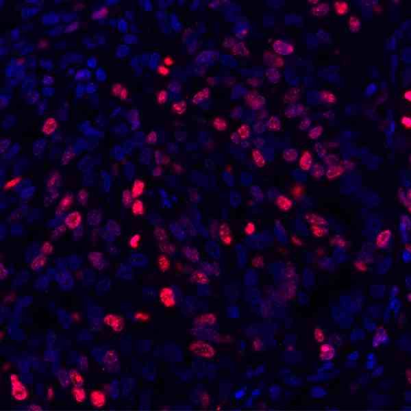
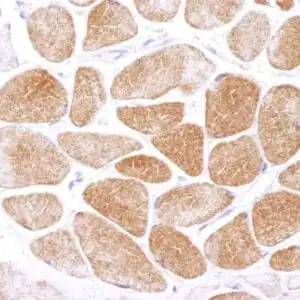
| Detection of human Ki-67 (red) by immunohistochemistry. Sample: FFPE section of human ovarian carcinoma. Antibody: Rabbit anti-Ki-67 recombinant monoclonal antibody used at 1:500. Secondary: HRP-conjugated goat anti-rabbit IgG . Substrate: Opal™. Counterstain: DAPI (blue) |
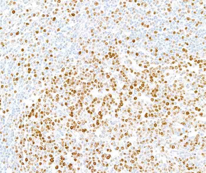 |
| Detection of human Ki-67 by immunohistochemistry. Sample: FFPE section of human tonsil. Antibody: Rabbit anti-Ki-67 recombinant monoclonal antibody. Secondary: HRP-conjugated goat anti-rabbit IgG. |
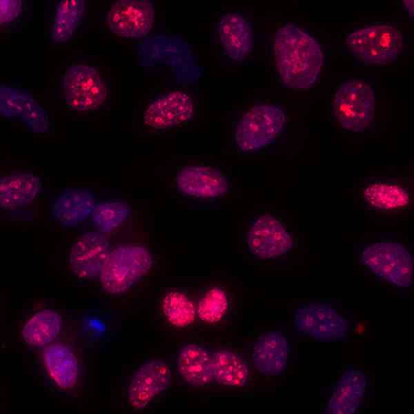 |
| Detection of human Ki-67 by immunocytochemistry. Sample: acetone/methanol fixed Hela cells. Antibody: Rabbit anti-Ki-67 recombinant monoclonal antibody used at 1:100. Secondary: DyLight® 594-conjugated goat anti-rabbit IgG. Counterstain: DAPI |
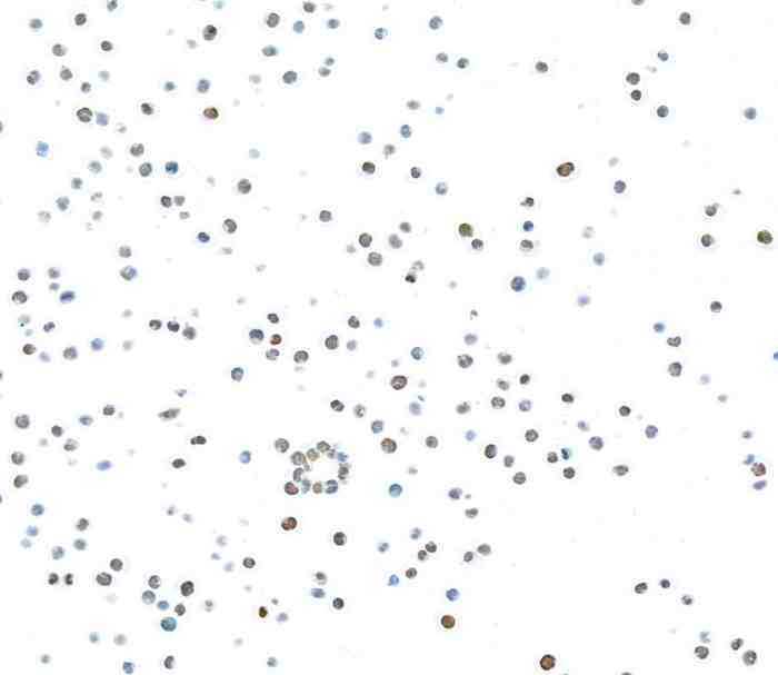 |
| Detection of human Ki-67 by immunocytochemistry. Sample: FFPE section of MOLT-4 cells. Antibody: Rabbit anti-Ki-67 recombinant monoclonal antibody. Secondary: HRP-conjugated goat anti-rabbit IgG. |
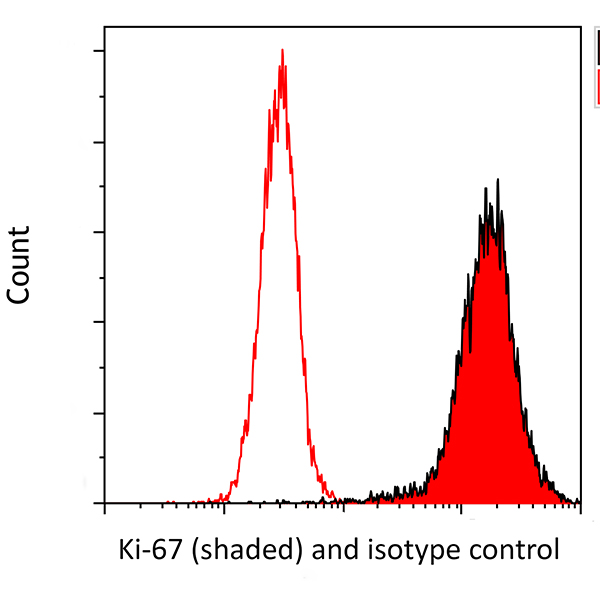 |
| Detection of human Ki-67 (shaded) in MOLT4 cells by flow cytometry. Antibody: Rabbit anti-Ki-67 recombinant monoclonal or isotype control (unshaded). Secondary: DyLight® 650-conjugated goat anti-rabbit IgG. |
FAQ & Publications
Frequently Asked Questions
What species does the rabbit anti-Ki67 polyclonal antibody 9056 react with, and in which applications has it been validated?
This antibody reacts with mouse and rat species. It has been validated for use in Western blotting (WB), immunocytochemistry/immunofluorescence (ICC/IF), immunohistochemistry (IHC), and flow cytometry (Flow). Recommended dilutions are 1:1000-5000 for WB and 1:2000-5000 for IF.
How should the rabbit anti-Ki67 polyclonal antibody 9056 be stored to maintain its stability and activity?
For short-term storage, keep the antibody at 2-8°C. For long-term preservation, store it at -20°C and avoid repeated freeze-thaw cycles. The antibody is supplied in a storage buffer containing PBS, 50% glycerol, and 0.04% sodium azide.
Publications
| pmid | title | authors | citation |
|---|---|---|---|
| We haven't added any publications to our database yet. | |||
Published literature highly relevant to the biological target of this product and referencing this antibody or clone are retrieved from the PubMed database provided by the United States National Library of Medicine at the National Institutes of Health.
Protocols
| relevant to this product |
|---|
| Western blot ICC |
Documents
| Batch Number | QC File | SDS |
|---|---|---|
| To view batch-specific Safety Datasheets and Quality Certificates associated with your account, please Log In. | ||
Only logged in customers who have purchased this product may leave a review.


Reviews
There are no reviews yet.