| Weight | 1 lbs |
|---|---|
| Dimensions | 9 × 5 × 2 in |
| host | mouse |
| isotype | IgG1 |
| clonality | monoclonal |
| concentration | 1 mg/mL |
| applications | ICC/IF, IHC, WB |
| available sizes | 100 µg |


mouse anti-Alpha-synuclein monoclonal antibody (211) 9067
$409.00
Antibody summary
- Mouse monoclonal to Alpha-synuclein
- Suitable for: WB, ICC/IF, IHC
- Reacts with: human, mouse, rat
- Isotype: IgG1
- 100 µg
mouse anti-Alpha-synuclein monoclonal antibody (211) 9067
| target relevance |
|---|
| Homo sapiens SNCA Alpha-synuclein |
| Protein names Alpha-synuclein |
| Alternative names Non-A beta component of AD amyloid, Non-A4 component of amyloid precursor |
| Gene names SNCA |
| Protein family Belongs to the synuclein family |
| Function Neuronal protein that plays several roles in synaptic activity such as regulation of synaptic vesicle trafficking and subsequent neurotransmitter release (PubMed:20798282, PubMed:26442590, PubMed:28288128, PubMed:30404828). Participates as a monomer in synaptic vesicle exocytosis by enhancing vesicle priming, fusion and dilation of exocytotic fusion pores (PubMed:28288128, PubMed:30404828). Mechanistically, acts by increasing local Ca(2+) release from microdomains which is essential for the enhancement of ATP-induced exocytosis (PubMed:30404828). Also acts as a molecular chaperone in its multimeric membrane-bound state, assisting in the folding of synaptic fusion components called SNAREs (Soluble NSF Attachment Protein REceptors) at presynaptic plasma membrane in conjunction with cysteine string protein-alpha/DNAJC5 (PubMed:20798282). This chaperone activity is important to sustain normal SNARE-complex assembly during aging (PubMed:20798282). Also plays a role in the regulation of the dopamine neurotransmission by associating with the dopamine transporter (DAT1) and thereby modulating its activity (PubMed:26442590) |
| Subcellular location Cytoplasm, Membrane, Nucleus, Synapse, Secreted, Cell projection, axon |
| Structure Soluble monomer. Homotetramer (PubMed:21841800). A dynamic intracellular population of tetramers and monomers coexists normally and the tetramer plays an essential role in maintaining homeostasis (PubMed:21841800). Interacts with UCHL1 (By similarity). Interacts with phospholipase D and histones. Interacts (via N-terminus) with synphilin-1/SNCAIP; this interaction promotes formation of SNCA inclusions in the cytoplasm (PubMed:19762560). Interacts with CALM1 (PubMed:23607618). Interacts with STXBP1; this interaction controls SNCA self-replicating aggregation (PubMed:27597756). Interacts with SNARE components VAMP2 and SNAP25; these interactions allows SNARE complex assembly and integrity (PubMed:20798282). Interacts with RPH3A and RAB3A (PubMed:15207266). Interacts with SERF1A; this interaction promotes the aggregation of SNCA (PubMed:22854022, PubMed:31034892). Interacts with SEPTIN4 (By similarity). Interacts with DDX10; this interaction causes DDX10 mislocalization to the nucleoplasm and cytoplasmic inclusions (PubMed:33657088) |
| Post-translational modification Phosphorylated, predominantly on serine residues. Phosphorylation by CK1 appears to occur on residues distinct from the residue phosphorylated by other kinases. Phosphorylation of Ser-129 is selective and extensive in synucleinopathy lesions. In vitro, phosphorylation at Ser-129 promoted insoluble fibril formation. Phosphorylated on Tyr-125 by a PTK2B-dependent pathway upon osmotic stress Hallmark lesions of neurodegenerative synucleinopathies contain alpha-synuclein that is modified by nitration of tyrosine residues and possibly by dityrosine cross-linking to generated stable oligomers Ubiquitinated. The predominant conjugate is the diubiquitinated form Acetylation at Met-1 seems to be important for proper folding and native oligomeric structure |
| Involvement in disease Parkinson disease 1, autosomal dominant A complex neurodegenerative disorder characterized by bradykinesia, resting tremor, muscular rigidity and postural instability. Additional features are characteristic postural abnormalities, dysautonomia, dystonic cramps, and dementia. The pathology of Parkinson disease involves the loss of dopaminergic neurons in the substantia nigra and the presence of Lewy bodies (intraneuronal accumulations of aggregated proteins), in surviving neurons in various areas of the brain. The disease is progressive and usually manifests after the age of 50 years, although early-onset cases (before 50 years) are known. The majority of the cases are sporadic suggesting a multifactorial etiology based on environmental and genetic factors. However, some patients present with a positive family history for the disease. Familial forms of the disease usually begin at earlier ages and are associated with atypical clinical features. Parkinson disease 4, autosomal dominant A complex neurodegenerative disorder with manifestations ranging from typical Parkinson disease to dementia with Lewy bodies. Clinical features include parkinsonian symptoms (resting tremor, rigidity, postural instability and bradykinesia), dementia, diffuse Lewy body pathology, autonomic dysfunction, hallucinations and paranoia. Dementia, Lewy body A neurodegenerative disorder characterized by mental impairment leading to dementia, parkinsonism, fluctuating cognitive function, visual hallucinations, falls, syncopal episodes, and sensitivity to neuroleptic medication. Brainstem or cortical intraneuronal accumulations of aggregated proteins (Lewy bodies) are the only essential pathologic features. Patients may also have hippocampal and neocortical senile plaques, sometimes in sufficient number to fulfill the diagnostic criteria for Alzheimer disease. |
| Keywords 3D-structure, Acetylation, Alternative splicing, Alzheimer disease, Amyloid, Cell projection, Copper, Cytoplasm, Direct protein sequencing, Disease variant, Membrane, Metal-binding, Neurodegeneration, Nucleus, Parkinson disease, Parkinsonism, Phosphoprotein, Proteomics identification, Reference proteome, Repeat, Secreted, Synapse, Ubl conjugation |
| Sequence MDVFMKGLSKAKEGVVAAAEKTKQGVAEAAGKTKEGVLYVGSKTKEGVVHGVATVAEKTK EQVTNVGGAVVTGVTAVAQKTVEGAGSIAAATGFVKKDQLGKNEEGAPQEGILEDMPVDP DNEAYEMPSEEGYQDYEPEA |
| UniProt accession: P37840 |
Data
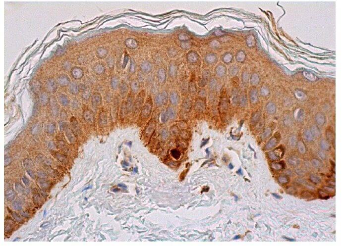 |
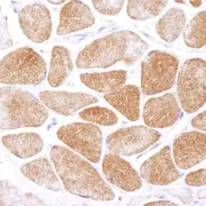
| Immunoperoxidase staining of formalin fixed, paraffin-embedded human skin tissue showing cytoplasmic staining of keratinocytes, fibroblasts and melanocytes. |
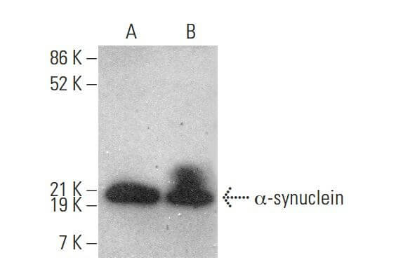 |
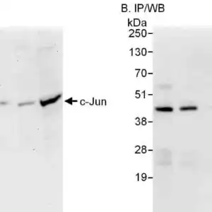
| Western blot analysis of α-synuclein expression in human brain (A) and human fetal brain (B) tissue extracts. |
 |
| Immunoperoxidase staining of formalin fixed, paraffin-embedded human bone marrow tissue showing cytoplasmic and nuclear staining of subset of hematopoietic cells. |
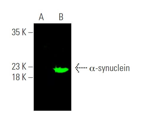 |
| Near-infrared western blot analysis of α-synuclein expression in non-transfected: sc-117752 (A) and human α-synuclein transfected: sc-111729 (B) whole cell lysates. |
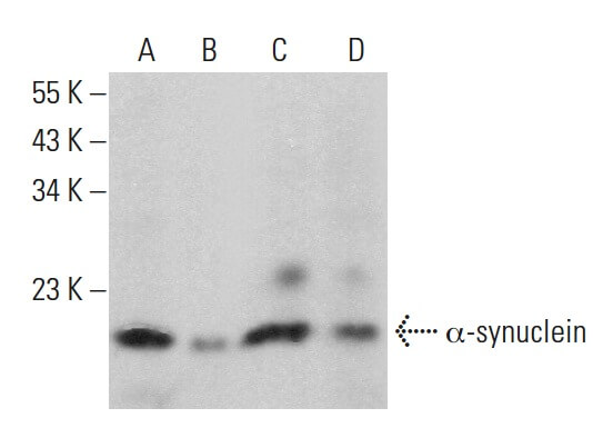 |
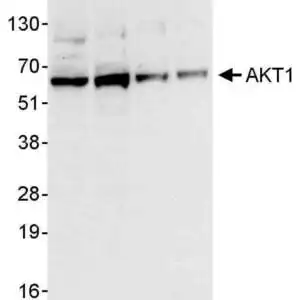
| Western blot analysis of α-synuclein expression in HEL 92.1.7 (A) and TF-1 (B) whole cell lysates and human brain (C) and human cerebellum (D) tissue extracts. |
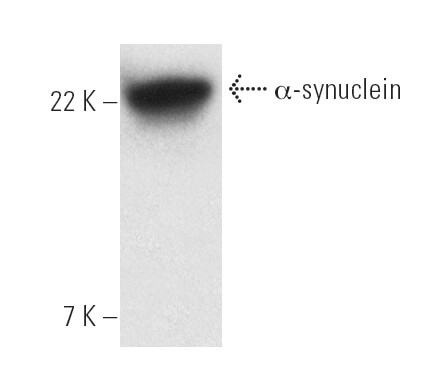 |
| Western blot analysis of α-synuclein expression in SH-SY5Y whole cell lysate |
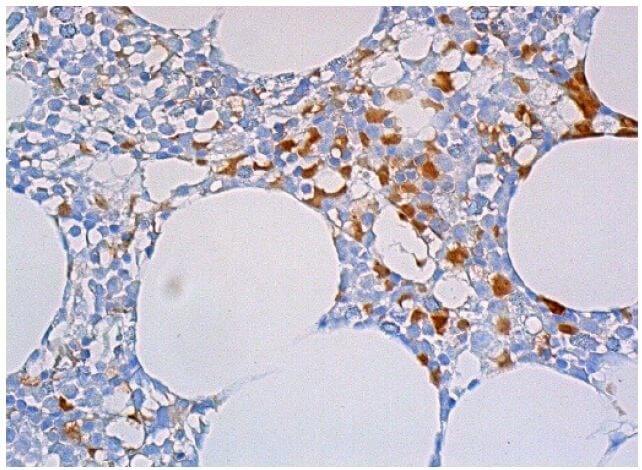 |
| Direct immunoperoxidase staining of formalin fixed, paraffin-embedded, human bone marrow tissue showing cytoplasmic staining of subset of hematopoietic cells. |
 |
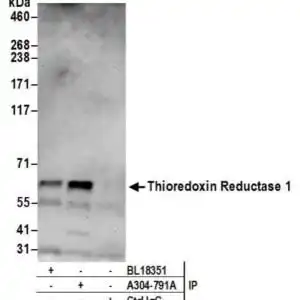
| Direct western blot analysis of α-synuclein expression in non-transfected(A) and human α-synuclein transfected(B) 293T whole cell lysates. |
 |
| Western blot analysis of α-synuclein expression in human cerebral cortex tissue extract |
FAQ & Publications
Frequently Asked Questions
What species does the mouse anti-Alpha-synuclein monoclonal antibody (211) 9067 react with?
This monoclonal antibody reacts with human, mouse, and rat Alpha-synuclein proteins.
Which applications has this antibody been validated for, and what are the recommended dilutions?
The antibody has been tested and is suitable for Western blotting (WB), immunocytochemistry/immunofluorescence (ICC/IF), and immunohistochemistry (IHC). Recommended dilutions are 1:1000 for WB and 1:1000 for IF/ICC.
How should the mouse anti-Alpha-synuclein monoclonal antibody (211) 9067 be stored to maintain stability?
For short-term storage, keep the antibody at 2-8°C. For long-term storage, it should be kept at -20°C. Avoid repeated freeze-thaw cycles to preserve antibody integrity.
What is the source and clonality of this Alpha-synuclein antibody, and what is its concentration and form?
The antibody is a mouse monoclonal of the IgG1 isotype, affinity purified, and provided as a liquid at 1 mg/mL concentration in a 100 µg size.
Publications
| pmid | title | authors | citation |
|---|---|---|---|
| We haven't added any publications to our database yet. | |||
Published literature highly relevant to the biological target of this product and referencing this antibody or clone are retrieved from the PubMed database provided by the United States National Library of Medicine at the National Institutes of Health.
Protocols
| relevant to this product |
|---|
| Western blot IHC ICC |
Documents
| Batch Number | QC File | SDS |
|---|---|---|
| To view batch-specific Safety Datasheets and Quality Certificates associated with your account, please Log In. | ||
Only logged in customers who have purchased this product may leave a review.


Reviews
There are no reviews yet.