| Weight | 1 lbs |
|---|---|
| Dimensions | 9 × 5 × 2 in |
| host | rabbit |
| isotype | IgG |
| clonality | monoclonal |
| concentration | 1 mg/mL |
| applications | ICC/IF, IHC, WB |
| available sizes | 100 µg |


rabbit anti-Vimentin monoclonal antibody (BLR100G) 9046
$409.00
Antibody summary
- Rabbit monoclonal to Vimentin
- Suitable for: WB, ICC/IF, IHC
- Reacts with: human, mouse, rat
- Isotype: IgG
- 100 µg
rabbit anti-Vimentin monoclonal antibody (BLR100G) 9046
| target relevance |
|---|
| Homo sapiens VIM Vimentin |
| Protein names Vimentin |
| Gene names VIM |
| Protein family Belongs to the intermediate filament family |
| Function Vimentins are class-III intermediate filaments found in various non-epithelial cells, especially mesenchymal cells. Vimentin is attached to the nucleus, endoplasmic reticulum, and mitochondria, either laterally or terminally. Plays a role in cell directional movement, orientation, cell sheet organization and Golgi complex polarization at the cell migration front (By similarity). Protects SCRIB from proteasomal degradation and facilitates its localization to intermediate filaments in a cell contact-mediated manner (By similarity). May promote axon outgrowth and motor fiber repair via DSP-mediated recruitment to outgrowth tips (By similarity) |
| Subcellular location Cytoplasm, Cytoplasm, cytoskeleton, Nucleus matrix, Cell membrane, Cell projection, axon |
| Structure (Microbial infection) Interacts with Chandipura virus glycoprotein; this interaction might facilitate the binding of the virus to the cells |
| Post-translational modification Filament disassembly during mitosis is promoted by phosphorylation at Ser-55 as well as by nestin (By similarity). One of the most prominent phosphoproteins in various cells of mesenchymal origin. Phosphorylation is enhanced during cell division, at which time vimentin filaments are significantly reorganized. Phosphorylation by PKN1 inhibits the formation of filaments. Phosphorylated at Ser-56 by CDK5 during neutrophil secretion in the cytoplasm (PubMed:21465480). Phosphorylated by STK33 (PubMed:18811945). Phosphorylated on tyrosine residues by SRMS (PubMed:29496907) O-glycosylated during cytokinesis at sites identical or close to phosphorylation sites, this interferes with the phosphorylation status S-nitrosylation is induced by interferon-gamma and oxidatively-modified low-densitity lipoprotein (LDL(ox)) possibly implicating the iNOS-S100A8/9 transnitrosylase complex |
| Involvement in disease Cataract 30, multiple types An opacification of the crystalline lens of the eye that frequently results in visual impairment or blindness. Opacities vary in morphology, are often confined to a portion of the lens, and may be static or progressive. In general, the more posteriorly located and dense an opacity, the greater the impact on visual function. |
| Keywords 3D-structure, Acetylation, Cataract, Cell membrane, Cell projection, Coiled coil, Cytoplasm, Cytoskeleton, Direct protein sequencing, Disease variant, Glycoprotein, Host-virus interaction, Intermediate filament, Isopeptide bond, Membrane, Nucleus, Phosphoprotein, Proteomics identification, Reference proteome, S-nitrosylation, Ubl conjugation |
| Sequence MSTRSVSSSSYRRMFGGPGTASRPSSSRSYVTTSTRTYSLGSALRPSTSRSLYASSPGGV YATRSSAVRLRSSVPGVRLLQDSVDFSLADAINTEFKNTRTNEKVELQELNDRFANYIDK VRFLEQQNKILLAELEQLKGQGKSRLGDLYEEEMRELRRQVDQLTNDKARVEVERDNLAE DIMRLREKLQEEMLQREEAENTLQSFRQDVDNASLARLDLERKVESLQEEIAFLKKLHEE EIQELQAQIQEQHVQIDVDVSKPDLTAALRDVRQQYESVAAKNLQEAEEWYKSKFADLSE AANRNNDALRQAKQESTEYRRQVQSLTCEVDALKGTNESLERQMREMEENFAVEAANYQD TIGRLQDEIQNMKEEMARHLREYQDLLNVKMALDIEIATYRKLLEGEESRISLPLPNFSS LNLRETNLDSLPLVDTHSKRTLLIKTVETRDGQVINETSQHHDDLE |
| UniProt accession: P08670 |
Data
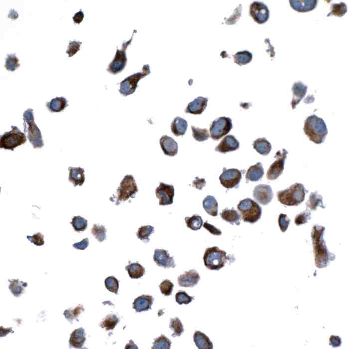 |
| Detection of human Vimentin in FFPE HeLa by ICC. Antibody: Rabbit anti-Vimentin recombinant monoclonal antibody. Secondary: HRP-conjugated goat anti-rabbit IgG. Substrate: DAB. |
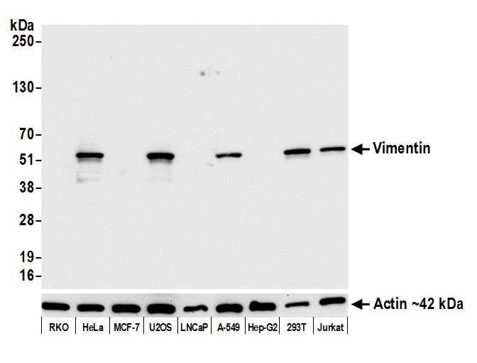 |
| Detection of human Vimentin by western blot. Samples: Whole cell lysate (5 µg) from RKO, HeLa, MCF-7, U2OS, LNCaP, A-549, Hep-G2, HEK293T, and Jurkat cells prepared using NETN lysis buffer. Antibody: Rabbit anti-Vimentin recombinant monoclonal antibody used at 1:1000. Secondary: HRP-conjugated goat anti-rabbit IgG. Detection: Chemiluminescence with an exposure time of 3 seconds. Lower Panel: Rabbit anti-Actin recombinant monoclonal antibody. |
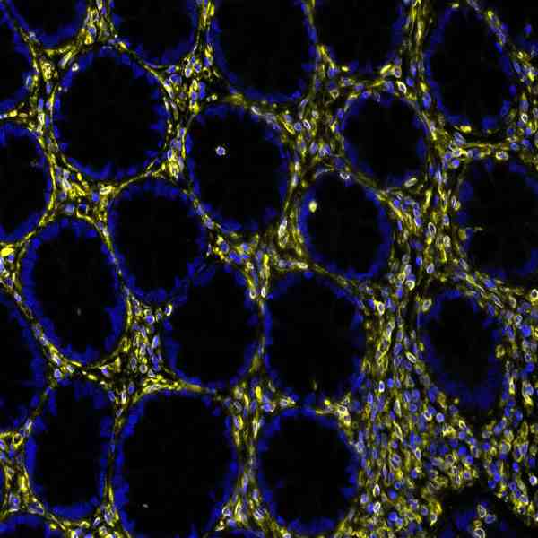 |
| Detection of human Vimentin in FFPE colon carcinoma by immunohistochemistry-IF. Antibody: Rabbit anti-Vimentin recombinant monoclonal antibody. Secondary: HRP-conjugated goat anti-rabbit IgG. Substrate: Opal™. Counterstain: DAPI. |
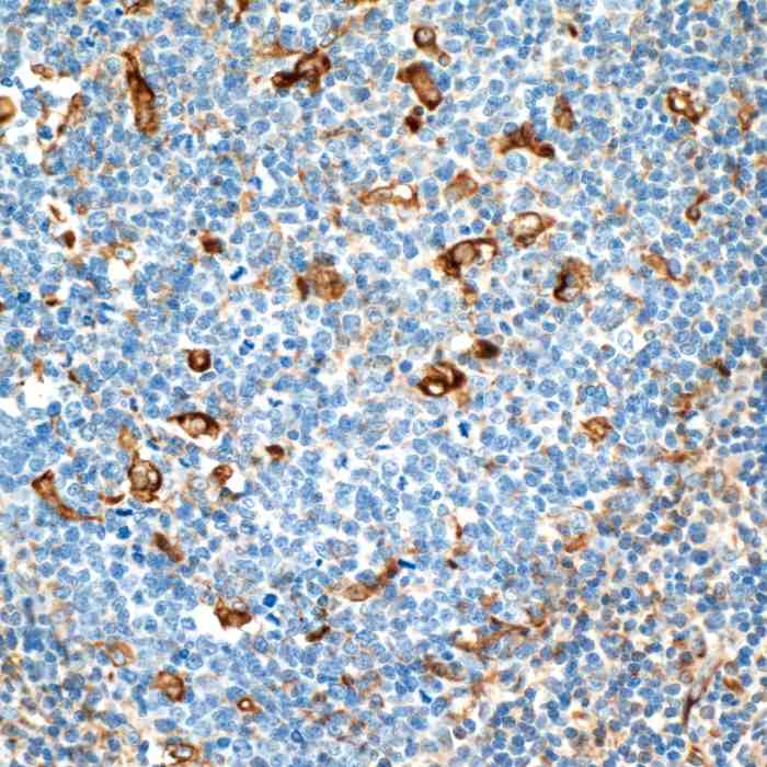 |
| Detection of human Vimentin in FFPE tonsil by IHC.Antibody: Rabbit anti-Vimentin recombinant monoclonal antibody. Secondary: HRP-conjugated goat anti-rabbit IgG. Substrate: DAB |
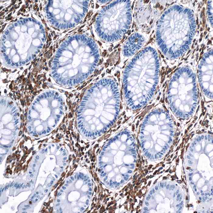 |
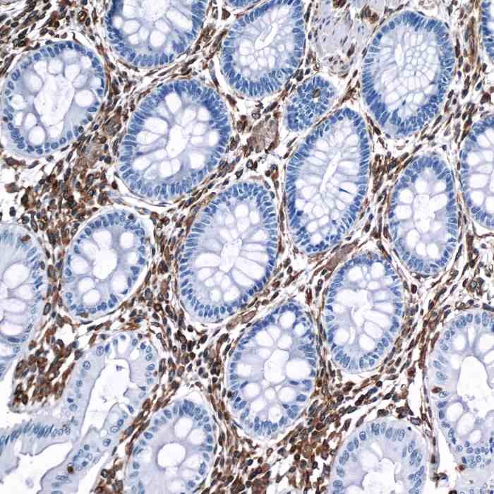
| Detection of human Vimentin in FFPE colon by IHC.Antibody: Rabbit anti-Vimentin recombinant monoclonal antibody. Secondary: HRP-conjugated goat anti-rabbit IgG. Substrate: DAB |
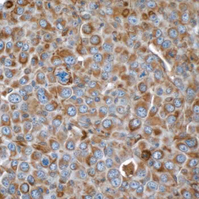 |
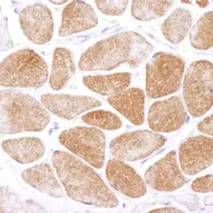
| Detection of mouse Vimentin in FFPE renal cell carcinoma by IHC.Antibody: Rabbit anti-Vimentin recombinant monoclonal antibody. Secondary: HRP-conjugated goat anti-rabbit IgG. Substrate: DAB. |
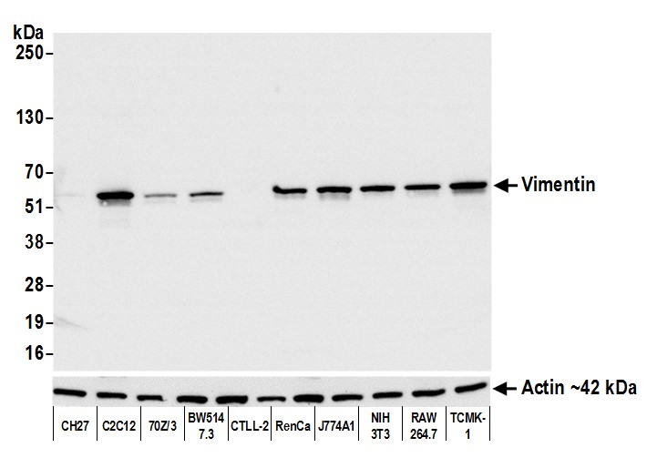 |
| Detection of mouse Vimentin by western blot. Samples: Whole cell lysate (5 µg) from CH27, C2C12, 70Z/3, BW5147.3, CTLL-2, RenCa, J774A1, NIH 3T3, RAW 264.7, and TCMK-1 cells prepared using NETN lysis buffer. Antibody: Rabbit anti-Vimentin recombinant monoclonal antibody used at 1:1000. Secondary: HRP-conjugated goat anti-rabbit IgG. Detection: Chemiluminescence with an exposure time of 1 second. Lower Panel: Rabbit anti-Actin recombinant monoclonal antibody. |
FAQ & Publications
Frequently Asked Questions
What species does the rabbit anti-Vimentin monoclonal antibody (BLR100G) 9046 react with?
This antibody reacts with human, mouse, and rat Vimentin proteins.
Which applications is this anti-Vimentin monoclonal antibody validated for?
The antibody is suitable for Western blot (WB), immunocytochemistry/immunofluorescence (ICC/IF), and immunohistochemistry (IHC) applications.
How should the rabbit anti-Vimentin monoclonal antibody be stored to maintain its stability?
For short-term storage, keep the antibody at 2-8°C. For long-term storage, it should be stored at -20°C while avoiding freeze/thaw cycles.
What is the recommended dilution for using this antibody in Western blot and immunofluorescence assays?
For Western blot, a dilution of 1:10,000 is recommended. For immunofluorescence (IF), immunocytochemistry (ICC), and immunohistochemistry (IHC), the recommended dilution is 1:5,000.
Publications
| pmid | title | authors | citation |
|---|---|---|---|
| We haven't added any publications to our database yet. | |||
Published literature highly relevant to the biological target of this product and referencing this antibody or clone are retrieved from the PubMed database provided by the United States National Library of Medicine at the National Institutes of Health.
Protocols
| relevant to this product |
|---|
| Western blot IHC ICC |
Documents
| Batch Number | QC File | SDS |
|---|---|---|
| To view batch-specific Safety Datasheets and Quality Certificates associated with your account, please Log In. | ||
Only logged in customers who have purchased this product may leave a review.


Reviews
There are no reviews yet.