Showing all 4 results
GFP antibodies
The GFP tag (Green Fluorescent Protein tag) is a powerful tool in molecular biology, derived from the jellyfish Aequorea victoria, where the GFP protein naturally emits green fluorescence when exposed to blue or ultraviolet light. When fused to a protein of interest, GFP acts as a reporter, allowing researchers to visually track protein expression, localization, and dynamics in real-time within living cells or tissues without the need for additional staining or detection steps.
GFP (Green Fluorescent Protein) fluoresces due to the unique structure of its chromophore, a light-absorbing molecule formed from specific amino acids within the protein. The fluorescence process occurs in several steps:
Chromophore Formation: The GFP chromophore is created through an autocatalytic reaction involving three key amino acids�serine (Ser65), tyrosine (Tyr66), and glycine (Gly67)�which are part of the protein's backbone. This reaction leads to the formation of a cyclic structure within the central part of the protein. This chromophore is capable of absorbing light and re-emitting it at a different wavelength, which is what causes fluorescence.
Absorption of Light: When GFP is exposed to blue or ultraviolet light (with an excitation wavelength around 395-475 nm), the electrons in the chromophore become excited, moving to a higher energy state.
Emission of Light: As the excited electrons return to their ground state, energy is released in the form of green light (with an emission wavelength of around 509 nm), which is what we see as fluorescence.
In Western blotting (WB), GFP-tagged proteins can still be detected using specific anti-GFP antibodies for confirmation or quantification of protein expression levels. The GFP antibodies available on your website are useful for Western blotting, immunofluorescence (IF), and immunohistochemistry (IHC), providing versatility in both real-time observation and post-experiment validation of GFP-tagged proteins.
| target | type | reactivity | applications | ||||
|---|---|---|---|---|---|---|---|
| 1777 | GFP | chicken polyclonal | other | WB ICC IHC | |||
| 1778 | GFP | rabbit polyclonal | other | WB ICC IHC | |||
| 7414 | GFP | mouse monoclonal (GF28R) | other | WB ICC ELISA | |||
| 9005 | GFP | rabbit monoclonal | other | WB ICC | |||

WB with antibody [1777] : Immunofluorescence cell staining- chicken anti-GFP polyclonal antibody 1777
Immunofluorescent analysis of 4% paraformaldehyde (PFA) - fixed, permeabilized with 0.1% triton X-100 CHO cells transfected with GFP constructs (CHO-GFP) anti-GFP polyclonal antibody 1777 at 1/100 dilution (10µg/mL), followed by Goat Anti-chicken IgY, Alexa 594 conjugate at 1/400 (5µg/mL) (red). The image also shows GFP (green), nucleus counterstained with DAPI (blue). Also shown lack of staining in parental cell line and from secondary only controls.

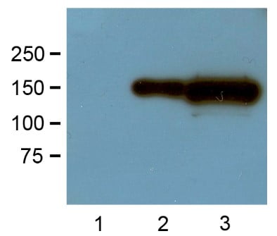
WB with antibody [7414] : 1:1,000 (1µg/mL) Ab dilution probed against HEK293 cells transfected with GFP-tagged protein vector: untransfected control (1), 1µg (2) and 10µg (3) of cell lysates used.

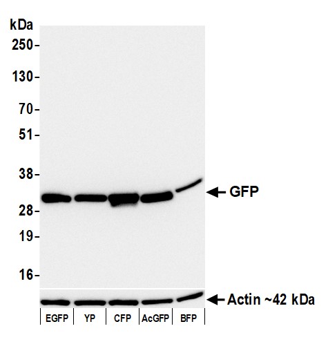
WB with antibody [9005] : Whole cell lysate (5 �g) from 293T cells transfected with EGFP, YP, CFP, AcGFP, or BFP. Antibody: Rabbit anti-GFP recombinant monoclonal antibody 9005 used at 1:1000. Secondary: HRP-conjugated goat anti-rabbit IgG. Detection: Chemiluminescence with an exposure time of 3 seconds. Lower Panel: Rabbit anti-Actin recombinant monoclonal antibody.


