| Weight | 1 lbs |
|---|---|
| Dimensions | 9 × 5 × 2 in |
| host | mouse |
| isotype | IgG1 |
| clonality | monoclonal |
| concentration | 1 mg/mL |
| applications | ICC/IF, IHC, WB |
| available sizes | 100 µg |


mouse anti-GFAP monoclonal antibody (2E1) 9024
$409.00
Antibody summary
- Mouse monoclonal to GFAP
- Suitable for: WB, ICC/IF, IHC
- Reacts with: human, mouse, rat
- Isotype: IgG2b
- 100 µg
mouse anti-GFAP monoclonal antibody (2E1) 9024
| target relevance |
|---|
| Homo sapiens GFAP Glial fibrillary acidic protein |
| Protein names Glial fibrillary acidic protein |
| Gene names GFAP |
| Protein family Belongs to the intermediate filament family |
| Function GFAP, a class-III intermediate filament, is a cell-specific marker that, during the development of the central nervous system, distinguishes astrocytes from other glial cells |
| Subcellular location Cytoplasm |
| Structure Interacts with PSEN1 (via N-terminus) |
| Post-translational modification Phosphorylated by PKN1 |
| Involvement in disease Alexander disease A rare disorder of the central nervous system. The most common form affects infants and young children, and is characterized by progressive failure of central myelination, usually leading to death within the first decade. Infants with Alexander disease develop a leukodystrophy with macrocephaly, seizures, and psychomotor retardation. Patients with juvenile or adult forms typically experience ataxia, bulbar signs and spasticity, and a more slowly progressive course. Histologically, Alexander disease is characterized by Rosenthal fibers, homogeneous eosinophilic inclusions in astrocytes. |
| Keywords 3D-structure, Alternative splicing, Citrullination, Coiled coil, Cytoplasm, Direct protein sequencing, Disease variant, Intermediate filament, Leukodystrophy, Methylation, Phosphoprotein, Proteomics identification, Reference proteome |
| Sequence MERRRITSAARRSYVSSGEMMVGGLAPGRRLGPGTRLSLARMPPPLPTRVDFSLAGALNA GFKETRASERAEMMELNDRFASYIEKVRFLEQQNKALAAELNQLRAKEPTKLADVYQAEL RELRLRLDQLTANSARLEVERDNLAQDLATVRQKLQDETNLRLEAENNLAAYRQEADEAT LARLDLERKIESLEEEIRFLRKIHEEEVRELQEQLARQQVHVELDVAKPDLTAALKEIRT QYEAMASSNMHEAEEWYRSKFADLTDAAARNAELLRQAKHEANDYRRQLQSLTCDLESLR GTNESLERQMREQEERHVREAASYQEALARLEEEGQSLKDEMARHLQEYQDLLNVKLALD IEIATYRKLLEGEENRITIPVQTFSNLQIRETSLDTKSVSEGHLKRNIVVKTVEMRDGEV IKESKQEHKDVM |
| UniProt accession: P14136 |
Data
 |
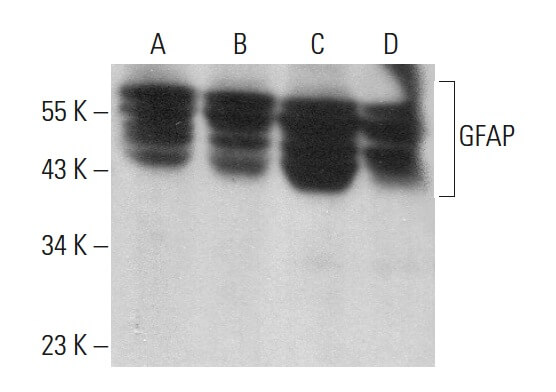
| Western blot analysis of GFAP expression in C6 (A), U-87 MG (B), SK-N-SH (C) and U-251-MG (D) whole cell lysates. Detection reagent used: m-IgGκ BP-HRP. |
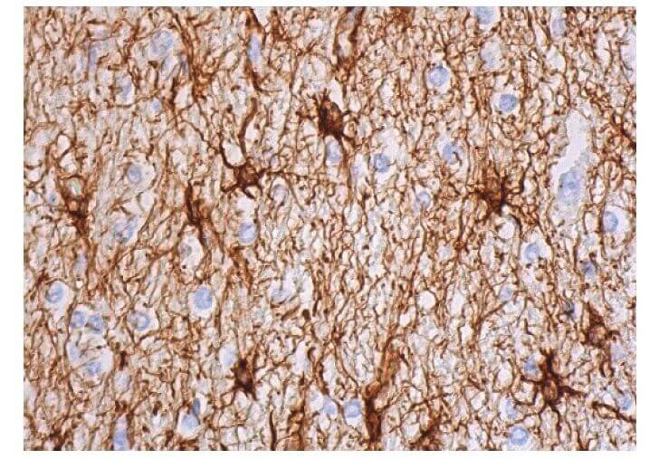 |
| Immunoperoxidase staining of formalin fixed, paraffin-embedded human cerebral cortex tissue showing cytoplasmic staining of astrocytes and neuropil staining. |
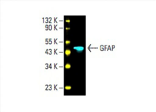 |
| Direct fluorescent western blot analysis of GFAP expression in U-251-MG whole cell lysate. |
 |
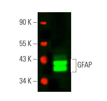
| Western blot analysis of GFAP expression in U-87 MG (A), U-251-MG (B), C2C12 (C) and Neuro-2A (D) whole cell lysates. |
 |
| Direct near-infrared western blot analysis of GFAP expression in human brain tissue extract. |
 |
| Immunoperoxidase staining of formalin fixed, paraffin-embedded human brain showing staining of astrocytes. |
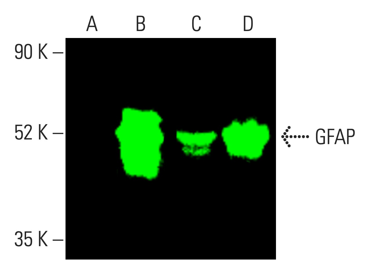 |
| Near-Infrared western blot analysis of GFAP expression in non-transfected 293T (A), human GFAP transfected 293T (B), C6 (C) and U-251-MG (D) whole cell lysates. |
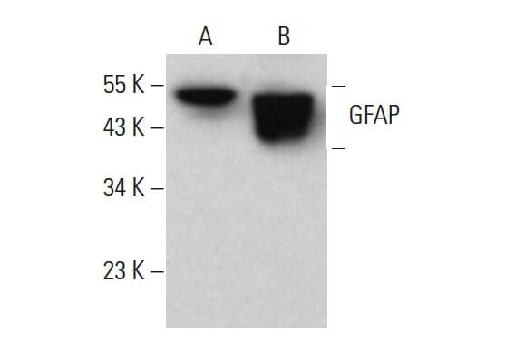 |
| Western blot analysis of GFAP expression in C6 whole cell lysate (A) and mouse brain tissue extract (B). |
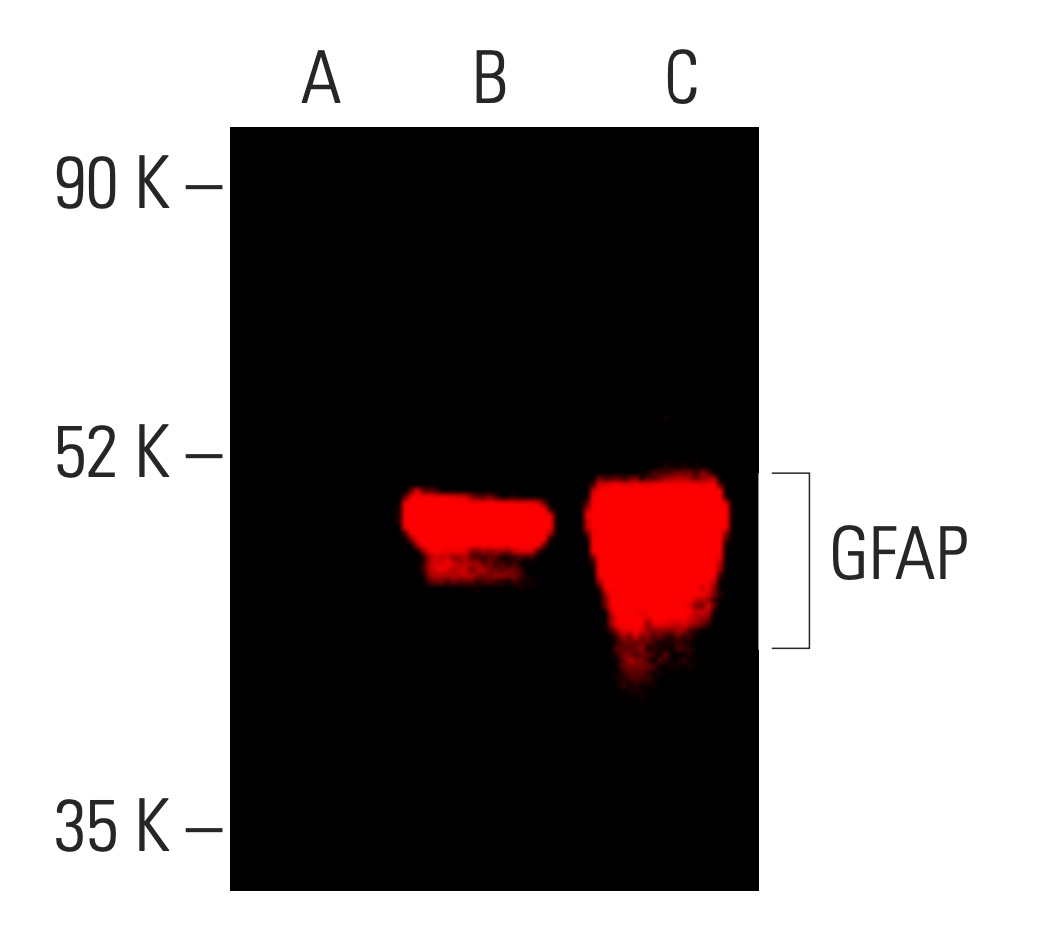 |
| Near-Infrared western blot analysis of GFAP expression in non-transfected (A) human GFAP transfected 293T (B) and human GFAP transfected 293T (C) whole cell lysates. |
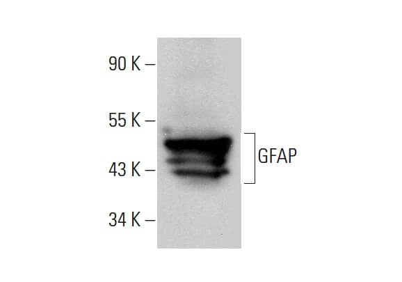 |
| Western blot analysis of GFAP expression in rat brain tissue extract. |
FAQ & Publications
Frequently Asked Questions
What species does the mouse anti-GFAP monoclonal antibody (2E1) 9024 react with?
This antibody reacts with human, mouse, and rat GFAP proteins.
Which applications has the mouse anti-GFAP monoclonal antibody (2E1) been validated for?
It has been tested and is suitable for Western blot (WB), immunocytochemistry/immunofluorescence (ICC/IF), and immunohistochemistry (IHC) applications.
How should the mouse anti-GFAP monoclonal antibody be stored to maintain stability?
For short-term storage, keep the antibody at 2-8°C. For long-term storage, it should be stored at -20°C, avoiding freeze/thaw cycles to preserve antibody integrity.
What is the immunogen used to generate the mouse anti-GFAP monoclonal antibody (2E1) 9024?
The immunogen is recombinant full-length human GFAP isotype 1 expressed in and purified from Escherichia coli (E. coli).
Publications
| pmid | title | authors | citation |
|---|---|---|---|
| 37759938 | Neuroimmune Support of Neuronal Regeneration and Neuroplasticity following Cerebral Ischemia in Juvenile Mice. | Ricaurte A Marquez-Ortiz, Vesna Tesic, Daniel R Hernandez, Bilkis Akhter, Nibedita Aich, Porter M Boudreaux, Garrett A Clemons, Celeste Yin-Chieh Wu, Hung Wen Lin, Krista M Rodgers | Brain Sci 13:N/A |
| 37667307 | Metal-organic framework materials promote neural differentiation of dental pulp stem cells in spinal cord injury. | Heng Zhou, Shuili Jing, Wei Xiong, Yangzhi Zhu, Xingxiang Duan, Ruohan Li, Youjian Peng, Tushar Kumeria, Yan He, Qingsong Ye | J Nanobiotechnology 21:316 |
| 37605262 | Inhibition of acid sphingomyelinase reduces reactive astrocyte secretion of mitotoxic extracellular vesicles and improves Alzheimer's disease pathology in the 5xFAD mouse. | Simone M Crivelli, Zainuddin Quadri, Hemendra J Vekaria, Zhihui Zhu, Priyanka Tripathi, Ahmed Elsherbini, Liping Zhang, Patrick G Sullivan, Erhard Bieberich | Acta Neuropathol Commun 11:135 |
| 37587766 | USP25 Inhibits Neuroinflammatory Responses After Cerebral Ischemic Stroke by Deubiquitinating TAB2. | Zhongding Li, Baohua Liu, Kate Lykke Lambertsen, Bettina Hjelm Clausen, Zhenhu Zhu, Xue Du, Yanqi Xu, Frantz Rom Poulsen, Bo Halle, Christian Bonde, Meng Chen, Xue Wang, Dirk Schlüter, Jingyong Huang, Ari Waisman, Weihong Song, Xu Wang | Adv Sci (Weinh) 10:e2301641 |
| 37513944 | EYE-503: A Novel Retinoic Acid Drug for Treating Retinal Neurodegeneration. | Sha Liu, Yuke Ji, Huan Li, Ling Ren, Junya Zhu, Tianjing Yang, Xiumiao Li, Jin Yao, Xin Cao, Biao Yan | Pharmaceuticals (Basel) 16:N/A |
Published literature highly relevant to the biological target of this product and referencing this antibody or clone are retrieved from the PubMed database provided by the United States National Library of Medicine at the National Institutes of Health.
Protocols
| relevant to this product |
|---|
| Western blot IHC ICC |
Documents
| Batch Number | QC File | SDS |
|---|---|---|
| To view batch-specific Safety Datasheets and Quality Certificates associated with your account, please Log In. | ||
Only logged in customers who have purchased this product may leave a review.

Reviews
There are no reviews yet.