| Weight | 1 lbs |
|---|---|
| Dimensions | 9 × 5 × 2 in |
| host | rabbit |
| isotype | IgG |
| clonality | polyclonal |
| concentration | 1 mg/mL |
| applications | ELISA |
| reactivity | tagged fusion proteins |
| available sizes | 1 mg, 100 µg, 25 µg |
rabbit anti-myc tag polyclonal antibody 8059
Price range: $100.00 through $2,600.00
Antibody summary
- Rabbit polyclonal to myc tag
- Suitable for: ELISA
- Reacts with: tagged fusion proteins
- Isotype: IgG
- 100 µg, 25 µg, 1 mg
rabbit anti-myc tag polyclonal antibody 8059
| antibody |
|---|
| Database link: Sequence -Glu-Gln-Lys-Leu-Ile-Ser-Glu-Glu-Asp-Leu Derived from c-myc 425-434 P01106 |
| Tested applications ELISA |
| Recommended dilutions user optimized |
| Application Notes This antibody reacts with EQKLISEEDL as determined by IEP and ELISA techniques. It is suitable for blotting, ELISA and IHC applications. Optimal working dilutions should be determined experimentally by the investigator. |
| Immunogen Highly purified EQKLISEEDL conjugated with KLH |
| Size and concentration 25, 100, 1000µg and 1 mg/mL |
| Form liquid |
| Storage Instructions 2-8°C |
| Storage buffer PBS, pH 7.2, 0.09% NaN3 |
| Purity affinity purified |
| Clonality polyclonal |
| Isotype IgG |
| Compatible secondaries goat anti-rabbit IgG, H&L chain specific, peroxidase conjugated, conjugated polyclonal antibody 9512 goat anti-rabbit IgG, H&L chain specific, biotin conjugated polyclonal antibody 2079 goat anti-rabbit IgG, H&L chain specific, FITC conjugated polyclonal antibody 7863 goat anti-rabbit IgG, H&L chain specific, Cross Absorbed polyclonal antibody 2371 goat anti-rabbit IgG, H&L chain specific, biotin conjugated polyclonal antibody, crossabsorbed 1715 goat anti-rabbit IgG, H&L chain specific, FITC conjugated polyclonal antibody, crossabsorbed 1720 |
| Isotype control Rabbit polyclonal - Isotype Control |
| target relevance |
|---|
| Myc tag is a small protein tag that minimally impacts protein structure. This antibody can be used to confirm expression and quantify Myc tag recombinant proteins in Western blotting and for their purification/copurification. When imaging in situ, Myc tag proteins can be identified by this antibody when used in conjunction with a suitable secondary antibody. Click for more on: epitope tags and myc tag |
| Protein names Myc proto-oncogene protein (Class E basic helix-loop-helix protein 39) (bHLHe39) (Proto-oncogene c-Myc) (Transcription factor p64) |
| Gene names MYC,MYC BHLHE39 |
| Mass 50565Da |
| Function FUNCTION: Transcription factor that binds DNA in a non-specific manner, yet also specifically recognizes the core sequence 5'-CAC[GA]TG-3' (PubMed:24940000, PubMed:25956029). Activates the transcription of growth-related genes (PubMed:24940000, PubMed:25956029). Binds to the VEGFA promoter, promoting VEGFA production and subsequent sprouting angiogenesis (PubMed:24940000, PubMed:25956029). Regulator of somatic reprogramming, controls self-renewal of embryonic stem cells (By similarity). Functions with TAF6L to activate target gene expression through RNA polymerase II pause release (By similarity). Positively regulates transcription of HNRNPA1, HNRNPA2 and PTBP1 which in turn regulate splicing of pyruvate kinase PKM by binding repressively to sequences flanking PKM exon 9, inhibiting exon 9 inclusion and resulting in exon 10 inclusion and production of the PKM M2 isoform (PubMed:20010808). {ECO:0000250|UniProtKB:P01108, ECO:0000269|PubMed:20010808, ECO:0000269|PubMed:24940000, ECO:0000269|PubMed:25956029}. |
| Subellular location SUBCELLULAR LOCATION: Nucleus, nucleoplasm {ECO:0000269|PubMed:17558397}. Nucleus, nucleolus {ECO:0000269|PubMed:17558397, ECO:0000269|PubMed:25775507, ECO:0000269|PubMed:38225354}. Nucleus {ECO:0000269|PubMed:22719065, ECO:0000269|PubMed:38225354}. Cytoplasm {ECO:0000269|PubMed:22719065}. Note=Localization to the nucleolus is dependent on HEATR1. {ECO:0000269|PubMed:38225354}. |
| Structure SUBUNIT: Efficient DNA binding requires dimerization with another bHLH protein. Binds DNA as a heterodimer with MAX (PubMed:9680483). Interacts with TAF1C and SPAG9. Interacts with PARP10. Interacts with KDM5A and KDM5B. Interacts (when phosphorylated at Thr-73 and Ser-77) with FBXW7 (PubMed:17558397, PubMed:25775507). Interacts with PIM2. Interacts with RIOX1. The heterodimer MYC:MAX interacts with ABI1; the interaction may enhance MYC:MAX transcriptional activity. Interacts with TRIM6 (By similarity). Interacts with NPM1; the binary complex is recruited to the promoter of MYC target genes and enhances their transcription (PubMed:25956029). Interacts with CIP2A; leading to the stabilization of MYC (PubMed:17632056). Interacts with NUP205 (PubMed:22719065). Interacts with HEATR1; the interaction is required for localization of MYC to the nucleolus (PubMed:38225354). {ECO:0000250|UniProtKB:P01108, ECO:0000269|PubMed:15103331, ECO:0000269|PubMed:15674325, ECO:0000269|PubMed:15723054, ECO:0000269|PubMed:17308053, ECO:0000269|PubMed:17311883, ECO:0000269|PubMed:17558397, ECO:0000269|PubMed:17632056, ECO:0000269|PubMed:17873522, ECO:0000269|PubMed:22719065, ECO:0000269|PubMed:25775507, ECO:0000269|PubMed:25956029, ECO:0000269|PubMed:38225354, ECO:0000269|PubMed:9680483}. |
| Post-translational modification PTM: Phosphorylated by PRKDC (PubMed:1597196). Phosphorylation at Ser-344 by PIM2 leads to the stabilization of MYC (By similarity). Phosphorylation at Ser-77 by CDK2 prevents Ras-induced senescence (PubMed:19966300, PubMed:20713526). Phosphorylated at Ser-77 by DYRK2; this primes the protein for subsequent phosphorylation by GSK3B at Thr-73 (PubMed:22307329). Phosphorylation at Thr-73 and Ser-77 by GSK3 is required for ubiquitination and degradation by the proteasome (PubMed:15103331, PubMed:17558397, PubMed:8386367). Dephosphorylation at Ser-77 by protein phosphatase 2A (PPP2CA) promotes its degradation; interaction with PPP2CA is enhanced by AMBRA1 (PubMed:25438055, PubMed:25803737). {ECO:0000250|UniProtKB:P01108, ECO:0000269|PubMed:15103331, ECO:0000269|PubMed:1597196, ECO:0000269|PubMed:17558397, ECO:0000269|PubMed:19966300, ECO:0000269|PubMed:20713526, ECO:0000269|PubMed:22307329, ECO:0000269|PubMed:25438055, ECO:0000269|PubMed:25803737, ECO:0000269|PubMed:8386367}.; PTM: Ubiquitinated by the SCF(FBXW7) complex when phosphorylated at Thr-73 and Ser-77, leading to its degradation by the proteasome (PubMed:15103331, PubMed:17558397, PubMed:25775507). In the nucleoplasm, ubiquitination is counteracted by USP28, which interacts with isoform 1 of FBXW7 (FBW7alpha), leading to its deubiquitination and preventing degradation (PubMed:17558397, PubMed:17873522). In the nucleolus, however, ubiquitination is not counteracted by USP28 but by USP36, due to the lack of interaction between isoform 3 of FBXW7 (FBW7gamma) and USP28, explaining the selective MYC degradation in the nucleolus (PubMed:17558397, PubMed:25775507). Also polyubiquitinated by the DCX(TRPC4AP) complex (PubMed:20551172, PubMed:29779948). Ubiquitinated by UBR5 when not forming a heterodimer with another bHLH protein, leading to its degradation: UBR5 recognizes and binds a degron that is only available upon heterodimer dissociation (PubMed:33208877, PubMed:37478862). Ubiquitinated by TRIM6 in a phosphorylation-independent manner (By similarity). {ECO:0000250|UniProtKB:P01108, ECO:0000269|PubMed:15103331, ECO:0000269|PubMed:17558397, ECO:0000269|PubMed:17873522, ECO:0000269|PubMed:20551172, ECO:0000269|PubMed:25775507, ECO:0000269|PubMed:29779948, ECO:0000269|PubMed:33208877, ECO:0000269|PubMed:37478862}. |
| Domain DOMAIN: The 9aaTAD motif is a transactivation domain present in a large number of yeast and animal transcription factors. {ECO:0000269|PubMed:34342803}. |
| Biotechnology BIOTECHNOLOGY: POU5F1/OCT4, SOX2, MYC/c-Myc and KLF4 are the four Yamanaka factors. When combined, these factors are sufficient to reprogram differentiated cells to an embryonic-like state designated iPS (induced pluripotent stem) cells. iPS cells exhibit the morphology and growth properties of ES cells and express ES cell marker genes. {ECO:0000269|PubMed:18035408}. |
| Involvement in disease DISEASE: Note=A chromosomal aberration involving MYC may be a cause of a form of B-cell chronic lymphocytic leukemia. Translocation t(8;12)(q24;q22) with BTG1. {ECO:0000269|PubMed:2069907}.; DISEASE: Burkitt lymphoma (BL) [MIM:113970]: A form of undifferentiated malignant lymphoma commonly manifested as a large osteolytic lesion in the jaw or as an abdominal mass. {ECO:0000269|PubMed:2166998, ECO:0000269|PubMed:8220424}. Note=The gene represented in this entry is involved in disease pathogenesis. Chromosomal aberrations involving MYC are usually found in Burkitt lymphoma. Translocations t(8;14), t(8;22) or t(2;8) which juxtapose MYC to one of the heavy or light chain immunoglobulin gene loci. |
| Target Relevance information above includes information from UniProt accession: P01106 |
| The UniProt Consortium |
Data
 |
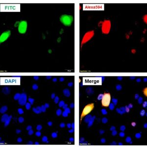
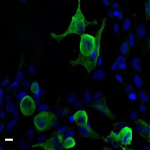
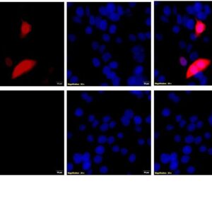
| Immunofluorescence cell staining rabbit anti-myc tag polyclonal antibody 8059 Immunofluorescent analysis of 4% paraformaldehyde (PFA)-fixed, permeabilized with 0.1% triton X-100 in CHO cells transfected with construct labeling with MYC antibody 8059 at 1:1000 dilution (1µg), followed by donkey-anti rabbit IgG (H&L), FITC conjugated antibody at 1:1000 dilution (1µg/mL) (green). Nucleus was counterstained with DAPI (blue). |
|
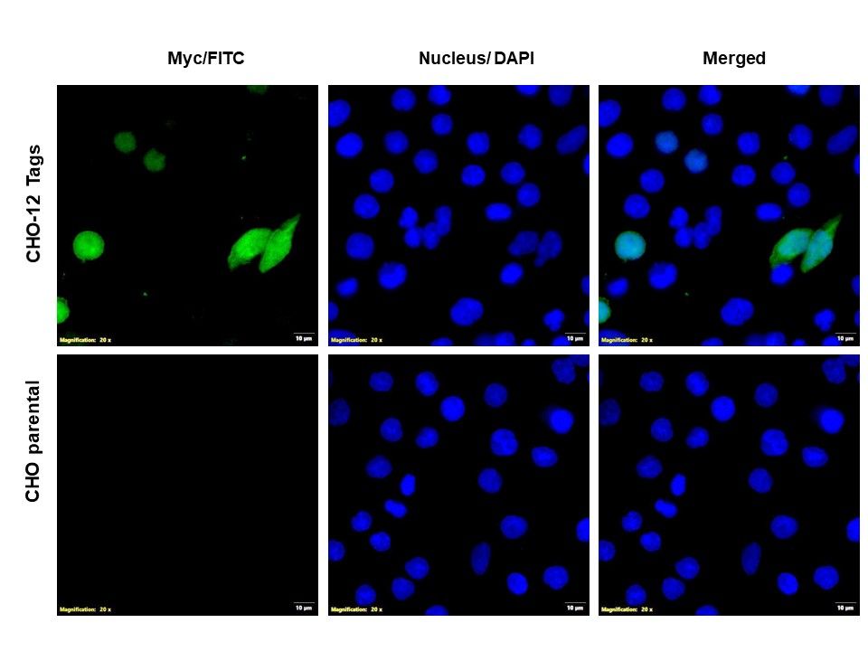
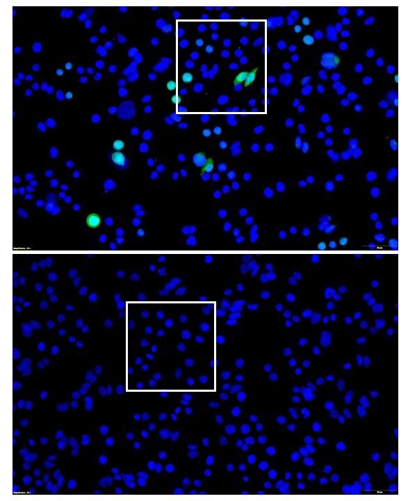
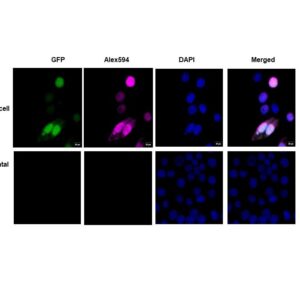
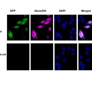
| Immunofluorescence cell staining rabbit anti-myc tag polyclonal antibody 8059 Immunofluorescent analysis of 4% paraformaldehyde (PFA)-fixed, permeabilized with 0.1% triton X-100 in CHO cells transfected with 12-tags constructs (named as CHO-12 tags, top) and parental CHO cells (bottom) labeling with MYC antibody 8059 at 1:1000 dilution (1µg), followed by donkey-anti rabbit IgG (H&L), FITC conjugated antibody at 1:1000 dilution (1µg/mL) (green). Nucleus was counterstained with DAPI (blue). |
Publications
| pmid | title | authors | citation |
|---|---|---|---|
| 37717261 | Structural features of thyroglobulin linked to protein trafficking. | Cintia E Citterio, Kookjoo Kim, Bhavana Rajesh, Kevin Pena, Oliver Biggs Clarke, Peter Arvan | Protein Sci 32:e4784 |
| 36581998 | RNF31 represses cell progression and immune evasion via YAP/PD-L1 suppression in triple negative breast Cancer. | Huijie Yang, Min Xue, Peng Su, Yan Zhou, Xin Li, Zhongbo Li, Yan Xia, Chenmiao Zhang, Mingxi Fu, Xiuxia Zheng, Guosheng Luo, Tian Wei, Xinxing Wang, Yinlu Ding, Jian Zhu, Ting Zhuang | J Exp Clin Cancer Res 41:364 |
| 36497344 | circRNF10 Regulates Tumorigenic Properties and Natural Killer Cell-Mediated Cytotoxicity against Breast Cancer through the miR-934/PTEN/PI3k-Akt Axis. | Fei Liu, Yang Sang, Yang Zheng, Lina Gu, Lingjiao Meng, Ziyi Li, Yuyang Dong, Zishuan Wei, Cuizhi Geng, Meixiang Sang | Cancers (Basel) 14:N/A |
| 36375840 | SMA-linked SMN mutants prevent phase separation properties and SMN interactions with FMRP family members. | Olivier Binda, Franceline Juillard, Julia Novion Ducassou, Constance Kleijwegt, Geneviève Paris, Andréanne Didillon, Faouzi Baklouti, Armelle Corpet, Yohann Couté, Jocelyn Côté, Patrick Lomonte | Life Sci Alliance 6:N/A |
| 36313802 | CasPlay provides a gRNA-barcoded CRISPR-based display platform for antibody repertoire profiling. | Karl W Barber, Ellen Shrock, Stephen J Elledge | Cell Rep Methods 2:100318 |
Protocols
| relevant to this product |
|---|
| ELISA |
Documents
| # | SDS | Certificate | |
|---|---|---|---|
| Please enter your product and batch number here to retrieve product datasheet, SDS, and QC information. | |||
39 reviews for rabbit anti-myc tag polyclonal antibody 8059
Only logged in customers who have purchased this product may leave a review.


verified user –
verified user –
verified user –
verified user –
verified user –
verified user –
verified user –
verified user –
verified user –
verified user –
verified user –
verified user –
verified user –
verified user –
verified user –
verified user –
verified user –
verified user –
verified user –
verified user –
verified user –
verified user –
verified user –
verified user –
verified user –
verified user –
verified user –
verified user –
verified user –
verified user –
verified user –
verified user –
verified user –
verified user –
verified user –
verified user –
verified user –
verified user –
verified user –