| Weight | 1 lbs |
|---|---|
| Dimensions | 9 × 5 × 2 in |
| accession | P48061-2 |
| express system | E.coli |
| product tag | biotin at C-terminal |
| purity | > 97% by SDS PAGE |
| molecular weight | Predicted Molecular Mass: 10.381kDa Extinction Coefficient: 14,180 M-1 cm-1 Actual Molecular Mass: 10.381kDa by ESI Mass Spec |
| available size | 100 µg, 20 µg, 5 µg, 50 µg |
| endotoxin | <0.01 EU per 1μg of the protein by the LAL method |


Biotinylated Stromal-Cell Derived Factor-1a (SDF-1a/CXCL12) 8026
Price range: $134.00 through $2,862.00
Summary
- Expression: E.coli
- Amino Acid Range: 22-89
Biotinylated Stromal-Cell Derived Factor-1a (SDF-1a/CXCL12) 8026
| antibody |
|---|
| Database link: human P48061-2 |
| Size and concentration 2, 10, 50, 100µg and lyophilized |
| Form Lyophilized |
| Storage Instructions Avoid repeated freeze-thaw cycles: • 12 months from date of receipt, -20 to -70 °C as supplied. • 1 month, 2 to 8 °C under sterile conditions after reconstitution. • 3 months, -20 to -70 °C under sterile conditions after reconstitution |
| Storage buffer Reconstitution: Spin sample prior to reconstitution. Recommended concentration of 100µg/mL in sterile water. Shipping: Room Temp |
| Purity > 97% by SDS PAGE and HPLC |
| target relevance |
|---|
| Homo sapiens CXCL12 Stromal cell-derived factor 1 |
| Protein names Stromal cell-derived factor 1 |
| Alternative names C-X-C motif chemokine 12, Intercrine reduced in hepatomas, Pre-B cell growth-stimulating factor |
| Gene names CXCL12 |
| Protein family Belongs to the intercrine alpha (chemokine CxC) family |
| Function Chemoattractant active on T-lymphocytes and monocytes but not neutrophils (PubMed:18802065, PubMed:39093700). Activates the C-X-C chemokine receptor CXCR4 to induce a rapid and transient rise in the level of intracellular calcium ions and chemotaxis (PubMed:8752281, PubMed:18802065, PubMed:39093700). Also binds to atypical chemokine receptor ACKR3, which activates the beta-arrestin pathway and acts as a scavenger receptor for CXCL12/SDF-1 (PubMed:16107333, PubMed:19255243). Binds to the allosteric site (site 2) of integrins and activates integrins ITGAV:ITGB3, ITGA4:ITGB1 and ITGA5:ITGB1 in a CXCR4-independent manner (PubMed:29301984). Acts as a positive regulator of monocyte migration and a negative regulator of monocyte adhesion via the LYN kinase (PubMed:18802065). Stimulates migration of monocytes and T-lymphocytes through its receptors, CXCR4 and ACKR3, and decreases monocyte adherence to surfaces coated with ICAM-1, a ligand for beta-2 integrins (PubMed:16107333, PubMed:18802065, PubMed:19255243, PubMed:39093700). CXCR4 signaling axis inhibits beta-2 integrin LFA-1 mediated adhesion of monocytes to ICAM-1 through LYN kinase (PubMed:18802065). Inhibits CXCR4-mediated infection by T-cell line-adapted HIV-1 (PubMed:8752281). Plays a protective role after myocardial infarction. Induces down-regulation and internalization of ACKR3 expressed in various cells. Has several critical functions during embryonic development; required for B-cell lymphopoiesis, myelopoiesis in bone marrow and heart ventricular septum formation (By similarity). Stimulates the proliferation of bone marrow-derived B-cell progenitors in the presence of IL7 as well as growth of stromal cell-dependent pre-B-cells (By similarity) |
| Subcellular location Secreted |
| Structure (Microbial infection) Interacts with molluscum contagiosum virus protein MC148 |
| Post-translational modification Processed forms SDF-1-beta(3-72) and SDF-1-alpha(3-67) are produced after secretion by proteolytic cleavage of isoforms Beta and Alpha, respectively. The N-terminal processing is probably achieved by DPP4. Isoform Alpha is first cleaved at the C-terminus to yield a SDF-1-alpha(1-67) intermediate before being processed at the N-terminus. The C-terminal processing of isoform Alpha is reduced by binding to heparin and, probably, cell surface proteoglycans |
| Keywords 3D-structure, Alternative splicing, Chemotaxis, Cytokine, Disulfide bond, Growth factor, Host-virus interaction, Proteomics identification, Reference proteome, Secreted, Signal |
| Sequence MNAKVVVVLVLVLTALCLSDGKPVSLSYRCPCRFFESHVARANVKHLKILNTPNCALQIV ARLKNNNRQVCIDPKLKWIQEYLEKALNKRFKM |
| UniProt accession: P48061 |
Data
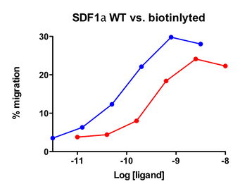 |
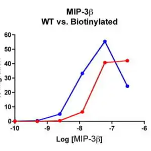
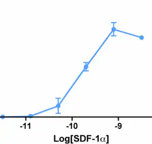
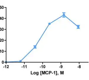
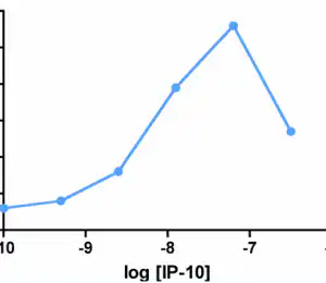
| Migration induced by SDF-1a Jurkat cells expressing endogenous CXCR4 were assayed for migration through the Transwell bare filter at various concentrations of SDF-1a. The response is expressed as the % of total input cells (Blue: wild type; Red: biotinylated). |
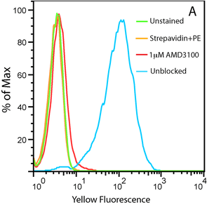 |
| Flow Cytometry Uptake of 20nM CXCL12-biotin by U937 cells in the presence (red trace) and absence (cyan) of a CXCR4 inhibitor, AMD 3100. U937 cells are not stained by streptavidin-PE in the absence of CXCL12-biotin (orange). |
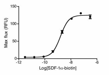 |
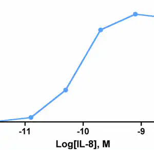
| Dose Dependent Ca2+ Flux HEK293 cells expressing CXCR4 were stimulated with SDF-1a-biotinylated. Calcium flux was measured with calcium dye from Molecular Devices. A dose response curve was obtained and an EC50 of 3.5nM was recorded |
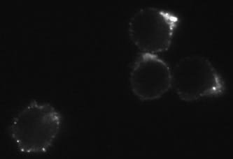 |
| CXCR4 Surface Detection Confocal microscopy images of surface stained U937 cells with SDF-1a-biotin conjugated to streptavidin-Cy3. |
 |
| CXCR4 Receptor Internalization Studies Distribution of the endogenous CXCR4 in Jurkat cells at 4°C (left image) and their internalization upon stimulation at 37°C (right image) were revealed by the biotinylated SDF-1a bound by streptavidin-Cy3 (red). |
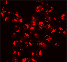 |
| CXCR4 Receptor Internalization Studies Distribution of the endogenous CXCR4 in Jurkat cells at 4°C (left image) and their internalization upon stimulation at 37°C (right image) were revealed by the biotinylated SDF-1a bound by streptavidin-Cy3 (red). |
FAQ & Publications
Frequently Asked Questions
What is the recommended storage condition and stability for the Biotinylated SDF-1a (CXCL12) protein after reconstitution?
After reconstitution, the Biotinylated SDF-1a (CXCL12) protein should be stored under sterile conditions at 2 to 8 °C for up to 1 month or at -20 to -70 °C for up to 3 months. It is important to avoid repeated freeze-thaw cycles to maintain protein integrity.
How does the biotinylation method used for this CXCL12 protein affect its biological activity in functional assays?
The protein is enzymatically biotinylated at a specific lysine residue with nearly 100% efficiency, preserving its biological activity. Functional assays such as calcium flux and migration demonstrate that the biotinylated CXCL12 retains comparable functionality to the unmodified chemokine, making it suitable for receptor identification, binding studies, and cellular assays.
Publications
| pmid | title | authors | citation |
|---|---|---|---|
| We haven't added any publications to our database yet. | |||
Published literature highly relevant to the biological target of this product and referencing this antibody or clone are retrieved from the PubMed database provided by the United States National Library of Medicine at the National Institutes of Health.
Protocols
| relevant to this product |
|---|
| Migration assay |
Documents
| # | ||
|---|---|---|
| Please enter your product and batch number here. | ||
Only logged in customers who have purchased this product may leave a review.
Reviews
There are no reviews yet.