| Weight | 1 lbs |
|---|---|
| Dimensions | 9 × 5 × 2 in |
| accession | P10147 |
| express system | E.coli |
| product tag | biotin at C-terminal |
| purity | > 97% by SDS PAGE |
| molecular weight | Predicted Molecular Mass: 10,237.4973 Da Extinction Coefficient: 18,020 M-1 cm-1 Actual Molecular Mass: 10,237.4973 Da by ESI Mass Spec |
| available size | 100 µg, 20 µg, 5 µg, 50 µg |
| endotoxin | <0.01 EU per 1μg of the protein by the LAL method |

Biotinylated Macrophage inflammatory proteins 1-a (MIP1a) 8017
Price range: $134.00 through $3,368.00
Summary
- Expression: E.coli
- Amino Acid Range: 24-92
Biotinylated Macrophage inflammatory proteins 1-a (MIP1a) 8017
| antibody |
|---|
| Database link: human P10147 |
| Size and concentration 2, 10, 50, 100µg and lyophilized |
| Form Lyophilized |
| Storage Instructions Avoid repeated freeze-thaw cycles: • 12 months from date of receipt, -20 to -70 °C as supplied. • 1 month, 2 to 8 °C under sterile conditions after reconstitution. • 3 months, -20 to -70 °C under sterile conditions after reconstitution |
| Storage buffer Reconstitution: Spin sample prior to reconstitution. Recommended concentration of 100µg/mL in sterile water. Shipping: Room Temp |
| Purity > 97% by SDS PAGE and HPLC |
| target relevance |
|---|
| Homo sapiens CCL3 C-C motif chemokine 3 |
| Protein names C-C motif chemokine 3 |
| Alternative names G0/G1 switch regulatory protein 19-1, Macrophage inflammatory protein 1-alpha, PAT 464.1, SIS-beta, Small-inducible cytokine A3, Tonsillar lymphocyte LD78 alpha protein |
| Gene names CCL3 |
| Protein family Belongs to the intercrine beta (chemokine CC) family |
| Function Monokine with inflammatory and chemokinetic properties. Binds to CCR1, CCR4 and CCR5. One of the major HIV-suppressive factors produced by CD8+ T-cells. Recombinant MIP-1-alpha induces a dose-dependent inhibition of different strains of HIV-1, HIV-2, and simian immunodeficiency virus (SIV) |
| Subcellular location Secreted |
| Structure Self-associates. Also heterodimer of MIP-1-alpha(4-69) and MIP-1-beta(3-69) (PubMed:12070155). Interacts with CCR1 (PubMed:15905581) |
| Post-translational modification N-terminal processed form LD78-alpha(4-69) is produced by proteolytic cleavage after secretion from HTLV1-transformed T-cells |
| Keywords 3D-structure, Chemotaxis, Cytokine, Direct protein sequencing, Disulfide bond, Inflammatory response, Proteomics identification, Reference proteome, Secreted, Signal |
| Sequence MQVSTAALAVLLCTMALCNQFSASLAADTPTACCFSYTSRQIPQNFIADYFETSSQCSKP GVIFLTKRSRQVCADPSEEWVQKYVSDLELSA |
| UniProt accession: P10147 |
Data
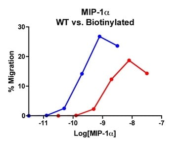 |
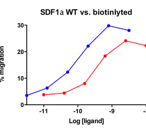
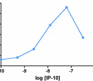
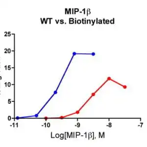
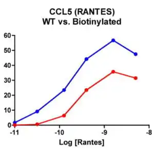
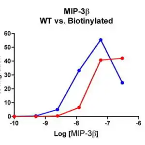
| Migration Assay: Cells expressing recombinant CCR5 were assayed for migration through a transwell filter at various concentrations of WT or Biotinylated MIP-1α. Responses are expressed as the % of total input cells (Blue: wild type; Red: biotinylated). |
FAQ & Publications
Frequently Asked Questions
What is the recommended storage condition for the Biotinylated Macrophage Inflammatory Protein 1-a (MIP1a) to maintain its stability?
The lyophilized product should be stored at -20 to -70 °C and is stable for 12 months from the date of receipt. After reconstitution, it can be stored at 2 to 8 °C for 1 month or at -20 to -70 °C for up to 3 months under sterile conditions. Avoid repeated freeze-thaw cycles to maintain protein integrity.
What expression system is used to produce this Biotinylated MIP1a protein, and what is the purity level?
This Biotinylated MIP1a protein is expressed in Escherichia coli (E.coli) cells. The purity of the protein is greater than 97%, confirmed by SDS-PAGE and HPLC analysis.
How is the biotin conjugation performed on MIP1a, and what are the advantages of this method?
The biotinylation of MIP1a is achieved enzymatically at a specific lysine residue located at the C-terminal, resulting in a biotinylated protein with functional properties comparable to the unmodified chemokine. This method has advantages over chemical biotinylation as it preserves biological activity and allows for precise labeling, facilitating receptor identification, chemokine binding studies, and cellular assays without the need for radioactive labeling.
What applications is the Biotinylated Macrophage Inflammatory Protein 1-a (MIP1a) suitable for in research?
Biotinylated MIP1a is useful in studies involving receptor identification and distribution, chemokine binding, and various cellular assays including migration assays. It can be combined with avidin analogues conjugated to fluorescent labels for visualization and quantification of chemokine-receptor interactions, making it a valuable tool in immunological and cell signaling research.
Publications
| pmid | title | authors | citation |
|---|---|---|---|
| We haven't added any publications to our database yet. | |||
Published literature highly relevant to the biological target of this product and referencing this antibody or clone are retrieved from the PubMed database provided by the United States National Library of Medicine at the National Institutes of Health.
Protocols
| relevant to this product |
|---|
| Migration assay |
Documents
| # | ||
|---|---|---|
| Please enter your product and batch number here. | ||
Only logged in customers who have purchased this product may leave a review.
Reviews
There are no reviews yet.