| Weight | 1 lbs |
|---|---|
| Dimensions | 9 × 5 × 2 in |
| host | chicken |
| isotype | IgY |
| clonality | polyclonal |
| concentration | 1 mg/mL |
| applications | ICC/IF, IHC, WB |
| available sizes | 1 mg, 100 µg, 25 µg |


chicken anti-GFP polyclonal antibody 1777
Price range: $100.00 through $2,600.00
Antibody summary
- Chicken polyclonal to GFP
- Suitable for: WB, ICC/IF, IHC
- Reacts with: tagged fusion proteins
- Isotype: IgY
- 100 µg, 25 µg, 1 mg
chicken anti-GFP polyclonal antibody 1777
| antibody |
|---|
| Database link: P42212 |
| Tested applications WB,ICC/IF,IHC |
| Recommended dilutions WB: 1:1000-3000 ICC/IF: 1:500-2000 For best results with other assays (e.g.: Dot, ELISA, IP, etc), please determine optimal working dilution by titration test. |
| Immunogen Recombinant GFP expressed in and purified from E. coli |
| Size and concentration 100µg and 1 mg/mL |
| Form liquid |
| Storage Instructions 2-8°C for short term, for longer term at -20°C. Avoid freeze / thaw cycles. |
| Storage buffer PBS, pH 7.2, 0.09% NaN3 |
| Purity affinity purified |
| Clonality polyclonal |
| Isotype IgY |
| Compatible secondaries goat anti-chicken IgY, H&L chain specific, peroxidase conjugated polyclonal antibody 1688 goat anti-chicken IgY, H&L chain specific, biotin conjugated polyclonal antibody 8036 goat anti-chicken IgY, H&L chain specific, FITC conjugated, Conjugated polyclonal antibody 4317 goat anti-chicken IgY, H&L chain specific, peroxidase conjugated polyclonal antibody, crossabsorbed 1708 goat anti-chicken IgY, H&L chain specific, biotin conjugated polyclonal antibody, crossabsorbed 1718 goat anti-chicken IgY, H&L chain specific, FITC conjugated polyclonal antibody, crossabsorbed 1723 |
| Isotype control Chicken polyclonal - Isotype Control |
| target relevance |
|---|
| Aequorea victoria GFP Green fluorescent protein |
| Protein names Green fluorescent protein |
| Gene names GFP |
| Protein family Belongs to the GFP family |
| Function Energy-transfer acceptor. Its role is to transduce the blue chemiluminescence of the protein aequorin into green fluorescent light by energy transfer. Fluoresces in vivo upon receiving energy from the Ca(2+)-activated photoprotein aequorin |
| Structure Monomer |
| Post-translational modification Contains a chromophore consisting of modified amino acid residues. The chromophore is formed by autocatalytic backbone condensation between Ser-65 and Gly-67, and oxidation of Tyr-66 to didehydrotyrosine. Maturation of the chromophore requires nothing other than molecular oxygen |
| Keywords 3D-structure, Chromophore, Direct protein sequencing, Luminescence, Photoprotein |
| Sequence MSKGEELFTGVVPILVELDGDVNGHKFSVSGEGEGDATYGKLTLKFICTTGKLPVPWPTL VTTFSYGVQCFSRYPDHMKQHDFFKSAMPEGYVQERTIFFKDDGNYKTRAEVKFEGDTLV NRIELKGIDFKEDGNILGHKLEYNYNSHNVYIMADKQKNGIKVNFKIRHNIEDGSVQLAD HYQQNTPIGDGPVLLPDNHYLSTQSALSKDPNEKRDHMVLLEFVTAAGITHGMDELYK |
| UniProt accession: P42212 |
Data
 |
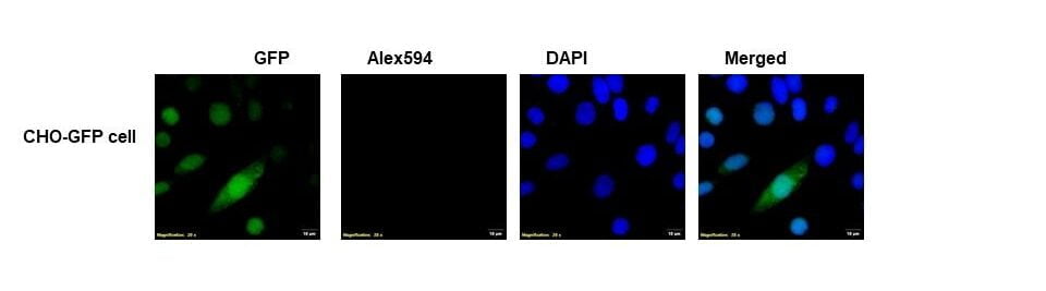
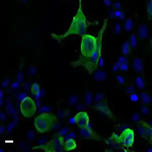
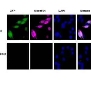
| Immunofluorescence cell staining- chicken anti-GFP polyclonal antibody 1777 Immunofluorescent analysis of 4% paraformaldehyde (PFA) - fixed, permeabilized with 0.1% triton X-100 CHO cells transfected with GFP constructs (CHO-GFP) anti-GFP polyclonal antibody 1777 at 1/100 dilution (10µg/mL), followed by Goat Anti-chicken IgY, Alexa 594 conjugate at 1/400 (5µg/mL) (red). The image also shows GFP (green), nucleus counterstained with DAPI (blue). Also shown lack of staining in parental cell line and from secondary only controls. |
 |
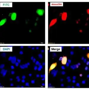
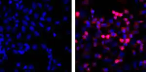
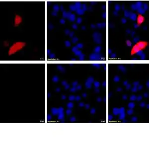
| Incubation with secondary antibody only. |
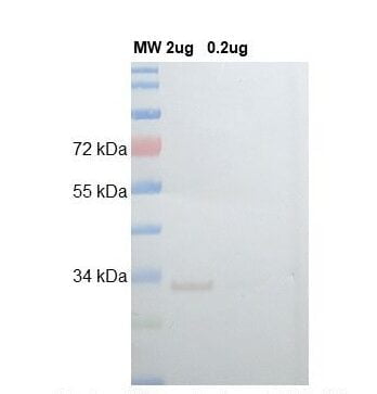 |
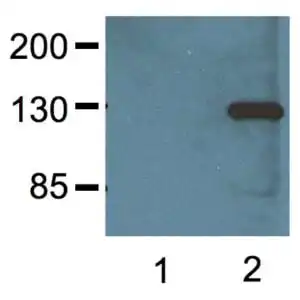
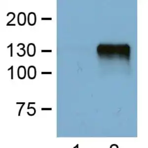
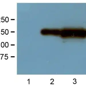
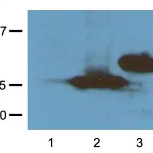
| Western blot analysis of chicken anti-GFP polyclonal antibody 1777 CHO cells were transfected with pcDNA3-GFP (as CHO-GFP). THE whole cell lysates were loaded at 2µg and 0.2µg per lane them incubated with chicken anti-GFP polyclonal antibody 1777 at 1:10,000 (01.%micro;g/mL) dilution for 1 hour and followed with anti-chicken IgY HRP conjugate at 1:10,000 (0.1µg/mL) at room temperature. |
FAQ & Publications
Frequently Asked Questions
What applications is the chicken anti-GFP polyclonal antibody 1777 validated for?
This antibody is validated for use in Western blotting (WB), immunocytochemistry/immunofluorescence (ICC/IF), and immunohistochemistry (IHC). Recommended dilutions are 1:1000-3000 for WB and 1:500-2000 for ICC/IF. For other applications such as Dot blot, ELISA, or immunoprecipitation, it is advised to optimize the working dilution by titration.
How should the chicken anti-GFP polyclonal antibody 1777 be stored to maintain stability?
For short-term storage, keep the antibody at 2-8°C. For long-term storage, store at -20°C and avoid repeated freeze/thaw cycles to preserve antibody integrity. The antibody is supplied in PBS buffer at pH 7.2 containing 0.09% sodium azide as a preservative.
Publications
| pmid | title | authors | citation |
|---|---|---|---|
| We haven't added any publications to our database yet. | |||
Published literature highly relevant to the biological target of this product and referencing this antibody or clone are retrieved from the PubMed database provided by the United States National Library of Medicine at the National Institutes of Health.
Protocols
| relevant to this product |
|---|
| Western blot IHC ICC |
Documents
| Batch Number | QC File | SDS |
|---|---|---|
| To view batch-specific Safety Datasheets and Quality Certificates associated with your account, please Log In. | ||
Only logged in customers who have purchased this product may leave a review.


Reviews
There are no reviews yet.