| Weight | 1 lbs |
|---|---|
| Dimensions | 9 × 5 × 2 in |
| host | rabbit |
| isotype | IgG |
| clonality | polyclonal |
| concentration | 1 mg/mL |
| applications | ICC/IF, IHC, WB |
| available sizes | 100 µg |


rabbit anti-Actin (pan) polyclonal antibody 9052
$409.00
Antibody summary
- Rabbit polyclonal to Actin (pan)
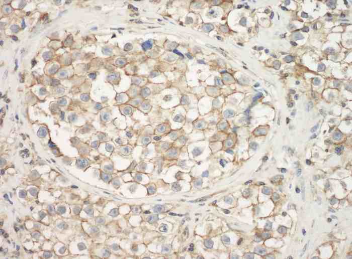
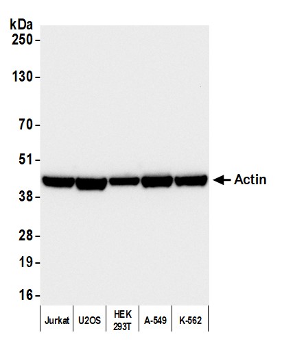
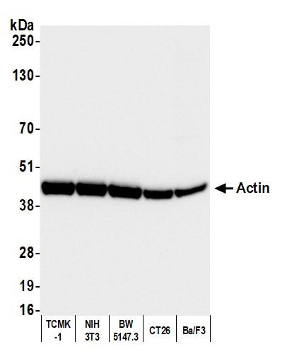
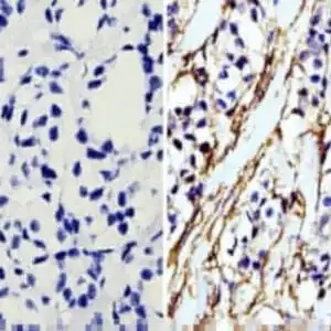
- Suitable for: WB, ICC/IF, IHC
- Reacts with: human, mouse, rat, cow, pig
- Isotype: IgG1
- 100 µg
rabbit anti-Actin (pan) polyclonal antibody 9052
| target relevance |
|---|
| Actin (pan) |
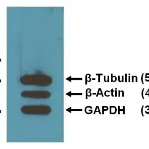
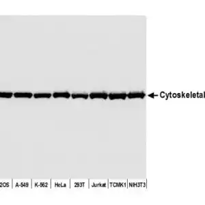
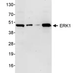
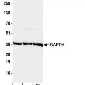
Data
 |
 |
 |
FAQ & Publications
Frequently Asked Questions
What species does the rabbit anti-Actin (pan) polyclonal antibody 9052 react with?
This antibody reacts with human, mouse, rat, cow, and pig actin isoforms.
Which applications is the rabbit anti-Actin (pan) polyclonal antibody 9052 validated for?
It is suitable for Western blotting (WB), immunocytochemistry/immunofluorescence (ICC/IF), and immunohistochemistry (IHC).
How should the rabbit anti-Actin (pan) polyclonal antibody 9052 be stored to maintain stability?
Store the antibody at 2-8°C for short term use, and for longer term storage, keep it at -20°C while avoiding freeze/thaw cycles.
What is the recommended dilution for using this antibody in Western blot and immunofluorescence?
For Western blot, a dilution of 1:1000 is recommended, and for immunofluorescence or immunohistochemistry, the recommended dilution ranges from 1:500 to 1:1000.
What is the immunogen used to generate the rabbit anti-Actin (pan) polyclonal antibody 9052?
The immunogen is an actin preparation derived from bovine brain.
Publications
| pmid | title | authors | citation |
|---|---|---|---|
| We haven't added any publications to our database yet. | |||
Published literature highly relevant to the biological target of this product and referencing this antibody or clone are retrieved from the PubMed database provided by the United States National Library of Medicine at the National Institutes of Health.
Protocols
| relevant to this product |
|---|
| Western blot IHC ICC |
Documents
| Batch Number | QC File | SDS |
|---|---|---|
| To view batch-specific Safety Datasheets and Quality Certificates associated with your account, please Log In. | ||
Only logged in customers who have purchased this product may leave a review.


Reviews
There are no reviews yet.