| Weight | 1 lbs |
|---|---|
| Dimensions | 9 × 5 × 2 in |
| host | mouse |
| isotype | IgG1 |
| clonality | monoclonal |
| concentration | 1 mg/mL |
| applications | WB |
| reactivity | human, mouse |
| available sizes | 1 mg, 100 µg, 25 µg |


mouse anti-ERK1 monoclonal antibody (E19) 3427
Price range: $100.00 through $2,600.00
Antibody summary
- Mouse monoclonal to ERK1
- Suitable for: WB
- Reacts with: human, mouse
- Isotype: IgG1
- 100 µg, 25 µg, 1 mg
mouse anti-ERK1 monoclonal antibody (E19) 3427
| antibody |
|---|
| Database link: human ERK1 P27361 |
| Tested applications WB |
| Recommended dilutions WB: 1:1000-10000 |
| Immunogen Recombinant full length ERK1 protein. |
| Form liquid |
| Storage Instructions -20°C for 2 years or more. Centrifuge after first thaw to maximize product recovery. Aliquot to avoid repeated freeze-thaw cycles. Store aliquots at 4°C for several days to weeks. |
| Storage buffer PBS, pH 7.2, 0.05% NaN3 |
| Purity affinity purified |
| Clonality monoclonal |
| Isotype IgG1 |
| Compatible secondaries goat anti-mouse IgG, H&L chain specific, peroxidase conjugated polyclonal antibody 5486 goat anti-mouse IgG, H&L chain specific, biotin conjugated, Conjugate polyclonal antibody 2685 goat anti-mouse IgG, H&L chain specific, FITC conjugated polyclonal antibody 7854 goat anti-mouse IgG, H&L chain specific, peroxidase conjugated polyclonal antibody, crossabsorbed 1706 goat anti-mouse IgG, H&L chain specific, biotin conjugated polyclonal antibody, crossabsorbed 1716 goat anti-mouse IgG, H&L chain specific, FITC conjugated polyclonal antibody, crossabsorbed 1721 |
| Isotype control Mouse monocolonal IgG1 - Isotype Control |
| Size and concentration 25, 100, 1000µg and 1 mg/mL |
| target relevance |
|---|
| Homo sapiens MAPK3 Mitogen-activated protein kinase 3 |
| Protein names Mitogen-activated protein kinase 3 |
| Gene names MAPK3 |
| Protein family Belongs to the protein kinase superfamily. CMGC Ser/Thr protein kinase family. MAP kinase subfamily |
| Function Serine/threonine kinase which acts as an essential component of the MAP kinase signal transduction pathway (PubMed:34497368). MAPK1/ERK2 and MAPK3/ERK1 are the 2 MAPKs which play an important role in the MAPK/ERK cascade. They participate also in a signaling cascade initiated by activated KIT and KITLG/SCF. Depending on the cellular context, the MAPK/ERK cascade mediates diverse biological functions such as cell growth, adhesion, survival and differentiation through the regulation of transcription, translation, cytoskeletal rearrangements. The MAPK/ERK cascade also plays a role in initiation and regulation of meiosis, mitosis, and postmitotic functions in differentiated cells by phosphorylating a number of transcription factors. About 160 substrates have already been discovered for ERKs. Many of these substrates are localized in the nucleus, and seem to participate in the regulation of transcription upon stimulation. However, other substrates are found in the cytosol as well as in other cellular organelles, and those are responsible for processes such as translation, mitosis and apoptosis. Moreover, the MAPK/ERK cascade is also involved in the regulation of the endosomal dynamics, including lysosome processing and endosome cycling through the perinuclear recycling compartment (PNRC); as well as in the fragmentation of the Golgi apparatus during mitosis. The substrates include transcription factors (such as ATF2, BCL6, ELK1, ERF, FOS, HSF4 or SPZ1), cytoskeletal elements (such as CANX, CTTN, GJA1, MAP2, MAPT, PXN, SORBS3 or STMN1), regulators of apoptosis (such as BAD, BTG2, CASP9, DAPK1, IER3, MCL1 or PPARG), regulators of translation (such as EIF4EBP1) and a variety of other signaling-related molecules (like ARHGEF2, DEPTOR, FRS2 or GRB10) (PubMed:35216969). Protein kinases (such as RAF1, RPS6KA1/RSK1, RPS6KA3/RSK2, RPS6KA2/RSK3, RPS6KA6/RSK4, SYK, MKNK1/MNK1, MKNK2/MNK2, RPS6KA5/MSK1, RPS6KA4/MSK2, MAPKAPK3 or MAPKAPK5) and phosphatases (such as DUSP1, DUSP4, DUSP6 or DUSP16) are other substrates which enable the propagation the MAPK/ERK signal to additional cytosolic and nuclear targets, thereby extending the specificity of the cascade. Phosphorylates GJA1 at 'Ser-279' and 'Ser-282' resulting in an increase in GJA1 ubiquitination and ultimately lysosomal degradation (By similarity) |
| Catalytic activity L-seryl-[protein] + ATP = O-phospho-L-seryl-[protein] + ADP + H(+) L-threonyl-[protein] + ATP = O-phospho-L-threonyl-[protein] + ADP + H(+) |
| Subcellular location Cytoplasm, Nucleus, Membrane, caveola, Cell junction, focal adhesion |
| Structure (Microbial infection) Binds to HIV-1 Nef through its SH3 domain. This interaction inhibits its tyrosine-kinase activity |
| Post-translational modification Phosphorylated upon KIT and FLT3 signaling (By similarity). Dually phosphorylated on Thr-202 and Tyr-204, which activates the enzyme. Ligand-activated ALK induces tyrosine phosphorylation. Dephosphorylated by PTPRJ at Tyr-204 Ubiquitinated by TRIM15 via 'Lys-63'-linked ubiquitination; leading to activation. Deubiquitinated by CYLD |
| UniProt accession: P27361 |
Data
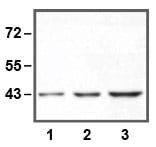 |
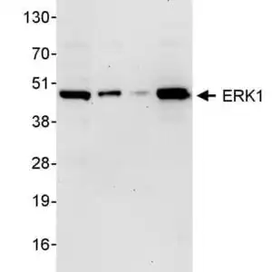
| 1:10,000 (0.1µg/mL) Ab dilution used in WB of HEK293 cell lysate; 1µg (1), 5µg (2), and 10µg (3) of cell lysate used. |
 |
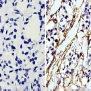
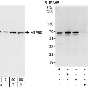
| IP of anti-ERK1 using 50µg of Jurkat cell lysate; 0.5µg (1), 1µg (2), and 2µg (3) of anti-ERK1 used. |
FAQ & Publications
Frequently Asked Questions
What species does the mouse anti-ERK1 monoclonal antibody (E19) 3427 react with?
This antibody reacts with human and mouse species.
What are the recommended storage conditions for the mouse anti-ERK1 monoclonal antibody (E19) 3427 to ensure stability?
Store the antibody at -20°C for up to 2 years. After the first thaw, centrifuge to maximize product recovery and aliquot to avoid repeated freeze-thaw cycles. Aliquots can be stored at 4°C for several days to weeks.
For which applications has the mouse anti-ERK1 monoclonal antibody (E19) 3427 been validated, and what are the recommended dilutions?
The antibody has been validated for use in Western blot (WB) applications. Recommended dilutions for WB range from 1:1000 to 1:10000.
Publications
| pmid | title | authors | citation |
|---|---|---|---|
| We haven't added any publications to our database yet. | |||
Published literature highly relevant to the biological target of this product and referencing this antibody or clone are retrieved from the PubMed database provided by the United States National Library of Medicine at the National Institutes of Health.
Protocols
| relevant to this product |
|---|
| Western blot |
Documents
| Batch Number | QC File | SDS |
|---|---|---|
| To view batch-specific Safety Datasheets and Quality Certificates associated with your account, please Log In. | ||
Only logged in customers who have purchased this product may leave a review.


Reviews
There are no reviews yet.