| Weight | 1 lbs |
|---|---|
| Dimensions | 9 × 5 × 2 in |
| host | rabbit |
| isotype | IgG |
| clonality | monoclonal |
| concentration | 1 mg/mL |
| applications | WB |
| available sizes | 100 µg |


rabbit anti-ERK1 monoclonal antibody 9031
$409.00
Antibody summary
- Rabbit monoclonal to ERK1
- Suitable for: WB
- Reacts with: human, mouse
- Isotype: IgG1
- 100 µg
rabbit anti-ERK1 monoclonal antibody 9031
| target relevance |
|---|
| ERK1 (Extracellular signal-Regulated Kinase 1) is a highly conserved member of the mitogen-activated protein kinase (MAPK) family, playing a crucial role in cellular signaling pathways. It is a serine/threonine kinases that becomes activated in response to a variety of extracellular stimuli, including growth factors, hormones, and stress. Upon activation, ERK1 translocates to the nucleus, where it phosphorylates numerous downstream targets, including transcription factors and other kinases. Through these phosphorylation events, ERK1 regulates a wide range of cellular processes, such as cell proliferation, differentiation, survival, and apoptosis. Dysregulation of ERK1 signaling has been implicated in various diseases, including cancer. neurodegenerative disorders, and inflammatory conditions. This antibody can be used as a loading control when evaluating post-translational modification of ERK1 and other cell singalling kinases and their modifications. Click for more on: loading controls and ERK1 |
| Protein names Mitogen-activated protein kinase 3 (MAP kinase 3) (MAPK 3) (EC 2.7.11.24) (ERT2) (Extracellular signal-regulated kinase 1) (ERK-1) (Insulin-stimulated MAP2 kinase) (MAP kinase isoform p44) (p44-MAPK) (Microtubule-associated protein 2 kinase) (p44-ERK1) |
| Gene names MAPK3,MAPK3 ERK1 PRKM3 |
| Protein family Protein kinase superfamily, CMGC Ser/Thr protein kinase family, MAP kinase subfamily |
| Mass 43136Da |
| Function FUNCTION: Serine/threonine kinase which acts as an essential component of the MAP kinase signal transduction pathway (PubMed:34497368). MAPK1/ERK2 and MAPK3/ERK1 are the 2 MAPKs which play an important role in the MAPK/ERK cascade. They participate also in a signaling cascade initiated by activated KIT and KITLG/SCF. Depending on the cellular context, the MAPK/ERK cascade mediates diverse biological functions such as cell growth, adhesion, survival and differentiation through the regulation of transcription, translation, cytoskeletal rearrangements. The MAPK/ERK cascade also plays a role in initiation and regulation of meiosis, mitosis, and postmitotic functions in differentiated cells by phosphorylating a number of transcription factors. About 160 substrates have already been discovered for ERKs. Many of these substrates are localized in the nucleus, and seem to participate in the regulation of transcription upon stimulation. However, other substrates are found in the cytosol as well as in other cellular organelles, and those are responsible for processes such as translation, mitosis and apoptosis. Moreover, the MAPK/ERK cascade is also involved in the regulation of the endosomal dynamics, including lysosome processing and endosome cycling through the perinuclear recycling compartment (PNRC); as well as in the fragmentation of the Golgi apparatus during mitosis. The substrates include transcription factors (such as ATF2, BCL6, ELK1, ERF, FOS, HSF4 or SPZ1), cytoskeletal elements (such as CANX, CTTN, GJA1, MAP2, MAPT, PXN, SORBS3 or STMN1), regulators of apoptosis (such as BAD, BTG2, CASP9, DAPK1, IER3, MCL1 or PPARG), regulators of translation (such as EIF4EBP1) and a variety of other signaling-related molecules (like ARHGEF2, DEPTOR, FRS2 or GRB10) (PubMed:35216969). Protein kinases (such as RAF1, RPS6KA1/RSK1, RPS6KA3/RSK2, RPS6KA2/RSK3, RPS6KA6/RSK4, SYK, MKNK1/MNK1, MKNK2/MNK2, RPS6KA5/MSK1, RPS6KA4/MSK2, MAPKAPK3 or MAPKAPK5) and phosphatases (such as DUSP1, DUSP4, DUSP6 or DUSP16) are other substrates which enable the propagation the MAPK/ERK signal to additional cytosolic and nuclear targets, thereby extending the specificity of the cascade. {ECO:0000269|PubMed:10393181, ECO:0000269|PubMed:10617468, ECO:0000269|PubMed:12110590, ECO:0000269|PubMed:12356731, ECO:0000269|PubMed:12974390, ECO:0000269|PubMed:15788397, ECO:0000269|PubMed:15952796, ECO:0000269|PubMed:16581800, ECO:0000269|PubMed:19265199, ECO:0000269|PubMed:34497368, ECO:0000269|PubMed:35216969, ECO:0000269|PubMed:8325880, ECO:0000269|PubMed:9155018, ECO:0000269|PubMed:9480836}. |
| Catalytic activity CATALYTIC ACTIVITY: Reaction=L-seryl-[protein] + ATP = O-phospho-L-seryl-[protein] + ADP + H(+); Xref=Rhea:RHEA:17989, Rhea:RHEA-COMP:9863, Rhea:RHEA-COMP:11604, ChEBI:CHEBI:15378, ChEBI:CHEBI:29999, ChEBI:CHEBI:30616, ChEBI:CHEBI:83421, ChEBI:CHEBI:456216; EC=2.7.11.24; Evidence={ECO:0000269|PubMed:35216969}; CATALYTIC ACTIVITY: Reaction=L-threonyl-[protein] + ATP = O-phospho-L-threonyl-[protein] + ADP + H(+); Xref=Rhea:RHEA:46608, Rhea:RHEA-COMP:11060, Rhea:RHEA-COMP:11605, ChEBI:CHEBI:15378, ChEBI:CHEBI:30013, ChEBI:CHEBI:30616, ChEBI:CHEBI:61977, ChEBI:CHEBI:456216; EC=2.7.11.24; |
| Subellular location SUBCELLULAR LOCATION: Cytoplasm {ECO:0000250|UniProtKB:P21708}. Nucleus. Membrane, caveola {ECO:0000250|UniProtKB:P21708}. Cell junction, focal adhesion {ECO:0000250|UniProtKB:Q63844}. Note=Autophosphorylation at Thr-207 promotes nuclear localization (PubMed:19060905). PEA15-binding redirects the biological outcome of MAPK3 kinase-signaling by sequestering MAPK3 into the cytoplasm (By similarity). {ECO:0000250|UniProtKB:Q63844, ECO:0000269|PubMed:19060905}. |
| Structure SUBUNIT: Binds both upstream activators and downstream substrates in multimolecular complexes. Found in a complex with at least BRAF, HRAS, MAP2K1/MEK1, MAPK3 and RGS14 (By similarity). Interacts with ADAM15, ARRB2, CANX, DAPK1 (via death domain), HSF4, IER3, MAP2K1/MEK1, MORG1, NISCH, and SGK1. Interacts with PEA15 and MKNK2 (By similarity). MKNK2 isoform 1 binding prevents from dephosphorylation and inactivation (By similarity). Interacts with TPR. Interacts with CDKN2AIP. Interacts with HSF1 (via D domain and preferentially with hyperphosphorylated form); this interaction occurs upon heat shock (PubMed:10747973). Interacts with CAVIN4 (By similarity). Interacts with GIT1; this interaction is necessary for MAPK3 localization to focal adhesions (By similarity). Interacts with ZNF263 (PubMed:32051553). Interacts with EBF4. {ECO:0000250|UniProtKB:P21708, ECO:0000250|UniProtKB:Q63844, ECO:0000269|PubMed:10393181, ECO:0000269|PubMed:10521408, ECO:0000269|PubMed:10747973, ECO:0000269|PubMed:11912194, ECO:0000269|PubMed:12356731, ECO:0000269|PubMed:15616583, ECO:0000269|PubMed:16581800, ECO:0000269|PubMed:18296648, ECO:0000269|PubMed:18435604, ECO:0000269|PubMed:18794356, ECO:0000269|PubMed:19060905, ECO:0000269|PubMed:19447520, ECO:0000269|PubMed:24825908, ECO:0000269|PubMed:32051553, ECO:0000269|PubMed:35939714}.; SUBUNIT: (Microbial infection) Binds to HIV-1 Nef through its SH3 domain. This interaction inhibits its tyrosine-kinase activity. {ECO:0000269|PubMed:8794306}. |
| Post-translational modification PTM: Phosphorylated upon KIT and FLT3 signaling (By similarity). Dually phosphorylated on Thr-202 and Tyr-204, which activates the enzyme. Ligand-activated ALK induces tyrosine phosphorylation. Dephosphorylated by PTPRJ at Tyr-204. {ECO:0000250, ECO:0000269|PubMed:17274988, ECO:0000269|PubMed:18983981, ECO:0000269|PubMed:19060905, ECO:0000269|PubMed:19265199, ECO:0000269|PubMed:19494114}.; PTM: Ubiquitinated by TRIM15 via 'Lys-63'-linked ubiquitination; leading to activation. Deubiquitinated by CYLD. {ECO:0000269|PubMed:34497368}. |
| Domain DOMAIN: The TXY motif contains the threonine and tyrosine residues whose phosphorylation activates the MAP kinases. {ECO:0000269|PubMed:10521408}. |
| Target Relevance information above includes information from UniProt accession: P27361 |
| The UniProt Consortium |
Data
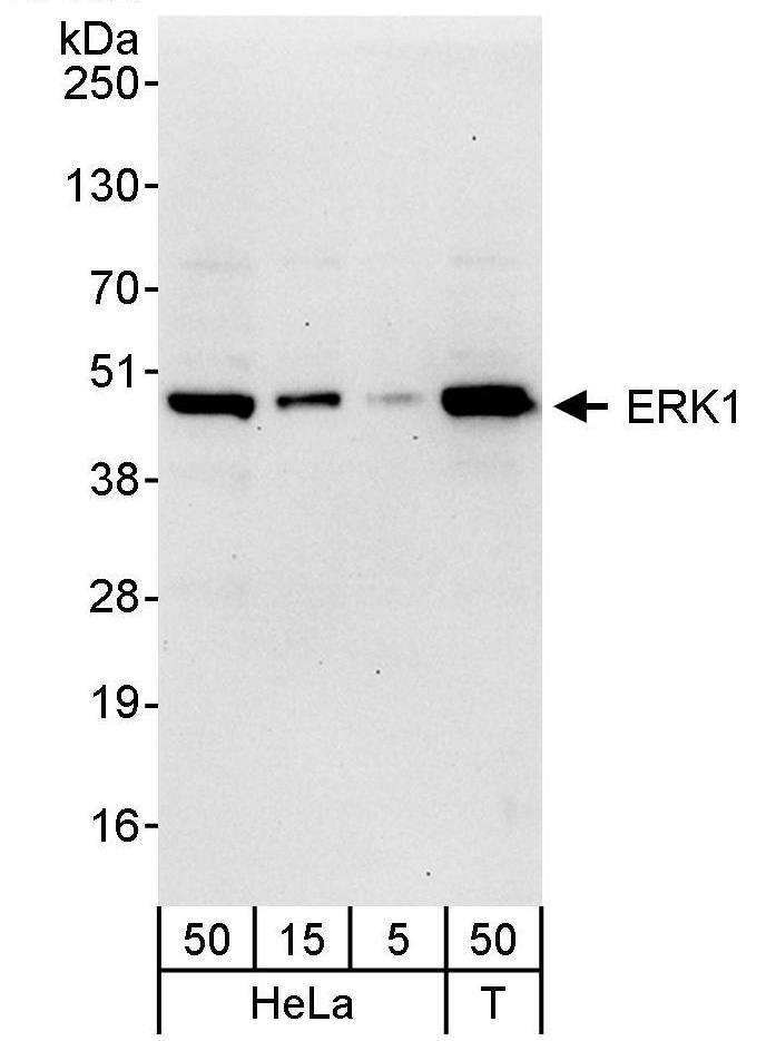 |
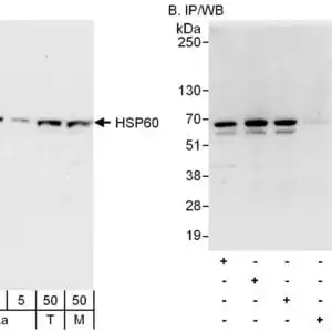
| Detection of human ERK1 by western blot. Samples: Whole cell lysate from HeLa (5, 15 and 50 µg) and HEK293T (T; 50 µg) cells. Antibodies: Affinity purified rabbit anti-ERK1 antibody used for WB at 0.04 µg/ml. Detection: Chemiluminescence with exposure time of 30 seconds. |
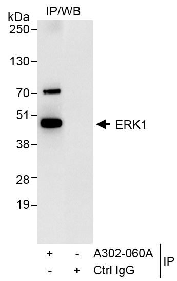 |
| Detection of human ERK1 by western blot of immunoprecipitates. Samples: Whole cell lysate (1 mg for IP, 20% of IP loaded) from HeLa cells. Antibodies: Affinity purified rabbit anti-ERK1 antibody used for IP at 3 µg/mg lysate. For blotting immunoprecipitated ERK1, the antibody was used at 1.0 µg/ml. Detection: Chemiluminescence with exposure time of 3 seconds. |
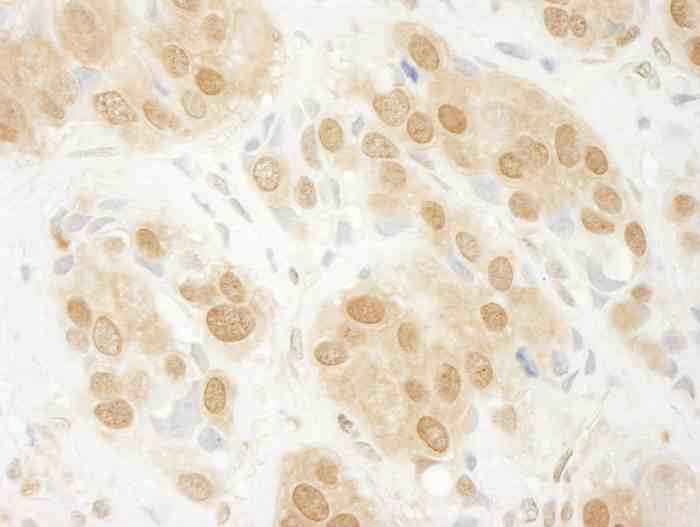 |
| Detection of human ERK1 by immunohistochemistry. Sample: FFPE section of human breast carcinoma. Antibody: Affinity purified rabbit anti-ERK1 antibody used at a dilution of 1:200 (1µg/ml). Detection: DAB |
Publications
| pmid | title | authors | citation |
|---|---|---|---|
| We haven't added any publications to our database yet. | |||
Protocols
| relevant to this product |
|---|
| Western blot |
Documents
| Batch Number | QC File | SDS |
|---|---|---|
| To view batch-specific Safety Datasheets and Quality Certificates associated with your account, please Log In. | ||
Only logged in customers who have purchased this product may leave a review.


Reviews
There are no reviews yet.