| Weight | 1 lbs |
|---|---|
| Dimensions | 9 × 5 × 2 in |
| host | mouse |
| isotype | IgG |
| clonality | monoclonal |
| concentration | concentrate, predilute |
| applications | IHC |
| reactivity | human |
| available size | 0.1 mL, 0.5 mL, 1 mL concentrated, 7 mL prediluted |

rabbit anti-CD3 monoclonal antibody (ZR414) 6081
Price range: $160.00 through $528.00
Antibody summary
- Rabbit monoclonal to CD3
- Suitable for: Immunohistochemistry (formalin-fixed, paraffin-embedded tissues)
- Reacts with: Human
- Isotype:IgG
- Control: Tonsil
- Visualization: Membrane
- 0.1, 0.5, 1.0 mL concentrated, 7 mL prediluted
rabbit anti-CD3 monoclonal antibody ZR414 6081
| target relevance |
|---|
| Homo sapiens CD3E T-cell surface glycoprotein CD3 epsilon chain |
| Protein names T-cell surface glycoprotein CD3 epsilon chain |
| Alternative names T-cell surface antigen T3/Leu-4 epsilon chain |
| Gene names CD3E |
| Function Part of the TCR-CD3 complex present on T-lymphocyte cell surface that plays an essential role in adaptive immune response (PubMed:15294938, PubMed:15546002, PubMed:2470098, PubMed:40592325, PubMed:8490660). When antigen presenting cells (APCs) activate T-cell receptor (TCR), TCR-mediated signals are transmitted across the cell membrane by the CD3 chains CD3D, CD3E, CD3G and CD247/CD3Z (PubMed:2470098, PubMed:40592325). All CD3 chains contain immunoreceptor tyrosine-based activation motifs (ITAMs) in their cytoplasmic domain (PubMed:2470098, PubMed:40592325). Upon TCR engagement, these motifs become phosphorylated by Src family protein tyrosine kinases LCK and FYN, resulting in the activation of downstream signaling pathways (PubMed:2470098, PubMed:40592325). CD3E ITAM phosphorylation creates docking sites for the protein kinase ZAP70 leading to ZAP70 phosphorylation and its conversion into a catalytically active enzyme (By similarity). In addition of this role of signal transduction in T-cell activation, CD3E plays an essential role in correct T-cell development (By similarity). Also participates in internalization and cell surface down-regulation of TCR-CD3 complexes via endocytosis sequences present in CD3E cytosolic region (PubMed:10384095, PubMed:26507128). In addition to its role as a TCR coreceptor, it serves as a receptor for ITPRIPL1 (PubMed:38614099). Ligand recognition inhibits T-cell activation by promoting interaction with NCK1, which prevents CD3E-ZAP70 interaction and blocks the ERK-NFkB signaling cascade and calcium influx (PubMed:12110186, PubMed:38614099) |
| Subcellular location Cell membrane |
| Structure The TCR-CD3 complex is composed of a CD3D-CD3E and a CD3G-CD3E heterodimers that preferentially associate with TCRalpha and TCRbeta, respectively, to form TCRalpha-CD3E-CD3G and TCRbeta/CD3G-CD3E trimers (PubMed:15136729, PubMed:15534202, PubMed:40592325). In turn, the hexamer interacts with CD247/CD3Z homodimer to form the TCR-CD3 complex (PubMed:15136729, PubMed:15534202). Alternatively, TCRalpha and TCRbeta can be replaced by TCRgamma and TCRdelta. Interacts with CD6 (PubMed:15294938). Interacts (via Proline-rich sequence) with NCK1; the interaction is ligand dependent but independent of tyrosine kinase activation (PubMed:12110186, PubMed:15972658, PubMed:38614099). Interacts with NUMB; this interaction is important for TCR-CD3 internalization and subsequent degradation (PubMed:26507128). Interacts (when tyrosine phosphorylated) with LAG3; disrupting the association between CD3E and LCK and preventing TCR activation (PubMed:15534202) |
| Post-translational modification Phosphorylated on Tyr residues after T-cell receptor triggering by LCK in association with CD4/CD8 |
| Involvement in disease Immunodeficiency 18 An autosomal recessive primary immunodeficiency characterized by onset in infancy or early childhood of recurrent infections. The severity is variable, encompassing both a mild immunodeficiency and severe combined immunodeficiency (SCID), resulting in early death without bone marrow transplantation in some patients. Immunologic work-up of the IMD18 SCID patients shows a T cell-negative, B cell-positive, natural killer (NK) cell-positive phenotype, whereas T-cell development is not impaired in the mild form of IMD18. |
| Keywords 3D-structure, Adaptive immunity, Cell membrane, Disulfide bond, Immunity, Immunoglobulin domain, Membrane, Phosphoprotein, Proteomics identification, Receptor, Reference proteome, Signal, Transmembrane, Transmembrane helix |
| Sequence MQSGTHWRVLGLCLLSVGVWGQDGNEEMGGITQTPYKVSISGTTVILTCPQYPGSEILWQ HNDKNIGGDEDDKNIGSDEDHLSLKEFSELEQSGYYVCYPRGSKPEDANFYLYLRARVCE NCMEMDVMSVATIVIVDICITGGLLLLVYYWSKNRKAKAKPVTRGAGAGGRQRGQNKERP PPVPNPDYEPIRKGQRDLYSGLNQRRI |
| UniProt accession: P07766 |
Data
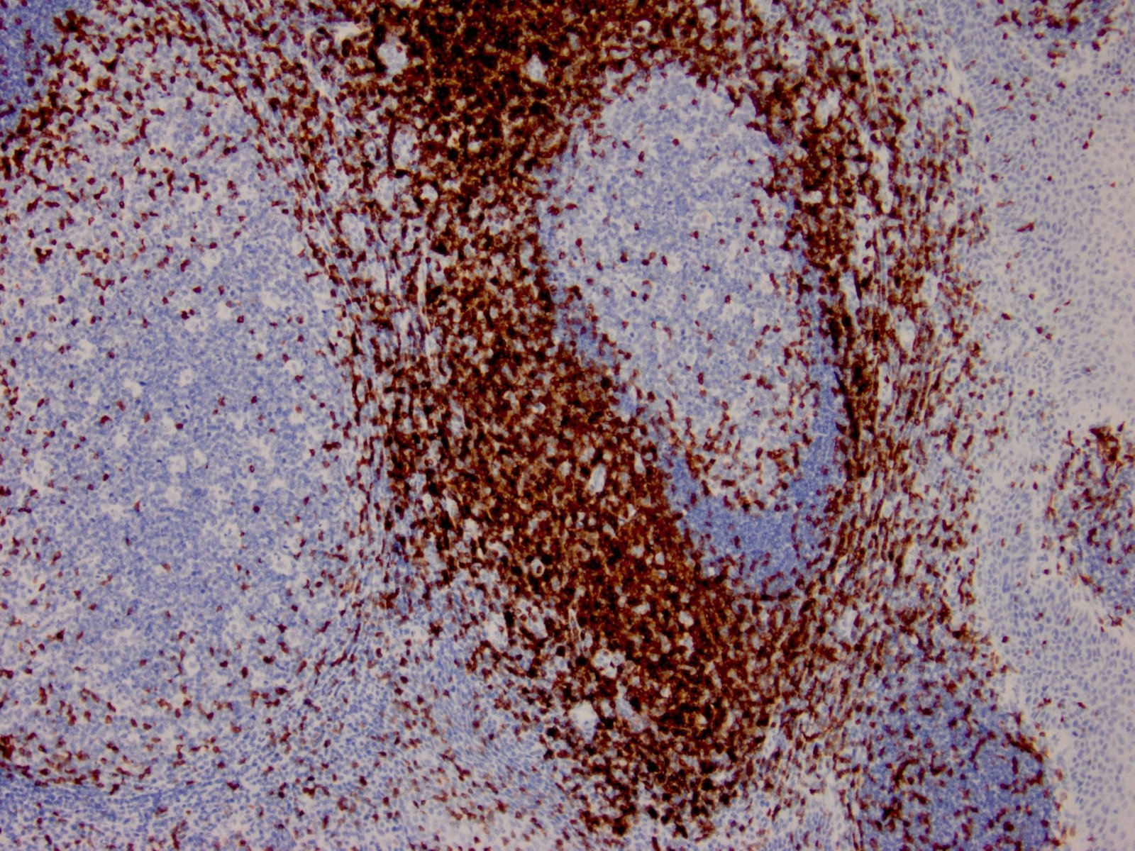 |
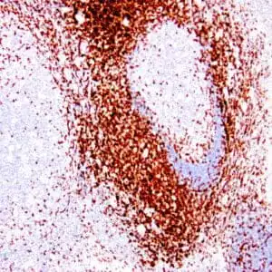
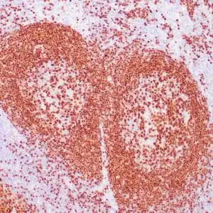
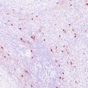
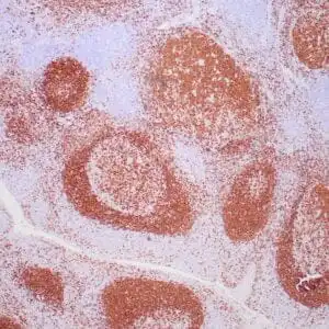
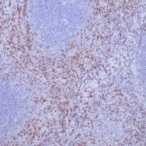
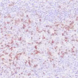
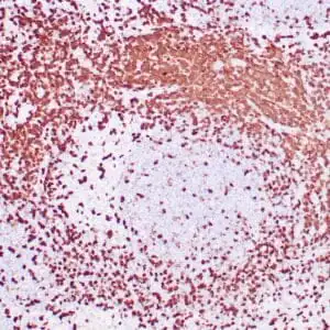
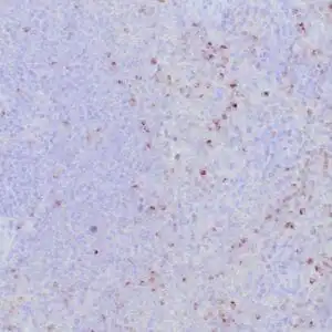
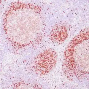
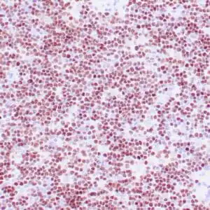
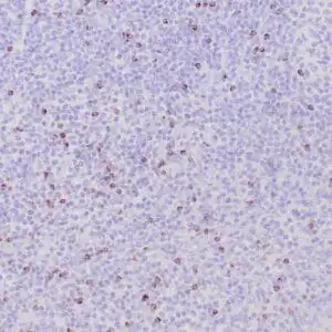
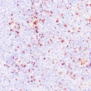
| Formalin-fixed, paraffin-embedded human lymph node stained with anti-CD3 antibody using peroxidase-conjugate and DAB chromogen. Note the membranous staining of perifollicular T-cells and no stain in B-cells |
FAQ & Publications
Frequently Asked Questions
What species does the rabbit anti-CD3 monoclonal antibody (ZR414) specifically react with?
This antibody specifically reacts with human CD3 protein.
What are the recommended storage conditions for the rabbit anti-CD3 monoclonal antibody (ZR414)?
For short-term storage, keep the antibody at 2-8°C. For long-term storage, it should be kept at -20°C, and freeze/thaw cycles should be avoided.
Which application and tissue type is the rabbit anti-CD3 monoclonal antibody (ZR414) validated for?
This antibody is suitable for immunohistochemistry (IHC) on formalin-fixed, paraffin-embedded human tissues, with tonsil tissue recommended as a positive control.
Publications
| pmid | title | authors | citation |
|---|---|---|---|
| We haven't added any publications to our database yet. | |||
Published literature highly relevant to the biological target of this product and referencing this antibody or clone are retrieved from the PubMed database provided by the United States National Library of Medicine at the National Institutes of Health.
Protocols
| relevant to this product |
|---|
| IHC |
Documents
| Batch Number | QC File | SDS |
|---|---|---|
| To view batch-specific Safety Datasheets and Quality Certificates associated with your account, please Log In. | ||
Only logged in customers who have purchased this product may leave a review.


Reviews
There are no reviews yet.