| Weight | 1 lbs |
|---|---|
| Dimensions | 9 × 5 × 2 in |
| host | mouse |
| isotype | IgG1 |
| clonality | monoclonal |
| concentration | concentrate, predilute |
| applications | IHC |
| reactivity | human |
| available size | 0.1 mL, 0.5 mL, 1 mL concentrated, 7 mL prediluted |

mouse anti-CD21 monoclonal antibody (ZM75) 6076
Price range: $160.00 through $528.00
Antibody summary
- Mouse monoclonal to CD21
- Suitable for: Immunohistochemistry (formalin-fixed, paraffin-embedded tissues)
- Reacts with: Human
- Isotype:IgG1
- Control: Tonsil, lymph node
- Visualization: Cell membrane
- 0.1, 0.5, 1.0 mL concentrated, 7 mL prediluted
mouse anti-CD21 monoclonal antibody ZM75 6076
| target relevance |
|---|
| Homo sapiens CR2 Complement receptor type 2 |
| Protein names Complement receptor type 2 |
| Alternative names Complement C3d receptor, Epstein-Barr virus receptor |
| Gene names CR2 |
| Protein family Belongs to the receptors of complement activation (RCA) family |
| Function Serves as a receptor for various ligands including complement component CD3d, HNRNPU OR IFNA1 (PubMed:1849076, PubMed:21527715, PubMed:7753047). When C3d is bound to antigens, attaches to C3d on B-cell surface and thereby facilitates the recognition and uptake of antigens by B-cells (PubMed:21527715). This interaction enhances B-cell activation and subsequent immune responses. Forms a complex with several partners on the surface of B-cells including CD19, FCRL5 and CD81, to form the B-cell coreceptor complex that plays a crucial role in B-cell activation and signaling (PubMed:1383329, PubMed:30107486). Also induces specific intracellular signaling separately from the BCR and CD19 by activating the tyrosine kinase SRC, which then phosphorylates nucleolin/NCL and triggers AKT and GSK3 kinase activities in a SYK/CD19-independent manner (PubMed:12938232). Acts as a ligand for CD23 (FcepsilonRII), a low-affinity receptor for IgE, which is expressed on B-cells and other immune cells, and thus participates in the regulation of IgE production (PubMed:1386409) |
| Subcellular location Cell membrane |
| Structure (Microbial infection) Interacts with Epstein-Barr virus gp350 protein |
| Involvement in disease Systemic lupus erythematosus 9 A chronic, relapsing, inflammatory, and often febrile multisystemic disorder of connective tissue, characterized principally by involvement of the skin, joints, kidneys and serosal membranes. It is of unknown etiology, but is thought to represent a failure of the regulatory mechanisms of the autoimmune system. The disease is marked by a wide range of system dysfunctions, an elevated erythrocyte sedimentation rate, and the formation of LE cells in the blood or bone marrow. Immunodeficiency, common variable, 7 A primary immunodeficiency characterized by antibody deficiency, hypogammaglobulinemia, recurrent bacterial infections and an inability to mount an antibody response to antigen. The defect results from a failure of B-cell differentiation and impaired secretion of immunoglobulins; the numbers of circulating B-cells is usually in the normal range, but can be low. |
| Keywords 3D-structure, Alternative splicing, Cell membrane, Complement pathway, Direct protein sequencing, Disulfide bond, Glycoprotein, Host cell receptor for virus entry, Host-virus interaction, Immunity, Innate immunity, Membrane, Proteomics identification, Receptor, Reference proteome, Repeat, Signal, Sushi, Systemic lupus erythematosus, Transmembrane, Transmembrane helix |
| Sequence MGAAGLLGVFLALVAPGVLGISCGSPPPILNGRISYYSTPIAVGTVIRYSCSGTFRLIGE KSLLCITKDKVDGTWDKPAPKCEYFNKYSSCPEPIVPGGYKIRGSTPYRHGDSVTFACKT NFSMNGNKSVWCQANNMWGPTRLPTCVSVFPLECPALPMIHNGHHTSENVGSIAPGLSVT YSCESGYLLVGEKIINCLSSGKWSAVPPTCEEARCKSLGRFPNGKVKEPPILRVGVTANF FCDEGYRLQGPPSSRCVIAGQGVAWTKMPVCEEIFCPSPPPILNGRHIGNSLANVSYGSI VTYTCDPDPEEGVNFILIGESTLRCTVDSQKTGTWSGPAPRCELSTSAVQCPHPQILRGR MVSGQKDRYTYNDTVIFACMFGFTLKGSKQIRCNAQGTWEPSAPVCEKECQAPPNILNGQ KEDRHMVRFDPGTSIKYSCNPGYVLVGEESIQCTSEGVWTPPVPQCKVAACEATGRQLLT KPQHQFVRPDVNSSCGEGYKLSGSVYQECQGTIPWFMEIRLCKEITCPPPPVIYNGAHTG SSLEDFPYGTTVTYTCNPGPERGVEFSLIGESTIRCTSNDQERGTWSGPAPLCKLSLLAV QCSHVHIANGYKISGKEAPYFYNDTVTFKCYSGFTLKGSSQIRCKADNTWDPEIPVCEKE TCQHVRQSLQELPAGSRVELVNTSCQDGYQLTGHAYQMCQDAENGIWFKKIPLCKVIHCH PPPVIVNGKHTGMMAENFLYGNEVSYECDQGFYLLGEKKLQCRSDSKGHGSWSGPSPQCL RSPPVTRCPNPEVKHGYKLNKTHSAYSHNDIVYVDCNPGFIMNGSRVIRCHTDNTWVPGV PTCIKKAFIGCPPPPKTPNGNHTGGNIARFSPGMSILYSCDQGYLLVGEALLLCTHEGTW SQPAPHCKEVNCSSPADMDGIQKGLEPRKMYQYGAVVTLECEDGYMLEGSPQSQCQSDHQ WNPPLAVCRSRSLAPVLCGIAAGLILLTFLIVITLYVISKHRARNYYTDTSQKEAFHLEA REVYSVDPYNPAS |
| UniProt accession: P20023 |
Data
 |
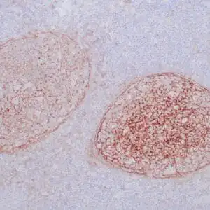
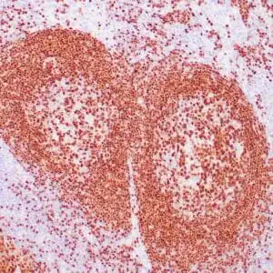
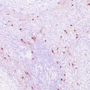
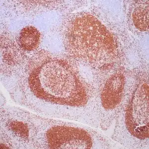
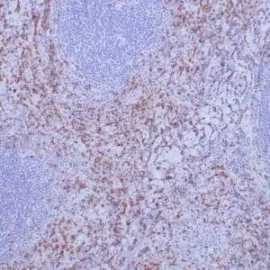
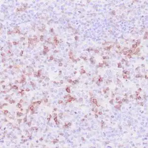
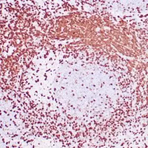
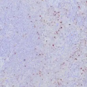
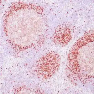
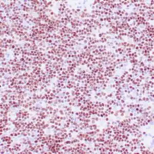
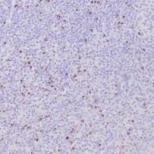
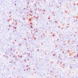
| Human tonsil stained with anti-CD21 antibodyusing peroxidase-conjugate and DAB chromogen. Note the membrane staining of follicular dendritic cells in follicles. |
FAQ & Publications
Frequently Asked Questions
What applications is the mouse anti-CD21 monoclonal antibody (ZM75) validated for?
This antibody is suitable for immunohistochemistry (IHC) on formalin-fixed, paraffin-embedded human tissue samples.
How should the mouse anti-CD21 monoclonal antibody (ZM75) be stored to maintain stability?
For short-term storage, keep the antibody at 2-8°C. For long-term storage, store it at -20°C and avoid repeated freeze-thaw cycles.
Publications
| pmid | title | authors | citation |
|---|---|---|---|
| We haven't added any publications to our database yet. | |||
Published literature highly relevant to the biological target of this product and referencing this antibody or clone are retrieved from the PubMed database provided by the United States National Library of Medicine at the National Institutes of Health.
Protocols
| relevant to this product |
|---|
| IHC |
Documents
| Batch Number | QC File | SDS |
|---|---|---|
| To view batch-specific Safety Datasheets and Quality Certificates associated with your account, please Log In. | ||
Only logged in customers who have purchased this product may leave a review.


Reviews
There are no reviews yet.