| Weight | 1 lbs |
|---|---|
| Dimensions | 9 × 5 × 2 in |
| host | mouse |
| isotype | IgG2a |
| clonality | monoclonal |
| concentration | concentrate, predilute |
| applications | IHC |
| reactivity | human |
| available size | 0.1 mL, 0.5 mL, 1 mL concentrated, 7 mL prediluted |

mouse anti-Annexin A1 monoclonal antibody (ZM211) 6023
Price range: $160.00 through $528.00
Antibody summary
- Mouse monoclonal to Annexin A1
- Suitable for: Immunohistochemistry (formalin-fixed, paraffin-embedded tissues)
- Reacts with: Human
- Isotype:IgG2a
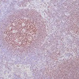
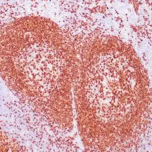
- Control: Lymph node or hairy cell leukemia
- Visualization: Nuclear, cytoplasmic, and membrane
- 0.1, 0.5, 1.0 mL concentrated, 7 mL prediluted
mouse anti-Annexin A1 monoclonal antibody ZM211 6023
| target relevance |
|---|
| Homo sapiens ANXA1 Annexin A1 |
| Protein names Annexin A1 |
| Alternative names Annexin I, Annexin-1, Calpactin II, Calpactin-2, Chromobindin-9, Lipocortin I, Phospholipase A2 inhibitory protein, p35 |
| Gene names ANXA1 |
| Protein family Belongs to the annexin family |
| Function Plays important roles in the innate immune response as effector of glucocorticoid-mediated responses and regulator of the inflammatory process. Has anti-inflammatory activity (PubMed:8425544). Plays a role in glucocorticoid-mediated down-regulation of the early phase of the inflammatory response (By similarity). Contributes to the adaptive immune response by enhancing signaling cascades that are triggered by T-cell activation, regulates differentiation and proliferation of activated T cells (PubMed:17008549). Promotes the differentiation of T cells into Th1 cells and negatively regulates differentiation into Th2 cells (PubMed:17008549). Has no effect on unstimulated T cells (PubMed:17008549). Negatively regulates hormone exocytosis via activation of the formyl peptide receptors and reorganization of the actin cytoskeleton (PubMed:19625660). Has high affinity for Ca(2+) and can bind up to eight Ca(2+) ions (By similarity). Displays Ca(2+)-dependent binding to phospholipid membranes (PubMed:2532504, PubMed:8557678). Plays a role in the formation of phagocytic cups and phagosomes. Plays a role in phagocytosis by mediating the Ca(2+)-dependent interaction between phagosomes and the actin cytoskeleton (By similarity). In the context of antitumor immunity, interacts with FPR1 on dendritic cells allowing for tumor-associated antigens uptake and cross-presentation to T cells to mount an antitumor specific T cell response |
| Subcellular location Nucleus, Cytoplasm, Cell projection, cilium, Cell membrane, Membrane, Endosome membrane, Basolateral cell membrane, Apical cell membrane, Lateral cell membrane, Secreted, Secreted, extracellular space, Cell membrane, Secreted, extracellular exosome, Cytoplasmic vesicle, secretory vesicle lumen, Cell projection, phagocytic cup, Early endosome, Cytoplasmic vesicle membrane |
| Structure Homodimer; non-covalently linked (By similarity). Homodimer; linked by transglutamylation (PubMed:2532504). Homodimers linked by transglutamylation are observed in placenta, but not in other tissues (PubMed:2532504). Interacts with S100A11 (PubMed:10673436, PubMed:8557678). Heterotetramer, formed by two molecules each of S100A11 and ANXA1 (PubMed:10673436). Interacts with DYSF (By similarity). Interacts with EGFR (By similarity) |
| Post-translational modification Phosphorylated by protein kinase C, EGFR and TRPM7 (PubMed:15485879, PubMed:2457390). Phosphorylated in response to EGF treatment (PubMed:2532504) Sumoylated Proteolytically cleaved by cathepsin CTSG to release the active N-terminal peptide Ac2-26 |
| Keywords 3D-structure, Acetylation, Adaptive immunity, Annexin, Calcium, Calcium/phospholipid-binding, Cell membrane, Cell projection, Cilium, Cytoplasm, Cytoplasmic vesicle, Direct protein sequencing, Disulfide bond, Endosome, Immunity, Inflammatory response, Innate immunity, Isopeptide bond, Membrane, Metal-binding, Nucleus, Pharmaceutical, Phospholipase A2 inhibitor, Phosphoprotein, Proteomics identification, Reference proteome, Repeat, Secreted, Ubl conjugation |
| Sequence MAMVSEFLKQAWFIENEEQEYVQTVKSSKGGPGSAVSPYPTFNPSSDVAALHKAIMVKGV DEATIIDILTKRNNAQRQQIKAAYLQETGKPLDETLKKALTGHLEEVVLALLKTPAQFDA DELRAAMKGLGTDEDTLIEILASRTNKEIRDINRVYREELKRDLAKDITSDTSGDFRNAL LSLAKGDRSEDFGVNEDLADSDARALYEAGERRKGTDVNVFNTILTTRSYPQLRRVFQKY TKYSKHDMNKVLDLELKGDIEKCLTAIVKCATSKPAFFAEKLHQAMKGVGTRHKALIRIM VSRSEIDMNDIKAFYQKMYGISLCQAILDETKGDYEKILVALCGGN |
| UniProt accession: P04083 |
Data
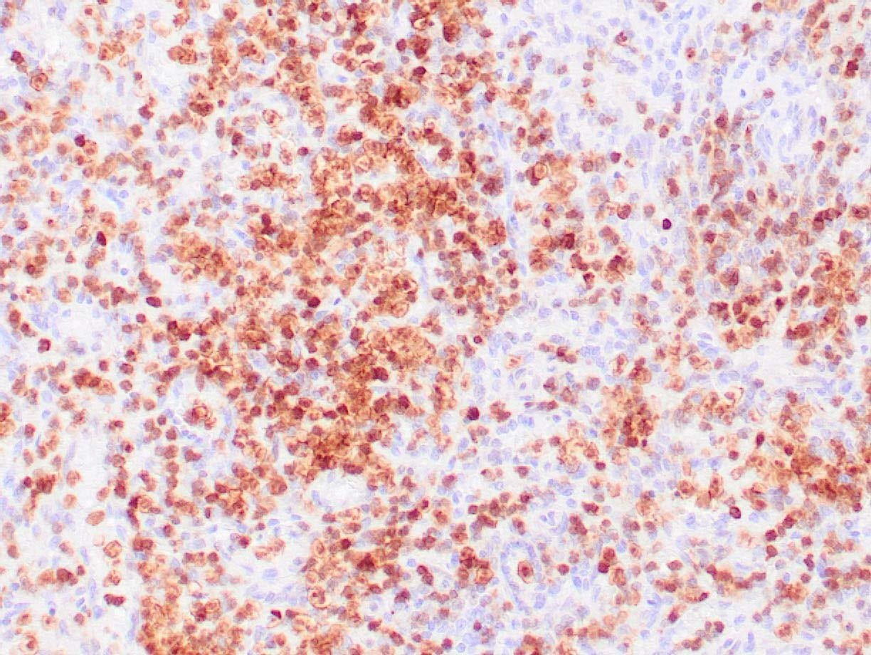 |
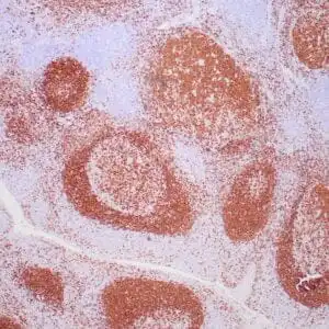
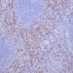
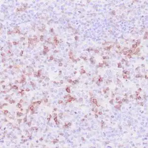
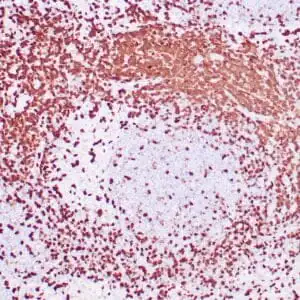
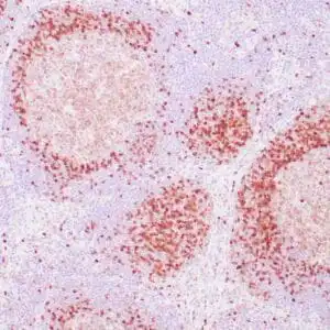
| Human lymph node involved by hairy cell leukemia stained with anti-Annexin A1 antibody using peroxidase-conjugate and DAB chromogen. Note nuclear/cytoplasmic staining of tumor cells. |
FAQ & Publications
Frequently Asked Questions
What species reactivity does the mouse anti-Annexin A1 monoclonal antibody (ZM211) 6023 exhibit?
This antibody specifically reacts with human Annexin A1 protein.
What are the recommended storage conditions for the mouse anti-Annexin A1 monoclonal antibody (ZM211) 6023?
For short-term storage, keep the antibody at 2-8°C. For longer-term storage, it should be kept at -20°C, and freeze/thaw cycles should be avoided.
Which applications is the mouse anti-Annexin A1 monoclonal antibody (ZM211) 6023 validated for, and what are the suggested dilutions?
This antibody is suitable for immunohistochemistry (IHC) on formalin-fixed, paraffin-embedded tissues. The recommended dilution for the concentrated antibody is 1:100 to 1:200.
Publications
| pmid | title | authors | citation |
|---|---|---|---|
| We haven't added any publications to our database yet. | |||
Published literature highly relevant to the biological target of this product and referencing this antibody or clone are retrieved from the PubMed database provided by the United States National Library of Medicine at the National Institutes of Health.
Protocols
| relevant to this product |
|---|
| IHC |
Documents
| Batch Number | QC File | SDS |
|---|---|---|
| To view batch-specific Safety Datasheets and Quality Certificates associated with your account, please Log In. | ||
Only logged in customers who have purchased this product may leave a review.


Reviews
There are no reviews yet.