| Weight | 1 lbs |
|---|---|
| Dimensions | 9 × 5 × 2 in |
| host | mouse |
| isotype | IgG |
| clonality | monoclonal |
| concentration | concentrate, predilute |
| applications | IHC |
| reactivity | human |
| available size | 0.1 mL, 0.5 mL, 1 mL concentrated, 7 mL prediluted |
rabbit anti-Helicobacterpylori monoclonal antibody (Poly) 6206
Price range: $160.00 through $528.00
Antibody summary
- Rabbit monoclonal to Helicobacterpylori
- Suitable for: Immunohistochemistry (formalin-fixed, paraffin-embedded tissues)
- Reacts with: Human
- Isotype:IgG
- Control: H. pylori infected tissue
- Visualization: Organisms
- 0.1, 0.5, 1.0 mL concentrated, 7 mL prediluted
rabbit anti-Helicobacterpylori monoclonal antibody Poly 6206
| target relevance |
|---|
| Protein names Helicobacter pylori |
| Structure Helicobacter pylori, previously known as Campylobacter pylori, is a gram-negative, flagellated, helical bacterium. Mutants can have a rod or curved rod shape, and these are less effective. Its helical body (from which the genus name, Helicobacter, derives) is thought to have evolved in order to penetrate the mucous lining of the stomach, helped by its flagella, and thereby establish infection. The bacterium was first identified as the causal agent of gastric ulcers in 1983 by the Australian doctors Barry Marshall and Robin Warren. |
| Biotechnology In the diagnosis of H. pylori infections, a distinction is made between non-invasive and invasive methods. Invasive procedures contain histology, urease rapid test and microbiological techniques such as cultivation and PCR. The C13-breath test, the Helicobacter pylori antigen detection in stool samples and serological antibody detection based on ELISA or immunoblot belong to the group of non-invasive methods. The detection of serum antibodies can be used for therapy control after eradication therapy |
Data
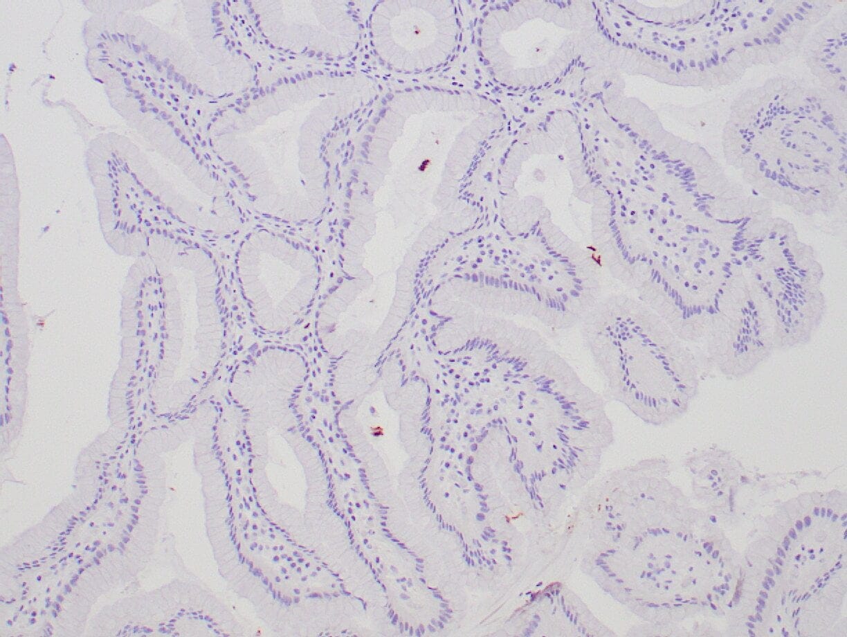 |
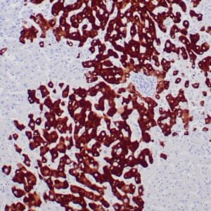
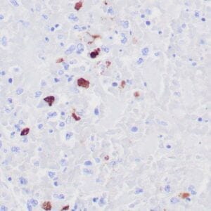
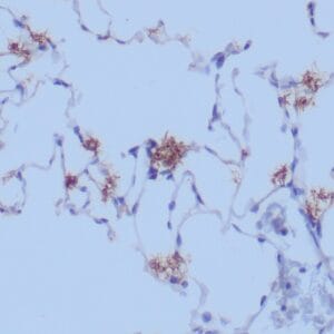
| Human stomach stained with anti-H. pylori antibody using peroxidase-conjugate and DAB chromogen. Note intense staining of H. Pylori organisms on the surface of gastric epithelial cells. |
Publications
| pmid | title | authors | citation |
|---|---|---|---|
| We haven't added any publications to our database yet. | |||
Protocols
| relevant to this product |
|---|
| IHC |
Documents
| # | SDS | Certificate | |
|---|---|---|---|
| Please enter your product and batch number here to retrieve product datasheet, SDS, and QC information. | |||
Only logged in customers who have purchased this product may leave a review.






Reviews
There are no reviews yet.