| Weight | 1 lbs |
|---|---|
| Dimensions | 9 × 5 × 2 in |
| host | mouse |
| isotype | IgG1 |
| clonality | monoclonal |
| concentration | concentrate, predilute |
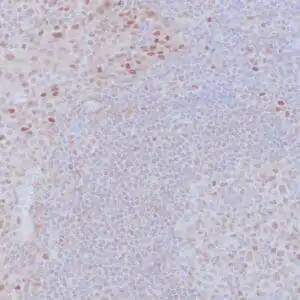
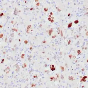
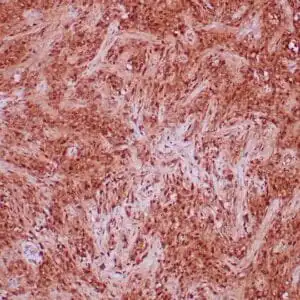
| applications | IHC |
| reactivity | human |
| available size | 0.1 mL, 0.5 mL, 1 mL concentrated, 7 mL prediluted |

mouse anti-Rb monoclonal antibody (1F8) 6352
Price range: $160.00 through $528.00
Antibody summary
- Mouse monoclonal to Rb
- Suitable for: Immunohistochemistry (formalin-fixed, paraffin-embedded tissues)
- Reacts with: Human
- Isotype:IgG1
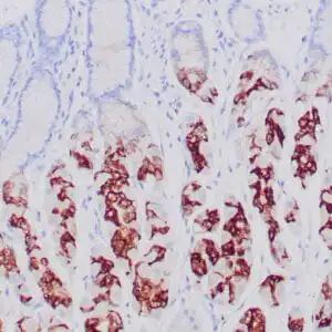
- Control: Colon cancer
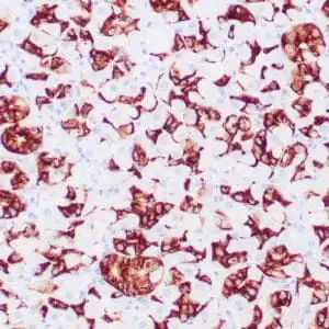
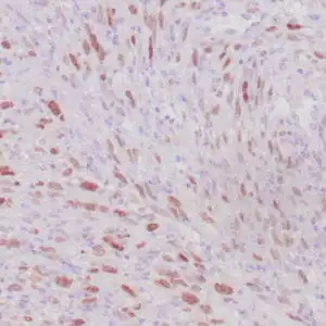
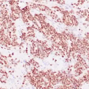
- Visualization: Nuclear
- 0.1, 0.5, 1.0 mL concentrated, 7 mL prediluted
mouse anti-Rb monoclonal antibody 1F8 6352
| target relevance |
|---|
| Homo sapiens RB1 Retinoblastoma-associated protein |
| Protein names Retinoblastoma-associated protein |
| Alternative names p105-Rb, p110-RB1, pRb, pp110 |
| Gene names RB1 |
| Protein family Belongs to the retinoblastoma protein (RB) family |
| Function Tumor suppressor that is a key regulator of the G1/S transition of the cell cycle (PubMed:10499802). The hypophosphorylated form binds transcription regulators of the E2F family, preventing transcription of E2F-responsive genes (PubMed:10499802). Both physically blocks E2Fs transactivating domain and recruits chromatin-modifying enzymes that actively repress transcription (PubMed:10499802). Cyclin and CDK-dependent phosphorylation of RB1 induces its dissociation from E2Fs, thereby activating transcription of E2F responsive genes and triggering entry into S phase (PubMed:10499802). RB1 also promotes the G0-G1 transition upon phosphorylation and activation by CDK3/cyclin-C (PubMed:15084261). Directly involved in heterochromatin formation by maintaining overall chromatin structure and, in particular, that of constitutive heterochromatin by stabilizing histone methylation. Recruits and targets histone methyltransferases SUV39H1, KMT5B and KMT5C, leading to epigenetic transcriptional repression. Controls histone H4 'Lys-20' trimethylation. Inhibits the intrinsic kinase activity of TAF1. Mediates transcriptional repression by SMARCA4/BRG1 by recruiting a histone deacetylase (HDAC) complex to the c-FOS promoter. In resting neurons, transcription of the c-FOS promoter is inhibited by BRG1-dependent recruitment of a phospho-RB1-HDAC1 repressor complex. Upon calcium influx, RB1 is dephosphorylated by calcineurin, which leads to release of the repressor complex (By similarity) |
| Subcellular location Nucleus, Cytoplasm |
| Structure (Microbial infection) Interacts with molluscum contagiosum virus protein MC007 |
| Post-translational modification Phosphorylated by CDK6 and CDK4, and subsequently by CDK2 at Ser-567 in G1, thereby releasing E2F1 which is then able to activate cell growth. Dephosphorylated at the late M phase. SV40 large T antigen, HPV E7 and adenovirus E1A bind to the underphosphorylated, active form of pRb. Phosphorylation at Thr-821 and Thr-826 promotes interaction between the C-terminal domain C and the Pocket domain, and thereby inhibits interactions with heterodimeric E2F/DP transcription factor complexes. Dephosphorylated at Ser-795 by calcineruin upon calcium stimulation. CDK3/cyclin-C-mediated phosphorylation at Ser-807 and Ser-811 is required for G0-G1 transition. Phosphorylated by CDK1 and CDK2 upon TGFB1-mediated apoptosis N-terminus is methylated by METTL11A/NTM1 (By similarity). Monomethylation at Lys-810 by SMYD2 enhances phosphorylation at Ser-807 and Ser-811, and promotes cell cycle progression. Monomethylation at Lys-860 by SMYD2 promotes interaction with L3MBTL1 Acetylated during keratinocyte differentiation. Acetylation at Lys-873 and Lys-874 regulates subcellular localization. Can be deacetylated by SIRT1 |
| Involvement in disease Childhood cancer retinoblastoma Congenital malignant tumor that arises from the nuclear layers of the retina. It occurs in about 1:20'000 live births and represents about 2% of childhood malignancies. It is bilateral in about 30% of cases. Although most RB appear sporadically, about 20% are transmitted as an autosomal dominant trait with incomplete penetrance. The diagnosis is usually made before the age of 2 years when strabismus or a gray to yellow reflex from pupil ('cat eye') is investigated. Bladder cancer A malignancy originating in tissues of the urinary bladder. It often presents with multiple tumors appearing at different times and at different sites in the bladder. Most bladder cancers are transitional cell carcinomas that begin in cells that normally make up the inner lining of the bladder. Other types of bladder cancer include squamous cell carcinoma (cancer that begins in thin, flat cells) and adenocarcinoma (cancer that begins in cells that make and release mucus and other fluids). Bladder cancer is a complex disorder with both genetic and environmental influences. Osteogenic sarcoma A sarcoma originating in bone-forming cells, affecting the ends of long bones. |
| Keywords 3D-structure, Acetylation, Cell cycle, Chromatin regulator, Cytoplasm, Direct protein sequencing, Disease variant, DNA-binding, Host-virus interaction, Methylation, Nucleus, Phosphoprotein, Proteomics identification, Reference proteome, Repressor, Transcription, Transcription regulation, Tumor suppressor |
| Sequence MPPKTPRKTAATAAAAAAEPPAPPPPPPPEEDPEQDSGPEDLPLVRLEFEETEEPDFTAL CQKLKIPDHVRERAWLTWEKVSSVDGVLGGYIQKKKELWGICIFIAAVDLDEMSFTFTEL QKNIEISVHKFFNLLKEIDTSTKVDNAMSRLLKKYDVLFALFSKLERTCELIYLTQPSSS ISTEINSALVLKVSWITFLLAKGEVLQMEDDLVISFQLMLCVLDYFIKLSPPMLLKEPYK TAVIPINGSPRTPRRGQNRSARIAKQLENDTRIIEVLCKEHECNIDEVKNVYFKNFIPFM NSLGLVTSNGLPEVENLSKRYEEIYLKNKDLDARLFLDHDKTLQTDSIDSFETQRTPRKS NLDEEVNVIPPHTPVRTVMNTIQQLMMILNSASDQPSENLISYFNNCTVNPKESILKRVK DIGYIFKEKFAKAVGQGCVEIGSQRYKLGVRLYYRVMESMLKSEEERLSIQNFSKLLNDN IFHMSLLACALEVVMATYSRSTSQNLDSGTDLSFPWILNVLNLKAFDFYKVIESFIKAEG NLTREMIKHLERCEHRIMESLAWLSDSPLFDLIKQSKDREGPTDHLESACPLNLPLQNNH TAADMYLSPVRSPKKKGSTTRVNSTANAETQATSAFQTQKPLKSTSLSLFYKKVYRLAYL RLNTLCERLLSEHPELEHIIWTLFQHTLQNEYELMRDRHLDQIMMCSMYGICKVKNIDLK FKIIVTAYKDLPHAVQETFKRVLIKEEEYDSIIVFYNSVFMQRLKTNILQYASTRPPTLS PIPHIPRSPYKFPSSPLRIPGGNIYISPLKSPYKISEGLPTPTKMTPRSRILVSIGESFG TSEKFQKINQMVCNSDRVLKRSAEGSNPPKPLKKLRFDIEGSDEADGSKHLPGESKFQQK LAEMTSTRTRMQKQKMNDSMDTSNKEEK |
| UniProt accession: P06400 |
Data
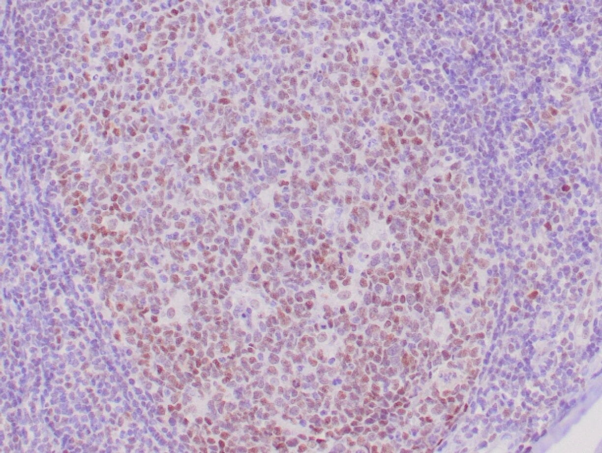 |
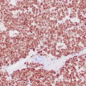
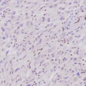
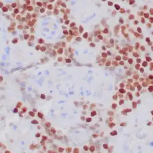
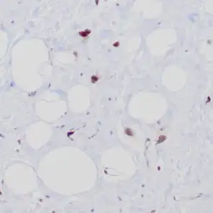
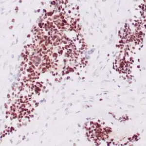
| Human tonsil stained with anti-retinoblastoma (Rb) antibody using peroxidase-conjugate and DAB chromogen. Note the nuclear staining of follicular center B cells. |
FAQ & Publications
Frequently Asked Questions
What is the recommended dilution for using the mouse anti-Rb monoclonal antibody (1F8) in immunohistochemistry?
For immunohistochemistry applications, the concentrated form of the mouse anti-Rb monoclonal antibody (1F8) is recommended to be diluted at 1:100 to 1:200.
Which species does the mouse anti-Rb monoclonal antibody (1F8) specifically react with?
This antibody specifically reacts with human species and is suitable for detecting human retinoblastoma protein in tissue samples.
How should the mouse anti-Rb monoclonal antibody (1F8) be stored to maintain stability?
For short-term storage, keep the antibody at 2-8°C. For long-term storage, it should be stored at -20°C, while avoiding repeated freeze-thaw cycles to preserve antibody integrity.
Publications
| pmid | title | authors | citation |
|---|---|---|---|
| We haven't added any publications to our database yet. | |||
Published literature highly relevant to the biological target of this product and referencing this antibody or clone are retrieved from the PubMed database provided by the United States National Library of Medicine at the National Institutes of Health.
Protocols
| relevant to this product |
|---|
| IHC |
Documents
| Batch Number | QC File | SDS |
|---|---|---|
| To view batch-specific Safety Datasheets and Quality Certificates associated with your account, please Log In. | ||
Only logged in customers who have purchased this product may leave a review.

Reviews
There are no reviews yet.