Skip to content

mouse anti-p53 monoclonal antibody (D01) 8319

$503.00

Antibody summary

  • Mouse monoclonal to p53
  • Suitable for: WB,ICC/IF,IHC-P,IHC-Fr,FACS,IP,ELISA
  • Isotype: IgG2a
  • 100 µg
SKU: 8319parent Categories: , , Tags: , ,
Weight1 lbs
Dimensions9 × 5 × 2 in
host

mouse

isotype

IgG2a

clonality

monoclonal

concentration

1 mg/mL

applications

ICC/IF, WB

reactivity

p53

available sizes

100 µg

mouse anti-p53 monoclonal antibody (D01) 8319

antibody
Tested applications
WB,IHC,IHC,ICC/IF
Recommended dilutions
IHC (frozen and paraffin), Immunoblotting.

IHC: use at 1-10ug/ml. Antigen retrieval: citrate buffer, pH 6.0, 15 minutes. #3339 on paraffin-embedded human breast carcinoma. #3339 on paraffin-embedded human colon carcinoma.

Immunoblotting: use at 1-5ug/ml. A band of 53kDa is detected.
Immunogen
Recombinant full length protein corresponding to Human p53 (N terminal). epitope is within aa 20-25
Size and concentration
100µg and lot specific
Form
liquid
Storage Instructions
This antibody is stable for at least one (1) year at -20°C. Avoid multiple freeze-thaw cycles.
Storage buffer
PBS, pH 7.4
Purity
protein affinty purification
Clonality
monoclonal
Isotype
IgG2a
Compatible secondaries
goat anti-mouse IgG, H&L chain specific, peroxidase conjugated polyclonal antibody 5486
goat anti-mouse IgG, H&L chain specific, biotin conjugated, Conjugate polyclonal antibody 2685
goat anti-mouse IgG, H&L chain specific, FITC conjugated polyclonal antibody 7854
goat anti-mouse IgG, H&L chain specific, peroxidase conjugated polyclonal antibody, crossabsorbed 1706
goat anti-mouse IgG, H&L chain specific, biotin conjugated polyclonal antibody, crossabsorbed 1716
goat anti-mouse IgG, H&L chain specific, FITC conjugated polyclonal antibody, crossabsorbed 1721
Isotype control
Mouse monocolonal IgG2a - Isotype Control
No information
target relevance

Data

benchmark-antibodies_anti-p53_antibody_8319_1.jpg
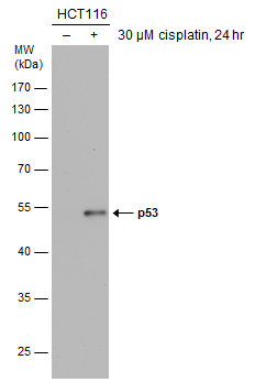
Untreated (?) and treated (+) HCT116 whole cell extracts (30 µg) were separated by 10% SDS-PAGE, and the membrane was blotted with p53 antibody [DO1] (8319) diluted at 1:1000. The HRP-conjugated anti-mouse IgG antibody was used to detect the primary antibody.
benchmark-antibodies_anti-p53_antibody_8319_2.jpg
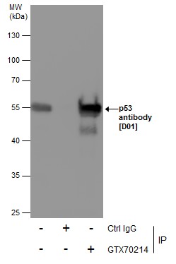
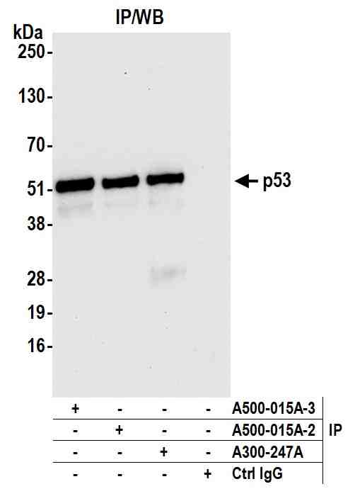
Immunoprecipitation of p53 protein from 293T whole cell extracts using 5 µg of p53 antibody [DO1] (8319).
Western blot analysis was performed using p53 antibody [D01] (8319).
EasyBlot anti-Mouse IgG \was used as a secondary reagent.
benchmark-antibodies_anti-p53_antibody_8319_3.jpg
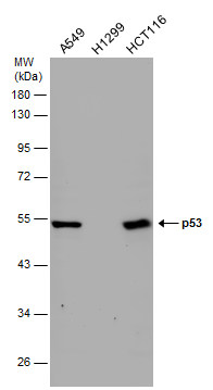
Various whole cell extracts (30 µg) were separated by 10% SDS-PAGE, and the membrane was blotted with p53 antibody [DO1] (8319) diluted at 1:1000. The HRP-conjugated anti-mouse IgG antibody was used to detect the primary antibody.
benchmark-antibodies_anti-p53_antibody_8319_4.jpg
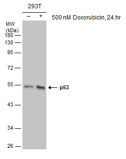
Untreated (?) and treated (+) 293T whole cell extracts (30 µg) were separated by 10% SDS-PAGE, and the membrane was blotted with p53 antibody [DO1] (8319) diluted at 1:1000. The HRP-conjugated anti-mouse IgG antibody was used to detect the primary antibody.
benchmark-antibodies_anti-p53_antibody_8319_5.jpg
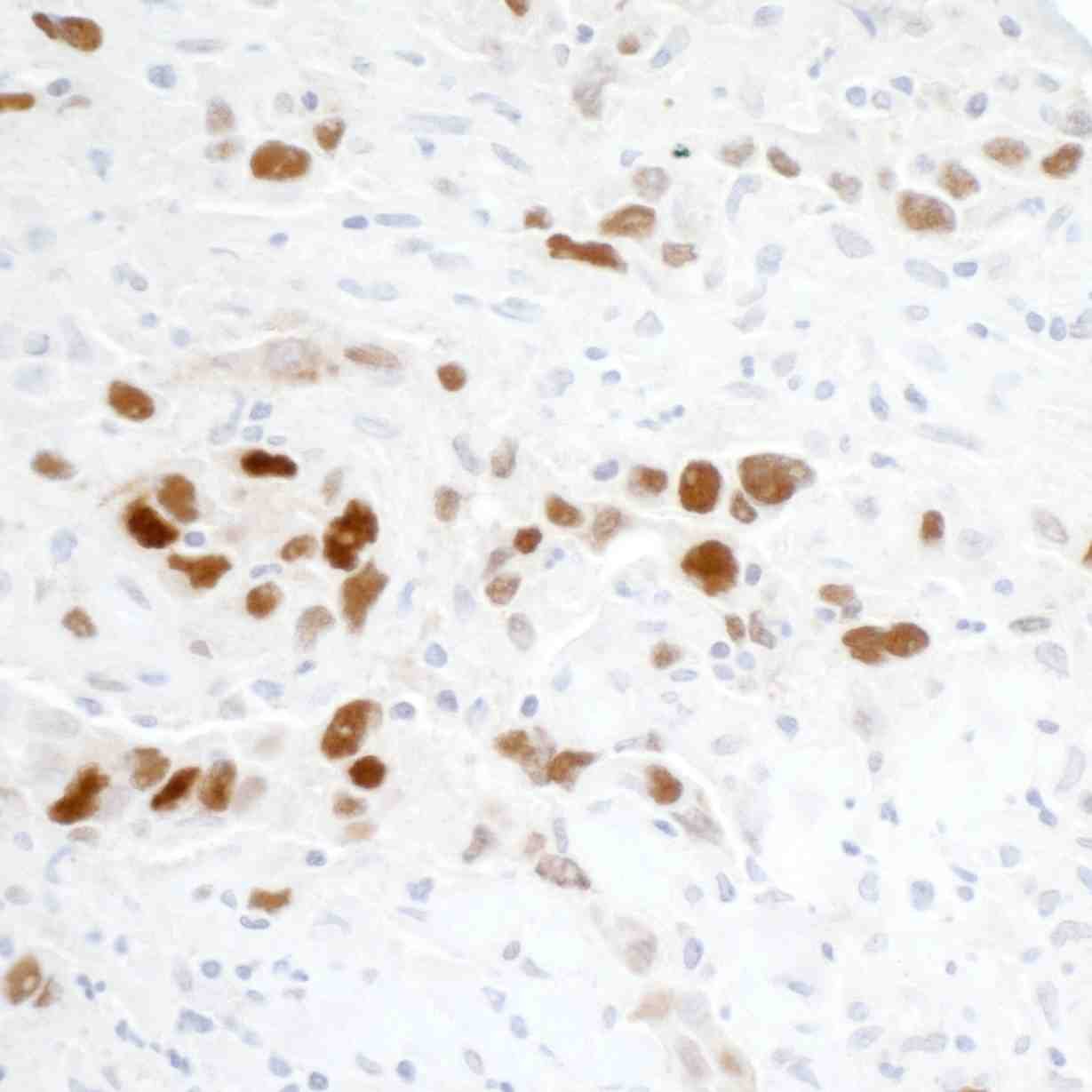
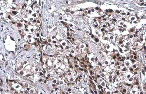
p53 antibody [DO1] detects p53 protein at cytoplasm and nucleus by immunohistochemical analysis.
Sample: Paraffin-embedded human breast carcinoma.
p53 stained by p53 antibody [DO1] (8319) diluted at 1:100.
Antigen Retrieval: Citrate buffer, pH 6.0, 15 min
benchmark-antibodies_anti-p53_antibody_8319_6.jpg
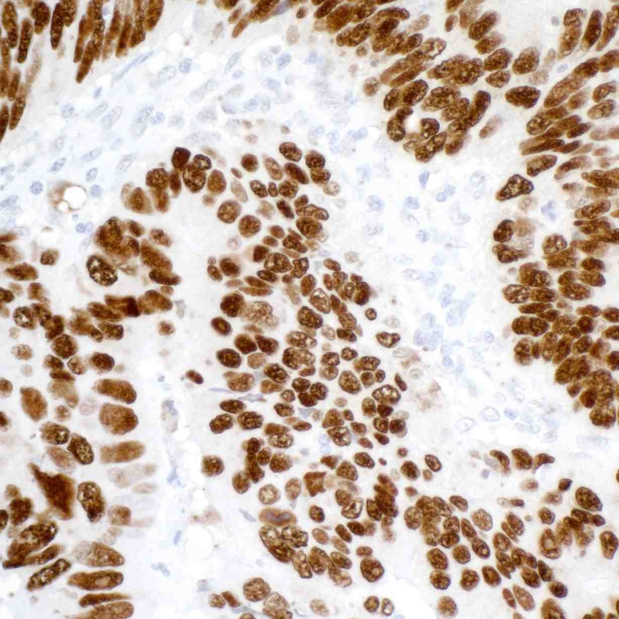
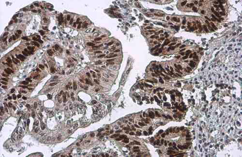
p53 antibody [DO1] detects p53 protein at cytoplasm and nucleus by immunohistochemical analysis.
Sample: Paraffin-embedded human colon cancer.
p53 stained by p53 antibody [DO1] (8319) diluted at 1:100.
Antigen Retrieval: Citrate buffer, pH 6.0, 15 min
benchmark-antibodies_anti-p53_antibody_8319_7.jpg
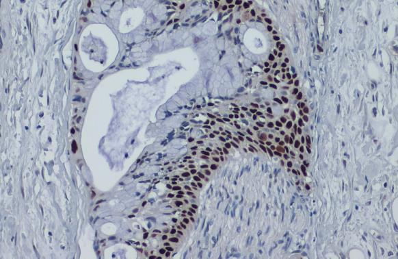
p53 antibody [DO1] detects p53 protein at nucleus by immunohistochemical analysis.
Sample: Paraffin-embedded human pancreatic cancer.
p53 stained by p53 antibody [DO1] (8319) diluted at 1:100.
Antigen Retrieval: Citrate buffer, pH 6.0, 15 min
benchmark-antibodies_anti-p53_antibody_8319_8.jpg
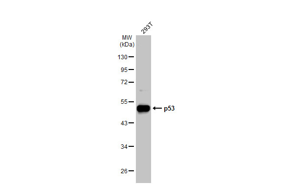
293T whole cell extracts (30 µg) was separated by 10% SDS-PAGE, and the membrane was blotted with p53 antibody [DO1] (8319) diluted at 1:40000. The HRP-conjugated anti-mouse IgG antibody was used to detect the primary antibody.
WB-image-anti-p53 antibody-8319_11
Detection of human p53 by western blot. Samples: Whole cell lysate from Hep-G2 (50 µg), A-549 (50 µg), and HEK293T (1 µg) cells prepared using NETN lysis buffer. Antibody: Mouse anti-p53 monoclonal antibody [DO-1] (#8319) used at 1:1000. Secondary: HRP-conjugated goat anti-mouse IgG. Detection: Chemiluminescence with an exposure time of 10 seconds.
IP-image-anti-p53 antibody-8319_12
Detection of human p53 by western blot of immunoprecipitates. Samples: Whole cell lysate (1.0 mg per IP reaction; 20% of IP loaded) from Hep-G2 cells prepared using NETN lysis buffer. Antibodies: Mouse anti-p53 monoclonal antibody [DO-1] (#8319) used for IP at 20 µl/mg lysate. p53 was also immunoprecipitated by a previous lot of this antibody (#8319) and rabbit anti-p53 antibody. For blotting immunoprecipitated p53 was used at 0.4 µg/ml. Detection: Chemiluminescence with an exposure time of 10 seconds.
IHC-image-anti-p53 antibody-8319_13
Detection of human p53 in FFPE colon carcinoma by immunohistochemsitry. Antibody: Mouse monoclonal anti-p53 [DO-1] (#8319). Secondary:HRP-conjugated goat anti-mouse IgG.Substrate:DAB.
IHC-image-anti-p53 antibody-8319_14
Detection of human p53 in FFPE breast carcinoma by immunohistochemsitry. Antibody: Mouse monoclonal anti-p53 [DO-1] (#8319). Secondary:HRP-conjugated goat anti-mouse IgG.Substrate:DAB.
Flow-image-anti-p53 antibody-8319_15
Detection of human p53 (shaded) in Ramos cells by flow cytometry. Antibody: Mouse anti-p53 recombinant monoclonal [DO-1] (#8319) or isotype control (unshaded). Secondary: DyLight? 488-conjugated goat anti-mouse IgG.

Publications

Published literature highly relevant to the biological target of this product and referencing this antibody or clone are retrieved from PubMed database provided by The United States National Library of Medicine at the National Institutes of Health.


pmidtitleauthorscitation

Protocols

relevant to this product
Western blot
IHC
ICC

Documents

#
No results found

Reviews

There are no reviews yet.

Only logged in customers who have purchased this product may leave a review.