| Weight | 1 lbs |
|---|---|
| Dimensions | 9 × 5 × 2 in |
| host | rabbit |
| isotype | IgG |
| clonality | monoclonal |
| concentration | 1 mg/mL |
| applications | ICC/IF, WB |
| available sizes | 100 µg |


rabbit anti-Tyrosine Hydroxylase monoclonal antibody 9009
$409.00
Antibody summary
- Rabbit monoclonal to Tyrosine Hydroxylase
- Suitable for: WB, ICC/IF
- Reacts with: human, mouse, rat
- Isotype: IgG
- 100 µg
rabbit anti-Tyrosine Hydroxylase monoclonal antibody 9009
| target relevance |
|---|
| Tyrosine hydroxylase (TH) is an enzyme critical for the synthesis of catecholamines, including dopamine, norepinephrine, and epinephrine. In research, TH has emerged as a vital cell marker, particularly for dopaminergic neurons. Dopaminergic neurons play essential roles in the brain's reward and motor systems and are implicated in various neurological disorders, such as Parkinson's disease and schizophrenia. TH immunohistochemistry, aided by specific antibodies targeting TH, allows researchers to identify and visualize dopaminergic neurons and assess their distribution and density in brain tissues. Additionally, these antibodies enable the quantification of TH levels, serving as a valuable tool for studying changes in dopaminergic function under different experimental conditions or in disease states. Furthermore, TH antibodies aid in the identification and analysis of TH-expressing cells in non-neuronal tissues, like the adrenal gland. Click for more on: cell markers and Tyrosine hydroxylase |
| Protein names Tyrosine 3-monooxygenase (EC 1.14.16.2) (Tyrosine 3-hydroxylase) (TH) |
| Gene names TH,TH TYH |
| Protein family Biopterin-dependent aromatic amino acid hydroxylase family |
| Mass 58600Da |
| Function FUNCTION: Catalyzes the conversion of L-tyrosine to L-dihydroxyphenylalanine (L-Dopa), the rate-limiting step in the biosynthesis of catecholamines, dopamine, noradrenaline, and adrenaline. Uses tetrahydrobiopterin and molecular oxygen to convert tyrosine to L-Dopa (PubMed:15287903, PubMed:1680128, PubMed:17391063, PubMed:24753243, PubMed:34922205, PubMed:8528210, Ref.18). In addition to tyrosine, is able to catalyze the hydroxylation of phenylalanine and tryptophan with lower specificity (By similarity). Positively regulates the regression of retinal hyaloid vessels during postnatal development (By similarity). {ECO:0000250|UniProtKB:P04177, ECO:0000250|UniProtKB:P24529, ECO:0000269|PubMed:15287903, ECO:0000269|PubMed:1680128, ECO:0000269|PubMed:17391063, ECO:0000269|PubMed:24753243, ECO:0000269|PubMed:34922205, ECO:0000269|PubMed:8528210, ECO:0000269|Ref.18}.; FUNCTION: [Isoform 5]: Lacks catalytic activity. {ECO:0000269|PubMed:17391063}.; FUNCTION: [Isoform 6]: Lacks catalytic activity. {ECO:0000269|PubMed:17391063}. |
| Catalytic activity CATALYTIC ACTIVITY: Reaction=(6R)-L-erythro-5,6,7,8-tetrahydrobiopterin + L-tyrosine + O2 = (4aS,6R)-4a-hydroxy-L-erythro-5,6,7,8-tetrahydrobiopterin + L-dopa; Xref=Rhea:RHEA:18201, ChEBI:CHEBI:15379, ChEBI:CHEBI:15642, ChEBI:CHEBI:57504, ChEBI:CHEBI:58315, ChEBI:CHEBI:59560; EC=1.14.16.2; Evidence={ECO:0000269|PubMed:15287903, ECO:0000269|PubMed:1680128, ECO:0000269|PubMed:17391063, ECO:0000269|PubMed:24753243, ECO:0000269|PubMed:34922205, ECO:0000269|PubMed:8528210, ECO:0000269|Ref.18}; PhysiologicalDirection=left-to-right; Xref=Rhea:RHEA:18202; Evidence={ECO:0000305|PubMed:17391063}; |
| Pathway PATHWAY: Catecholamine biosynthesis; dopamine biosynthesis; dopamine from L-tyrosine: step 1/2. {ECO:0000269|PubMed:17391063}. |
| Subellular location SUBCELLULAR LOCATION: Cytoplasm, perinuclear region {ECO:0000250|UniProtKB:P24529}. Nucleus {ECO:0000250|UniProtKB:P04177}. Cell projection, axon {ECO:0000250|UniProtKB:P24529}. Cytoplasm {ECO:0000250|UniProtKB:P04177}. Cytoplasmic vesicle, secretory vesicle, synaptic vesicle {ECO:0000250|UniProtKB:P04177}. Note=When phosphorylated at Ser-19 shows a nuclear distribution and when phosphorylated at Ser-31 as well at Ser-40 shows a cytosolic distribution (By similarity). Expressed in dopaminergic axons and axon terminals. {ECO:0000250|UniProtKB:P04177}. |
| Tissues TISSUE SPECIFICITY: Mainly expressed in the brain and adrenal glands. |
| Structure SUBUNIT: Homotetramer (PubMed:24947669, Ref.18). Interacts (when phosphorylated at Ser-19) with YWHAG; one YWHAG dimer binds to one TH tetramer and this interaction may influence the phosphorylation and dephosphorylation of other sites (PubMed:24947669). Interacts with NT5DC2; the interaction results in reduced phosphorylation and decreased catalytic activity of TH (By similarity). {ECO:0000250|UniProtKB:P04177, ECO:0000269|PubMed:24947669, ECO:0000269|Ref.18}. |
| Post-translational modification PTM: Phosphorylated on Ser-19, Ser-62 and Ser-71 by several protein kinases with different site specificities. Phosphorylation at Ser-62 and Ser-71 leads to an increase of TH activity (PubMed:7901013). Phosphorylation at Ser-71 activates the enzyme and also counteracts the feedback inhibition of TH by catecholamines (PubMed:15287903). Phosphorylation of Ser-19 and Ser-62 triggers the proteasomal degradation of TH through the ubiquitin-proteasome pathway (By similarity). Phosphorylation at Ser-62 facilitates transport of TH from the soma to the nerve terminals via the microtubule network (PubMed:28637871). Phosphorylation at Ser-19 induces the high-affinity binding to the 14-3-3 protein YWHAG; this interaction may influence the phosphorylation and dephosphorylation of other sites (PubMed:24947669). Ser-19 increases the phosphorylation at Ser-71 in a hierarchical manner, leading to increased activity (By similarity). {ECO:0000250|UniProtKB:P04177, ECO:0000269|PubMed:15287903, ECO:0000269|PubMed:24947669, ECO:0000269|PubMed:28637871, ECO:0000269|PubMed:7901013}. |
| Involvement in disease DISEASE: Segawa syndrome autosomal recessive (ARSEGS) [MIM:605407]: A form of DOPA-responsive dystonia presenting in infancy or early childhood. Dystonia is defined by the presence of sustained involuntary muscle contractions, often leading to abnormal postures. Some cases present with parkinsonian symptoms in infancy. Unlike all other forms of dystonia, it is an eminently treatable condition, due to a favorable response to L-DOPA. {ECO:0000269|PubMed:10585338, ECO:0000269|PubMed:11196107, ECO:0000269|PubMed:11246459, ECO:0000269|PubMed:15505183, ECO:0000269|PubMed:15747353, ECO:0000269|PubMed:16049992, ECO:0000269|PubMed:17696123, ECO:0000269|PubMed:18058633, ECO:0000269|PubMed:18554280, ECO:0000269|PubMed:19491146, ECO:0000269|PubMed:20056467, ECO:0000269|PubMed:20430833, ECO:0000269|PubMed:21940685, ECO:0000269|PubMed:22264700, ECO:0000269|PubMed:22815559, ECO:0000269|PubMed:23762320, ECO:0000269|PubMed:23939262, ECO:0000269|PubMed:24753243, ECO:0000269|PubMed:7814018, ECO:0000269|PubMed:8528210, ECO:0000269|PubMed:8817341, ECO:0000269|PubMed:9613851, ECO:0000269|PubMed:9703425}. Note=The disease is caused by variants affecting the gene represented in this entry.; DISEASE: Note=May play a role in the pathogenesis of Parkinson disease (PD). A genome-wide copy number variation analysis has identified a 34 kilobase deletion over the TH gene in a PD patient but not in any controls. {ECO:0000269|PubMed:20809526}. |
| Target Relevance information above includes information from UniProt accession: P07101 |
| The UniProt Consortium |
Data
 |
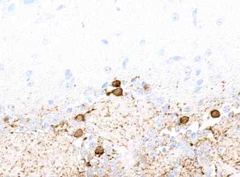
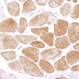
| Detection of human Tyrosine Hydroxylase in FFPE mouse brain by immunohistochemistry. Antibody: Rabbit anti-Tyrosine Hydroxylase recombinant monoclonal antibody. Secondary: HRP-conjugated goat anti-rabbit IgG. Substrate: DAB. |
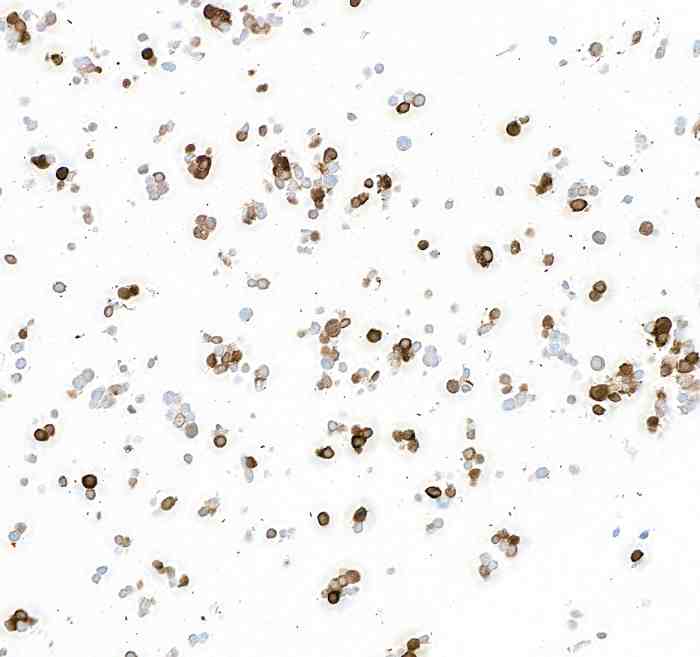 |
| Detection of human Tyrosine Hydroxylase in FFPE SK-N-BE(2) cells by immunocytochemistry. Antibody: Rabbit anti-Tyrosine Hydroxylase recombinant monoclonal antibody. Secondary: HRP-conjugated goat anti-rabbit IgG. Substrate: DAB. |
 |
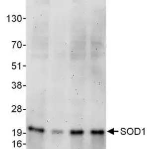
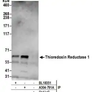
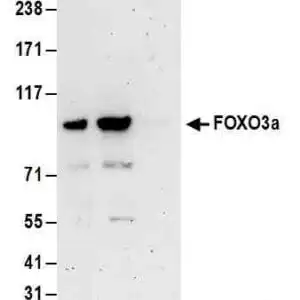
| Detection of human Tyrosine Hydroxylase by Western blot. Samples: Whole cell lysate (10 µg) from HEK293T, SK-N-SH, SK-N-BE(2), Jurkat, and SK-N-MC cells prepared using NETN lysis buffer. Antibody: Rabbit anti-Tyrosine Hydroxylase recombinant monoclonal antibody used at 1:1000. Secondary: HRP-conjugated goat anti-rabbit IgG. Detection: Chemiluminescence with an exposure time of 30 seconds. Lower Panel: Rabbit anti-COPB2 antibody. |
 |
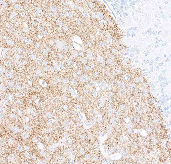
| Detection of human Tyrosine Hydroxylase in FFPE mouse olfactory bulb by immunohistochemistry. Antibody: Rabbit anti-Tyrosine Hydroxylase recombinant monoclonal antibody. Secondary: HRP-conjugated goat anti-rabbit IgG. Substrate: DAB. |
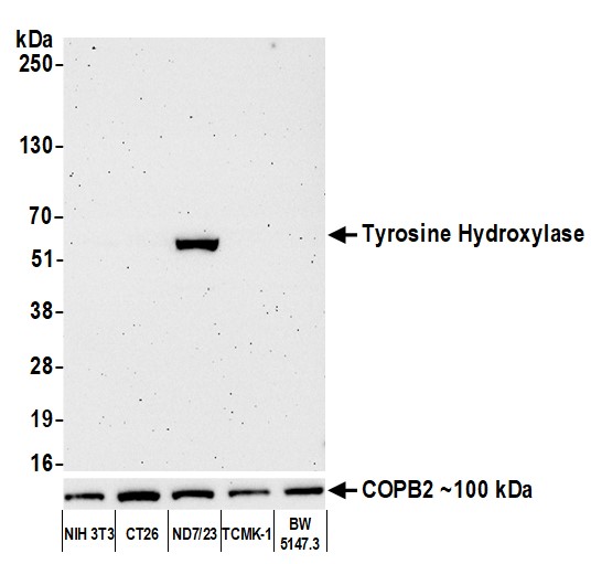 |
| Detection of human Tyrosine Hydroxylase by Western blot. Samples: Whole cell lysate (10 µg) from HEK293T, SK-N-SH, SK-N-BE(2), Jurkat, and SK-N-MC cells prepared using NETN lysis buffer. Antibody: Rabbit anti-Tyrosine Hydroxylase recombinant monoclonal antibody used at 1:1000. Secondary: HRP-conjugated goat anti-rabbit IgG. Detection: Chemiluminescence with an exposure time of 30 seconds. Lower Panel: Rabbit anti-COPB2 antibody. |
FAQ & Publications
Frequently Asked Questions
What species does the rabbit anti-Tyrosine Hydroxylase monoclonal antibody 9009 react with?
This antibody reacts with human, mouse, and rat samples.
For which applications has the rabbit anti-Tyrosine Hydroxylase monoclonal antibody 9009 been validated?
The antibody is suitable for Western blot (WB) and immunocytochemistry/immunofluorescence (ICC/IF) applications.
How should the rabbit anti-Tyrosine Hydroxylase monoclonal antibody 9009 be stored to maintain stability?
Store the antibody at 2-8°C for short term use and at -20°C for longer term storage, avoiding freeze/thaw cycles.
What is the concentration and form of the rabbit anti-Tyrosine Hydroxylase monoclonal antibody 9009 provided?
The antibody is supplied as a liquid at a concentration of 1 mg/mL.
What immunogen was used to generate the rabbit anti-Tyrosine Hydroxylase monoclonal antibody 9009?
The immunogen is full-length human Tyrosine Hydroxylase expressed in and purified from E. coli.
Publications
| pmid | title | authors | citation |
|---|---|---|---|
| We haven't added any publications to our database yet. | |||
Published literature highly relevant to the biological target of this product and referencing this antibody or clone are retrieved from the PubMed database provided by the United States National Library of Medicine at the National Institutes of Health.
Protocols
| relevant to this product |
|---|
| Western blot ICC |
Documents
| Batch Number | QC File | SDS |
|---|---|---|
| To view batch-specific Safety Datasheets and Quality Certificates associated with your account, please Log In. | ||
Only logged in customers who have purchased this product may leave a review.


Reviews
There are no reviews yet.