| Weight | 1 lbs |
|---|---|
| Dimensions | 9 × 5 × 2 in |
| host | rabbit |
| isotype | IgG |
| clonality | polyclonal |
| concentration | 1 mg/mL |
| applications | ICC/IF, WB |
| reactivity | TACE / ADAM17 |
| available sizes | 100 µg |


rabbit anti-TACE (CT) polyclonal antibody 8082
$469.00
Antibody summary
- Rabbit polyclonal to TACE (CT)
- Suitable for: ELISA,WB,ICC,IF
- Isotype: IgG
- 100 µg
rabbit anti-TACE (CT) polyclonal antibody 8082
| antibody |
|---|
| Tested applications WB,ICC/IF,ELISA |
| Recommended dilutions Immunoblotting: use at 1ug/mL. Positive control: HeLa or Jurkat cell lysate. Immunocytochemistry: use at 10ug/mL. These are recommended concentrations. Enduser should determine optimal concentrations for their applications. |
| Immunogen Peptide corresponding to aa 807-823 of human TACE (accession no. NP_003174). This sequence differs from mouse and rat TACE by one amino acid. |
| Size and concentration 100µg and lot specific |
| Form liquid |
| Storage Instructions This antibody is stable for at least one (1) year at -20°C. Avoid multiple freeze-thaw cycles. |
| Storage buffer PBS, pH 7.4. |
| Purity peptide affinity purification |
| Clonality polyclonal |
| Isotype IgG |
| Compatible secondaries goat anti-rabbit IgG, H&L chain specific, peroxidase conjugated, conjugated polyclonal antibody 9512 goat anti-rabbit IgG, H&L chain specific, biotin conjugated polyclonal antibody 2079 goat anti-rabbit IgG, H&L chain specific, FITC conjugated polyclonal antibody 7863 goat anti-rabbit IgG, H&L chain specific, Cross Absorbed polyclonal antibody 2371 goat anti-rabbit IgG, H&L chain specific, biotin conjugated polyclonal antibody, crossabsorbed 1715 goat anti-rabbit IgG, H&L chain specific, FITC conjugated polyclonal antibody, crossabsorbed 1720 |
| Isotype control Rabbit polyclonal - Isotype Control |
| target relevance |
|---|
| Protein names Disintegrin and metalloproteinase domain-containing protein 17 (ADAM 17) (EC 3.4.24.86) (Snake venom-like protease) (TNF-alpha convertase) (TNF-alpha-converting enzyme) (CD antigen CD156b) |
| Gene names ADAM17,ADAM17 CSVP TACE |
| Mass 93021Da |
| Function FUNCTION: Transmembrane metalloprotease which mediates the ectodomain shedding of a myriad of transmembrane proteins including adhesion proteins, growth factor precursors and cytokines important for inflammation and immunity (PubMed:24226769, PubMed:24227843, PubMed:28060820, PubMed:28923481). Cleaves the membrane-bound precursor of TNF-alpha to its mature soluble form (PubMed:36078095, PubMed:9034191). Responsible for the proteolytical release of soluble JAM3 from endothelial cells surface (PubMed:20592283). Responsible for the proteolytic release of several other cell-surface proteins, including p75 TNF-receptor, interleukin 1 receptor type II, p55 TNF-receptor, transforming growth factor-alpha, L-selectin, growth hormone receptor, MUC1 and the amyloid precursor protein (PubMed:12441351). Acts as an activator of Notch pathway by mediating cleavage of Notch, generating the membrane-associated intermediate fragment called Notch extracellular truncation (NEXT) (PubMed:24226769). Plays a role in the proteolytic processing of ACE2 (PubMed:24227843). Plays a role in hemostasis through shedding of GP1BA, the platelet glycoprotein Ib alpha chain (By similarity). Mediates the proteolytic cleavage of LAG3, leading to release the secreted form of LAG3 (By similarity). Mediates the proteolytic cleavage of IL6R, leading to the release of secreted form of IL6R (PubMed:26876177, PubMed:28060820). Mediates the proteolytic cleavage and shedding of FCGR3A upon NK cell stimulation, a mechanism that allows for increased NK cell motility and detachment from opsonized target cells. Cleaves TREM2, resulting in shedding of the TREM2 ectodomain (PubMed:28923481). {ECO:0000250|UniProtKB:Q9Z0F8, ECO:0000269|PubMed:12441351, ECO:0000269|PubMed:20592283, ECO:0000269|PubMed:24226769, ECO:0000269|PubMed:24227843, ECO:0000269|PubMed:24337742, ECO:0000269|PubMed:26876177, ECO:0000269|PubMed:28060820, ECO:0000269|PubMed:28923481, ECO:0000269|PubMed:36078095, ECO:0000269|PubMed:9034191}. |
| Catalytic activity CATALYTIC ACTIVITY: Reaction=Narrow endopeptidase specificity. Cleaves Pro-Leu-Ala-Gln-Ala-|-Val-Arg-Ser-Ser-Ser in the membrane-bound, 26-kDa form of tumor necrosis factor alpha (TNFalpha). Similarly cleaves other membrane-anchored, cell-surface proteins to 'shed' the extracellular domains.; EC=3.4.24.86; Evidence={ECO:0000269|PubMed:12441351, ECO:0000269|PubMed:20592283, ECO:0000269|PubMed:24227843, ECO:0000269|PubMed:28923481}; |
| Subellular location SUBCELLULAR LOCATION: Cell membrane {ECO:0000269|PubMed:36078095}; Single-pass type I membrane protein. |
| Tissues TISSUE SPECIFICITY: Ubiquitously expressed. Expressed at highest levels in adult heart, placenta, skeletal muscle, pancreas, spleen, thymus, prostate, testes, ovary and small intestine, and in fetal brain, lung, liver and kidney. Expressed in natural killer cells (at protein level) (PubMed:24337742). {ECO:0000269|PubMed:24337742}. |
| Structure SUBUNIT: Interacts with MAD2L1, MAPK14 and MUC1 (PubMed:12441351, PubMed:20188673). Interacts with iRhom1/RHBDF1 and iRhom2/RHBDF2 (PubMed:29897333). Interacts with FRMD8 via its interaction with iRhom1/RHBDF1 and iRhom2/RHBDF2 (PubMed:29897333). Interacts with TSPAN8 (PubMed:36078095). {ECO:0000269|PubMed:12441351, ECO:0000269|PubMed:20188673, ECO:0000269|PubMed:29897333, ECO:0000269|PubMed:36078095}. |
| Post-translational modification PTM: The precursor is cleaved by a furin endopeptidase. {ECO:0000250}.; PTM: Phosphorylated. Stimulation by growth factor or phorbol 12-myristate 13-acetate induces phosphorylation of Ser-819 but decreases phosphorylation of Ser-791. Phosphorylation at Thr-735 by MAPK14 is required for ADAM17-mediated ectodomain shedding. {ECO:0000269|PubMed:12058067, ECO:0000269|PubMed:12621058, ECO:0000269|PubMed:20188673}. |
| Domain DOMAIN: Must be membrane anchored to cleave the different substrates. The cytoplasmic domain is not required for the this activity. Only the catalytic domain is essential to shed TNF and p75 TNFR (By similarity). {ECO:0000250}.; DOMAIN: The conserved cysteine present in the cysteine-switch motif binds the catalytic zinc ion, thus inhibiting the enzyme. The dissociation of the cysteine from the zinc ion upon the activation-peptide release activates the enzyme. |
| Involvement in disease DISEASE: Inflammatory skin and bowel disease, neonatal, 1 (NISBD1) [MIM:614328]: A disorder characterized by inflammatory features with neonatal onset, involving the skin, hair, and gut. The skin lesions involve perioral and perianal erythema, psoriasiform erythroderma, with flares of erythema, scaling, and widespread pustules. Gastrointestinal symptoms include malabsorptive diarrhea that is exacerbated by intercurrent gastrointestinal infections. The hair is short or broken, and the eyelashes and eyebrows are wiry and disorganized. {ECO:0000269|PubMed:22010916}. Note=The disease is caused by variants affecting the gene represented in this entry. |
| Target Relevance information above includes information from UniProt accession: P78536 |
| The UniProt Consortium |
Data
 |
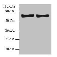
| Western Blot Validation of TACE in Human Cell Lines Loading: 15 µg of lysates per lane. Antibodies: TACE (1 µg /mL), 1h incubation at RT in 5% NFDM/TBST. Secondary: Goat anti-rabbit IgG HRP conjugate at 1:10000 dilution. Lanes: HeLa (A,D), Jurkat (B, E), Raji (C,F) in the absence (A-C) or presence (E-F) of blocking peptide. |
 |
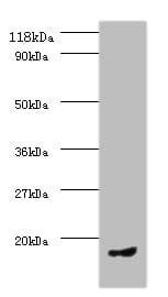
| KO Validation in HeLa Cells Loading: 10 µg of HeLa WT cell lysates or TACE KO cell lysates. Antibodies: TACE 8082 (0.25 µg/mL) and beta-actin 3779 (1 µg/mL), 1 h incubation at RT in 5% NFDM/TBST. Secondary: Goat Anti-Rabbit IgG HRP conjugate at 1:10000 dilution. |
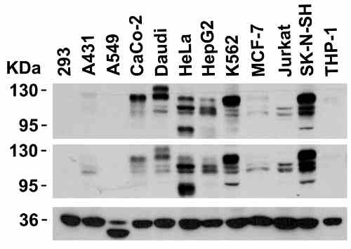 |
| Independent Antibody Validation (IAV) via Protein Expression Profile in Cell Lines Loading: 15 µg of lysates per lane. Antibodies: TACE 8082 (0.5 µg/mL), TACE 22-001 (2 µg/mL), and GAPDH (0.02 µg/mL), 1h incubation at RT in 5% NFDM/TBST. Secondary: Goat anti-rabbit IgG HRP conjugate at 1:10000 dilution. |
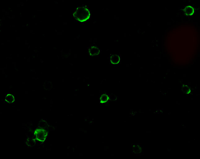 |
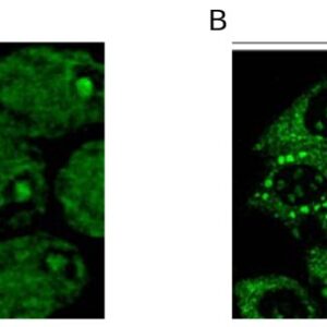
| Immunofluorescence Validation of TACE in HeLa Cells Immunofluorescent analysis of 4% paraformaldehyde-fixed HeLa cells labeling TACE with 8082 at 10 µg/mL, followed by goat anti-rabbit IgG secondary antibody at 1/500 dilution (green). |
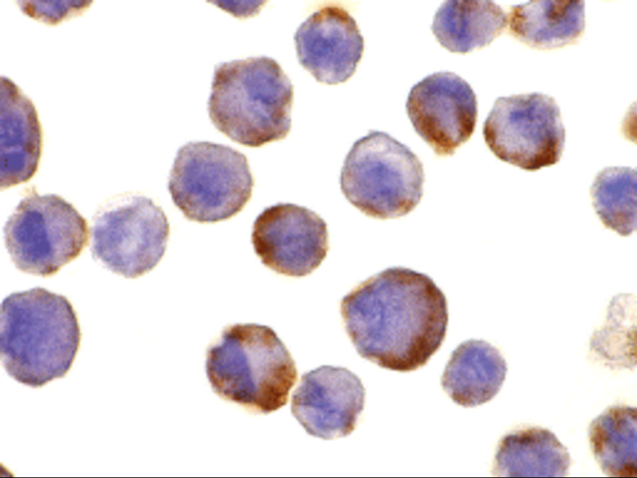 |
| Immunocytochemistry Validation of TACE in HeLa Cells Immunohistochemical analysis of HeLa cells using anti-TACE antibody (8082) at 10 µg/mL. Cells was fixed with formaldehyde and blocked with 10% serum for 1 h at RT; antigen retrieval was by heat mediation with a citrate buffer (pH6). Samples were incubated with primary antibody overnight at 4C. A goat anti-rabbit IgG H&L (HRP) at 1/250 was used as secondary. Counter stained with Hematoxylin. |
 |
| KD Validation of TACE in Monkey COS Cells. (Wang et al., 2006) COS cells stably expressing Pref-1A were transfected with control siRNA or TACE siRNA. TACE was detected in lysates by using the anti-TACE antibody (8082). TACE expression levels were markedly reduced in TACE knockdown cell lysate. |
 |
| KD Validation of TACE in MDA-MB-435 Cells. (McGowan et al., 2416) ADAM-17 protein expression, following transfection with ADAM-17 shRNA (two clones) or neomycin-resistant negative control vector, was examined by immunoblot analysis with anti-ADAM-17 antibodies (8082). |
 |
| Overexpression Validation of TACE in MCF-7 Cells. (McGowan et al., 2416) ADAM-17 (TACE) protein expression, following transfection of vector and ADAM-17 cDNA, was examined by immunoblot analysis with anti-ADAM-17 (8082) antibodies in MCF-7 cells. Increased ADAM-17 was detected in ADAM-17 transfected cells. |
 |
| Induced Expression Validation of TACE in Rat Cortical Neurons (Hurtado et al., 2002) Effect of oxygen-glucose deprivation(OGD) or glutamate on the levels of TACE/ADAM17 in rat cortical cultures. Western blot analysis of TACE in homogenates from control, glutamate, and OGD-exposed cultures from a representative experiment. |
Publications
| pmid | title | authors | citation |
|---|---|---|---|
| We haven't added any publications to our database yet. | |||
Protocols
| relevant to this product |
|---|
| Western blot IHC ICC |
Documents
| # | SDS | Certificate | |
|---|---|---|---|
| Please enter your product and batch number here to retrieve product datasheet, SDS, and QC information. | |||
Only logged in customers who have purchased this product may leave a review.

Reviews
There are no reviews yet.