| Weight | 1 lbs |
|---|---|
| Dimensions | 9 × 5 × 2 in |
| host | rabbit |
| isotype | IgG |
| clonality | polyclonal |
| concentration | 1 mg/mL |
| applications | ICC/IF, WB |
| reactivity | Survivin (CT) |
| available sizes | 100 µg |


rabbit anti-Survivin (CT) polyclonal antibody 4260
$445.00
Antibody summary
- Rabbit polyclonal to Survivin (CT)
- Suitable for: ELISA,WB,ICC,IF
- Isotype: IgG
- 100 µg
rabbit anti-Survivin (CT) polyclonal antibody 4260
| antibody |
|---|
| Tested applications WB,ICC/IF,ELISA |
| Recommended dilutions Immunoblotting: use at 1ug/mL. Positive control: MOLT4 cell lysate. Immunocytochemistry: use at 10ug/mL. These are recommended concentrations. Enduser should determine optimal concentrations for their applications. |
| Immunogen Peptide corresponding to aa 129- 142 of human Survivin (accession no. NP_001159). |
| Size and concentration 100µg and lot specific |
| Form liquid |
| Storage Instructions This antibody is stable for at least one (1) year at -20°C. Avoid multiple freeze-thaw cycles. |
| Storage buffer PBS, pH 7.4. |
| Purity peptide affinity purification |
| Clonality polyclonal |
| Isotype IgG |
| Compatible secondaries goat anti-rabbit IgG, H&L chain specific, peroxidase conjugated, conjugated polyclonal antibody 9512 goat anti-rabbit IgG, H&L chain specific, biotin conjugated polyclonal antibody 2079 goat anti-rabbit IgG, H&L chain specific, FITC conjugated polyclonal antibody 7863 goat anti-rabbit IgG, H&L chain specific, Cross Absorbed polyclonal antibody 2371 goat anti-rabbit IgG, H&L chain specific, biotin conjugated polyclonal antibody, crossabsorbed 1715 goat anti-rabbit IgG, H&L chain specific, FITC conjugated polyclonal antibody, crossabsorbed 1720 |
| Isotype control Rabbit polyclonal - Isotype Control |
| target relevance |
|---|
| Homo sapiens BIRC5 Baculoviral IAP repeat-containing protein 5 |
| Protein names Baculoviral IAP repeat-containing protein 5 |
| Alternative names Apoptosis inhibitor 4, Apoptosis inhibitor survivin |
| Gene names BIRC5 |
| Protein family Belongs to the IAP family |
| Function Multitasking protein that has dual roles in promoting cell proliferation and preventing apoptosis (PubMed:20627126, PubMed:21364656, PubMed:25778398, PubMed:28218735, PubMed:9859993). Component of a chromosome passage protein complex (CPC) which is essential for chromosome alignment and segregation during mitosis and cytokinesis (PubMed:16322459). Acts as an important regulator of the localization of this complex; directs CPC movement to different locations from the inner centromere during prometaphase to midbody during cytokinesis and participates in the organization of the center spindle by associating with polymerized microtubules (PubMed:20826784). Involved in the recruitment of CPC to centromeres during early mitosis via association with histone H3 phosphorylated at 'Thr-3' (H3pT3) during mitosis (PubMed:20929775). The complex with RAN plays a role in mitotic spindle formation by serving as a physical scaffold to help deliver the RAN effector molecule TPX2 to microtubules (PubMed:18591255). May counteract a default induction of apoptosis in G2/M phase (PubMed:9859993). The acetylated form represses STAT3 transactivation of target gene promoters (PubMed:20826784). May play a role in neoplasia (PubMed:10626797). Inhibitor of CASP3 and CASP7 (PubMed:21536684). Essential for the maintenance of mitochondrial integrity and function (PubMed:25778398). Isoform 2 and isoform 3 do not appear to play vital roles in mitosis (PubMed:12773388, PubMed:16291752). Isoform 3 shows a marked reduction in its anti-apoptotic effects when compared with the displayed wild-type isoform (PubMed:10626797) |
| Subcellular location Cytoplasm, Nucleus, Chromosome, Chromosome, centromere, Cytoplasm, cytoskeleton, spindle, Chromosome, centromere, kinetochore, Midbody |
| Structure (Microbial infection) Interacts with Epstein-Barr virus (EBV) EBNA1; this interaction is probably important for EBV episome maintenance in Burkitt's lymphoma cells |
| Post-translational modification Ubiquitinated by the Cul9-RING ubiquitin-protein ligase complex, leading to its degradation. Ubiquitination is required for centrosomal targeting. Deubiquitinated by USP35 or USP38; leading to stabilization (PubMed:34438346) In vitro phosphorylation at Thr-117 by AURKB prevents interaction with INCENP and localization to mitotic chromosomes (PubMed:14610074). Phosphorylation at Thr-48 by CK2 is critical for its mitotic and anti-apoptotic activities (PubMed:21252625). Phosphorylation at Thr-34 by CDK15 is critical for its anti-apoptotic activity (PubMed:24866247). Phosphorylation at Ser-20 by AURKC is critical for regulation of proper chromosome alignment and segregation, and possibly cytokinesis Acetylation at Lys-129 by CBP results in its homodimerization, while deacetylation promotes the formation of monomers which heterodimerize with XPO1/CRM1 which facilitates its nuclear export. The acetylated form represses STAT3 transactivation. The dynamic equilibrium between its acetylation and deacetylation at Lys-129 determines its interaction with XPO1/CRM1, its subsequent subcellular localization, and its ability to inhibit STAT3 transactivation |
| Keywords 3D-structure, Acetylation, Alternative splicing, Apoptosis, Cell cycle, Cell division, Centromere, Chromosome, Chromosome partition, Cytoplasm, Cytoskeleton, Host-virus interaction, Kinetochore, Metal-binding, Microtubule, Mitosis, Nucleus, Phosphoprotein, Protease inhibitor, Proteomics identification, Reference proteome, Repressor, Thiol protease inhibitor, Transcription, Transcription regulation, Ubl conjugation, Zinc |
| Sequence MGAPTLPPAWQPFLKDHRISTFKNWPFLEGCACTPERMAEAGFIHCPTENEPDLAQCFFC FKELEGWEPDDDPIEEHKKHSSGCAFLSVKKQFEELTLGEFLKLDRERAKNKIAKETNNK KKEFEETAKKVRRAIEQLAAMD |
| UniProt accession: O15392 |
Data
 |
| Western Blot Validation in Human MOLT4 Cell Lysate with the absence (A) or presence (B) of blocking peptide Loading: 15 µg of lysates per lane. Antibodies: Survivin 4260 (1 µg/mL), 1h incubation at RT in 5% NFDM/TBST.Secondary: Goat anti-rabbit IgG HRP conjugate at 1:10000 dilution. |
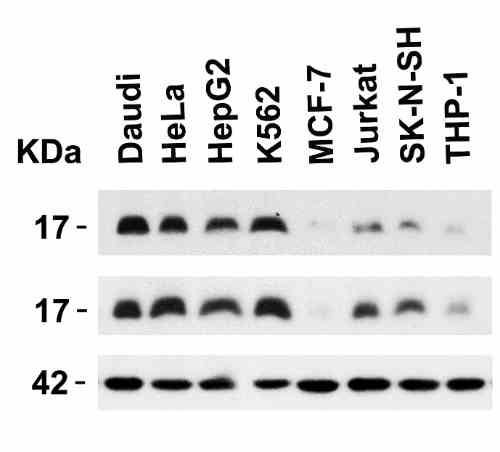 |
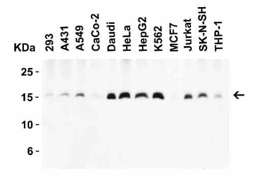
| Independent Antibody Validation (IAV) via Protein Expression Profile in Human Cell Lines Loading: 15 µg of lysates per lane. Antibodies: Survivin 2233 (5 µg/mL), Survivin 4260 (4 µg/mL) and beta-actin 3779 (1 µg/mL), 1h incubation at RT in 5% NFDM/TBST.Secondary: Goat anti-rabbit IgG HRP conjugate at 1:10000 dilution. |
 |
| Western Blot Validation in Human Cell Lines Loading: 15 µg of lysates per lane. Antibodies: Survivin 4260 (4 µg/mL), 1h incubation at RT in 5% NFDM/TBST.Secondary: Goat anti-rabbit IgG HRP conjugate at 1:10000 dilution. |
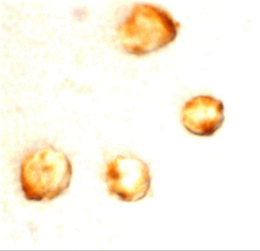 |
| Immunocytochemistry Validation of Survivin in MOLT4 Cells Immunocytochemical analysis of MOLT4 cells using anti-Survivin antibody (4260) at 10 µg/mL. Cells was fixed with formaldehyde and blocked with 10% serum for 1 h at RT; antigen retrieval was by heat mediation with a citrate buffer (pH6). Samples were incubated with primary antibody overnight at 4C. A goat anti-rabbit IgG H&L (HRP) at 1/250 was used as secondary. Counter stained with Hematoxylin. |
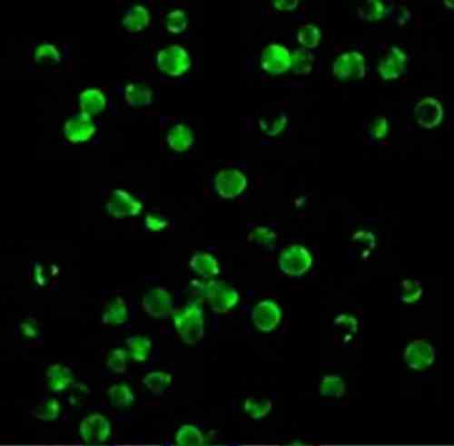 |
| Immunofluorescence Validation of Survivin in MOLT4 Cells Immunofluorescent analysis of 4% paraformaldehyde-fixed MOLT4 cells labeling Survivin with 4260 at 10 µg/mL, followed by goat anti-rabbit IgG secondary antibody at 1/500 dilution (green). |
 |
| Slot Blot Validation of Survivin in Hepatocellular Carcinoma (HCC) Patients (Zhang et al., 2416) Slot blot analysis of Survivin recombinant protein expression with anti-Survivin antibodies (4260) in the four representative HCC sera. Survivin (Lane 7) was detected in the fourth HCC serum (E), but not in the normal human serum (A) and the other three HCC sera (B-D). Phosphate bufferedsaline (PBS) (Lanes 9 and 10) was used as a negative control. |
 |
| Western Blot Validation of Survivin with Recombinant Protein (Megliorino et al., 2005) Lane 1: A 28 kDa peptide that corresponded to the size of ORF of survivin was detected in SDS-PAGE with Coomassie blue staining. Lane 2: WB analysis of Survivin recombinant protein expression with anti-Survivin antibodies (4260). |
 |
| Western Blot Validation of Survivin with Recombinant Protein in Sera from Cancer Patients (Megliorino et al., 2005) WB analysis of Survivin recombinant protein expression with anti-Survivin antibodies (4260) in six cancer sera. 6 x His-tagged Survivin recombinant protein (Lanes 4-9) was strongly detected at 28kD, but not in the normal human serum (Lane 3). |
FAQ & Publications
Frequently Asked Questions
What are the recommended applications and dilutions for the rabbit anti-Survivin (CT) polyclonal antibody 4260?
This antibody is suitable for ELISA, Western Blot (WB), Immunocytochemistry (ICC), and Immunofluorescence (IF). Recommended dilutions are 1 µg/mL for immunoblotting and 10 µg/mL for immunocytochemistry. Users should optimize concentrations for their specific applications.
How should I store the rabbit anti-Survivin (CT) polyclonal antibody 4260 to ensure stability?
The antibody should be stored at -20°C where it remains stable for at least one year. Avoid multiple freeze-thaw cycles to preserve antibody integrity.
What is the immunogen used to generate the rabbit anti-Survivin (CT) polyclonal antibody 4260 and what is the host species?
This antibody was raised in rabbit against a peptide corresponding to amino acids 129-142 of human Survivin (accession no. NP_001159).
Publications
| pmid | title | authors | citation |
|---|---|---|---|
| We haven't added any publications to our database yet. | |||
Published literature highly relevant to the biological target of this product and referencing this antibody or clone are retrieved from the PubMed database provided by the United States National Library of Medicine at the National Institutes of Health.
Protocols
| relevant to this product |
|---|
| Western blot IHC ICC |
Documents
| Batch Number | QC File | SDS |
|---|---|---|
| To view batch-specific Safety Datasheets and Quality Certificates associated with your account, please Log In. | ||
Only logged in customers who have purchased this product may leave a review.


Reviews
There are no reviews yet.