| Weight | 1 lbs |
|---|---|
| Dimensions | 9 × 5 × 2 in |
| host | rabbit |
| isotype | IgG |
| clonality | polyclonal |
| concentration | 1 mg/mL |
| applications | ICC/IF, WB |
| reactivity | Aurora A/STK15 |
| available sizes | 100 µg |

rabbit anti-STK15 polyclonal antibody 2579
$518.00
Antibody summary
- Rabbit polyclonal to STK15
- Suitable for: IP
- Isotype: Whole IgG
- 100 µg
rabbit anti-STK15 polyclonal antibody 2579
| antibody |
|---|
| Tested applications IP |
| Recommended dilutions Immunoprecipitation: 2-10ug/mg lysate. These are recommended concentrations. Endusers should determine optimal concentrations for their applications. |
| Immunogen Synthetic peptide representing a portion of the protein encoded within exon 5. |
| Size and concentration 100µg and lot specific |
| Form liquid |
| Storage Instructions This antibody is stable at 2 - 8°C for one (1) year from date of receipt. |
| Storage buffer Tris-citrate/phosphate buffer, pH 7 to 8 contai |
| Purity immunogen affinity purification |
| Clonality polyclonal |
| Isotype IgG |
| Compatible secondaries goat anti-rabbit IgG, H&L chain specific, peroxidase conjugated, conjugated polyclonal antibody 9512 goat anti-rabbit IgG, H&L chain specific, biotin conjugated polyclonal antibody 2079 goat anti-rabbit IgG, H&L chain specific, FITC conjugated polyclonal antibody 7863 goat anti-rabbit IgG, H&L chain specific, Cross Absorbed polyclonal antibody 2371 goat anti-rabbit IgG, H&L chain specific, biotin conjugated polyclonal antibody, crossabsorbed 1715 goat anti-rabbit IgG, H&L chain specific, FITC conjugated polyclonal antibody, crossabsorbed 1720 |
| Isotype control Rabbit polyclonal - Isotype Control |
| target relevance |
|---|
| Homo sapiens AURKA Aurora kinase A |
| Protein names Aurora kinase A |
| Alternative names Aurora 2, Aurora/IPL1-related kinase 1, Breast tumor-amplified kinase, Ipl1- and aurora-related kinase 1, Serine/threonine-protein kinase 15, Serine/threonine-protein kinase 6, Serine/threonine-protein kinase Ayk1, Serine/threonine-protein kinase aurora-A |
| Gene names AURKA |
| Protein family Belongs to the protein kinase superfamily. Ser/Thr protein kinase family. Aurora subfamily |
| Function Mitotic serine/threonine kinase that contributes to the regulation of cell cycle progression (PubMed:11039908, PubMed:12390251, PubMed:17125279, PubMed:17360485, PubMed:18615013, PubMed:26246606). Associates with the centrosome and the spindle microtubules during mitosis and plays a critical role in various mitotic events including the establishment of mitotic spindle, centrosome duplication, centrosome separation as well as maturation, chromosomal alignment, spindle assembly checkpoint, and cytokinesis (PubMed:14523000, PubMed:26246606). Required for normal spindle positioning during mitosis and for the localization of NUMA1 and DCTN1 to the cell cortex during metaphase (PubMed:27335426). Required for initial activation of CDK1 at centrosomes (PubMed:13678582, PubMed:15128871). Phosphorylates numerous target proteins, including ARHGEF2, BORA, BRCA1, CDC25B, DLGP5, HDAC6, KIF2A, LATS2, NDEL1, PARD3, PPP1R2, PLK1, RASSF1, TACC3, p53/TP53 and TPX2 (PubMed:11551964, PubMed:14702041, PubMed:15128871, PubMed:15147269, PubMed:15987997, PubMed:17604723, PubMed:18056443, PubMed:18615013). Phosphorylates MCRS1 which is required for MCRS1-mediated kinetochore fiber assembly and mitotic progression (PubMed:27192185). Regulates KIF2A tubulin depolymerase activity (PubMed:19351716). Important for microtubule formation and/or stabilization (PubMed:18056443). Required for normal axon formation (PubMed:19812038). Plays a role in microtubule remodeling during neurite extension (PubMed:19668197). Also acts as a key regulatory component of the p53/TP53 pathway, and particularly the checkpoint-response pathways critical for oncogenic transformation of cells, by phosphorylating and destabilizing p53/TP53 (PubMed:14702041). Phosphorylates its own inhibitors, the protein phosphatase type 1 (PP1) isoforms, to inhibit their activity (PubMed:11551964). Inhibits cilia outgrowth (By similarity). Required for cilia disassembly via phosphorylation of HDAC6 and subsequent deacetylation of alpha-tubulin (PubMed:17604723, PubMed:20643351). Regulates protein levels of the anti-apoptosis protein BIRC5 by suppressing the expression of the SCF(FBXL7) E3 ubiquitin-protein ligase substrate adapter FBXL7 through the phosphorylation of the transcription factor FOXP1 (PubMed:28218735) |
| Catalytic activity L-seryl-[protein] + ATP = O-phospho-L-seryl-[protein] + ADP + H(+) L-threonyl-[protein] + ATP = O-phospho-L-threonyl-[protein] + ADP + H(+) |
| Subcellular location Cytoplasm, cytoskeleton, microtubule organizing center, centrosome, Cytoplasm, cytoskeleton, spindle pole, Cytoplasm, cytoskeleton, microtubule organizing center, centrosome, centriole, Cell projection, neuron projection, Cell projection, cilium, Cytoplasm, cytoskeleton, cilium basal body, Basolateral cell membrane |
| Structure Part of a complex composed of NEDD9, AURKA and CTTN; within the complex NEDD9 acts as a scaffold protein and is required for complex formation (PubMed:24574519). Identified in a complex with AUNIP and NIN (PubMed:20596670). Interacts with FBXL7 (By similarity). Interacts with CPEB1, JTB, TACC1, TPX2, PPP2CA, as well as with the protein phosphatase type 1 (PP1) isoforms PPP1CA, PPP1CB and PPP1CC (PubMed:11551964, PubMed:14580337, PubMed:14603251, PubMed:15966895, PubMed:17229885, PubMed:18662907, PubMed:19357306, PubMed:19668197, PubMed:19801554, PubMed:21225229, PubMed:27837025). Also interacts with its substrates ARHGEF2, BORA, KIF2A, PARD3, and p53/TP53 (PubMed:14702041, PubMed:16890155, PubMed:17488622, PubMed:19351716, PubMed:19812038). Interaction with BORA promotes phosphorylation of PLK1 (By similarity). Interacts with CIMAP3 (PubMed:20643351). Interacts with GADD45A, competing with its oligomerization (PubMed:20460379). Interacts (via C-terminus) with AUNIP (via C-terminus) (PubMed:20596670). Interacts with FRY; this interaction facilitates AURKA-mediated PLK1 phosphorylation (PubMed:22753416). Interacts with SIRT2 (PubMed:17726514, PubMed:22014574). Interacts with MYCN; interaction is phospho-independent and triggers AURKA activation; AURKA competes with FBXW7 for binding to unphosphorylated MYCN but not for binding to phosphorylated MYCN (PubMed:27837025). Interacts with HNRNPU (PubMed:21242313, PubMed:25986610). Interacts with AAAS (PubMed:26246606). Interacts with KLHL18 and CUL3 (PubMed:23213400). Interacts with FOXP1 (PubMed:28218735). Interacts with HDAC6; AURKA-mediated phosphorylation of HDAC6 promotes deacetylation of alpha-tubulin (PubMed:17604723) |
| Post-translational modification Activated by phosphorylation at Thr-288; this brings about a change in the conformation of the activation segment. Phosphorylation at Thr-288 varies during the cell cycle and is highest during M phase. Autophosphorylated at Thr-288 upon TPX2 binding. Thr-288 can be phosphorylated by several kinases, including PAK and PKA. Protein phosphatase type 1 (PP1) binds AURKA and inhibits its activity by dephosphorylating Thr-288 during mitosis. Phosphorylation at Ser-342 decreases the kinase activity. PPP2CA controls degradation by dephosphorylating Ser-51 at the end of mitosis Ubiquitinated by the E3 ubiquitin-protein ligase complex SCF(FBXL7) during mitosis, leading to its degradation by the proteasome (By similarity). Ubiquitinated by CHFR, leading to its degradation by the proteasome (By similarity). Ubiquitinated by the anaphase-promoting complex (APC), leading to its degradation by the proteasome (PubMed:10851084, PubMed:11039908). Ubiquitinated by the CUL3-KLHL18 ligase leading to its activation at the centrosome which is required for initiating mitotic entry (PubMed:23213400). Ubiquitination mediated by CUL3-KLHL18 ligase does not lead to its degradation by the proteasome (PubMed:23213400) |
| Keywords 3D-structure, ATP-binding, Cell cycle, Cell division, Cell membrane, Cell projection, Cilium, Cilium biogenesis/degradation, Cytoplasm, Cytoskeleton, Isopeptide bond, Kinase, Membrane, Microtubule, Mitosis, Nucleotide-binding, Phosphoprotein, Proteomics identification, Proto-oncogene, Reference proteome, Serine/threonine-protein kinase, Transferase, Ubl conjugation |
| Sequence MDRSKENCISGPVKATAPVGGPKRVLVTQQFPCQNPLPVNSGQAQRVLCPSNSSQRIPLQ AQKLVSSHKPVQNQKQKQLQATSVPHPVSRPLNNTQKSKQPLPSAPENNPEEELASKQKN EESKKRQWALEDFEIGRPLGKGKFGNVYLAREKQSKFILALKVLFKAQLEKAGVEHQLRR EVEIQSHLRHPNILRLYGYFHDATRVYLILEYAPLGTVYRELQKLSKFDEQRTATYITEL ANALSYCHSKRVIHRDIKPENLLLGSAGELKIADFGWSVHAPSSRRTTLCGTLDYLPPEM IEGRMHDEKVDLWSLGVLCYEFLVGKPPFEANTYQETYKRISRVEFTFPDFVTEGARDLI SRLLKHNPSQRPMLREVLEHPWITANSSKPSNCQNKESASKQS |
| UniProt accession: O14965 |
Data
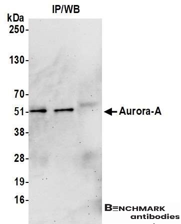 |
| Detection of human Aurora A by western blot of immunoprecipitates. Samples: Whole cell lysate (1.0 mg per IP reaction; 20% of IP loaded) from HeLa cells prepared using NETN lysis buffer. Antibodies: Affinity purified rabbit anti-Aurora A antibody 2579 (lot 2579-6) used for IP at 6 µg per reaction. Aurora A was also immunoprecipitated by a previous lot of this antibody (lot 2579-1). For blotting immunoprecipitated Aurora A, 13042 was used at 0.1 µg /ml. Detection: Chemiluminescence with an exposure time of 75 seconds. |
FAQ & Publications
Frequently Asked Questions
What is the recommended dilution range for immunoprecipitation using the rabbit anti-STK15 polyclonal antibody?
The recommended concentration for immunoprecipitation is 2-10 µg of antibody per mg of lysate. However, end users should optimize the concentration based on their specific experimental conditions.
How should the rabbit anti-STK15 polyclonal antibody be stored to maintain its stability?
This antibody should be stored at 2 to 8°C and is stable for one year from the date of receipt when kept under these conditions.
What is the immunogen used to generate the rabbit anti-STK15 polyclonal antibody?
The antibody was raised against a synthetic peptide representing a portion of the protein encoded within exon 5 of the STK15 gene.
Which species or protein does the rabbit anti-STK15 polyclonal antibody specifically react with?
This antibody specifically reacts with Aurora A kinase, also known as STK15.
What is the concentration and form of the rabbit anti-STK15 polyclonal antibody provided?
The antibody is supplied as a liquid at a concentration of 1 mg/mL, with a total amount of 100 µg per vial.
Publications
| pmid | title | authors | citation |
|---|---|---|---|
| We haven't added any publications to our database yet. | |||
Published literature highly relevant to the biological target of this product and referencing this antibody or clone are retrieved from the PubMed database provided by the United States National Library of Medicine at the National Institutes of Health.
Protocols
| relevant to this product |
|---|
| Western blot IHC ICC |
Documents
| Batch Number | QC File | SDS |
|---|---|---|
| To view batch-specific Safety Datasheets and Quality Certificates associated with your account, please Log In. | ||
Only logged in customers who have purchased this product may leave a review.


Reviews
There are no reviews yet.