| Weight | 1 lbs |
|---|---|
| Dimensions | 9 × 5 × 2 in |
| host | rabbit |
| isotype | IgG |
| clonality | polyclonal |
| concentration | 1 mg/mL |
| applications | ICC/IF, WB |
| reactivity | STAT1α |
| available sizes | 100 µg |


rabbit anti-STAT1 α polyclonal antibody 4141
$445.00
Antibody summary
- Rabbit polyclonal to STAT1 α
- Suitable for: ELISA,WB,ICC,IP,IHC-P,IF
- Isotype: IgG
- 100 µg
rabbit anti-STAT1 α polyclonal antibody 4141
| antibody |
|---|
| Tested applications WB,IHC,IHC,ICC/IF,ELISA,IP |
| Recommended dilutions Immunoblotting : use a1:1,000 dilution. Immunoprecipitation: use 2-4 ug antibody per sample. Positive control: Whole cell lysate from HeLa cells, Jurkat cells, or A431 cells. |
| Immunogen Peptide corresponding to aa 712-750 of human STAT1a. |
| Size and concentration 100µg and lot specific |
| Form liquid |
| Storage Instructions This antibody is stable for at least one (1) year at -20°C. Avoid multiple freeze- thaw cycles. |
| Storage buffer PBS, pH 7.4. |
| Purity peptide affinity purification |
| Clonality polyclonal |
| Isotype IgG |
| Compatible secondaries goat anti-rabbit IgG, H&L chain specific, peroxidase conjugated, conjugated polyclonal antibody 9512 goat anti-rabbit IgG, H&L chain specific, biotin conjugated polyclonal antibody 2079 goat anti-rabbit IgG, H&L chain specific, FITC conjugated polyclonal antibody 7863 goat anti-rabbit IgG, H&L chain specific, Cross Absorbed polyclonal antibody 2371 goat anti-rabbit IgG, H&L chain specific, biotin conjugated polyclonal antibody, crossabsorbed 1715 goat anti-rabbit IgG, H&L chain specific, FITC conjugated polyclonal antibody, crossabsorbed 1720 |
| Isotype control Rabbit polyclonal - Isotype Control |
| target relevance |
|---|
| Homo sapiens STAT1 Signal transducer and activator of transcription 1-alpha/beta |
| Protein names Signal transducer and activator of transcription 1-alpha/beta |
| Alternative names Transcription factor ISGF-3 components p91/p84 |
| Gene names STAT1 |
| Protein family Belongs to the transcription factor STAT family |
| Function Signal transducer and transcription activator that mediates cellular responses to interferons (IFNs), cytokine KITLG/SCF and other cytokines and other growth factors (PubMed:12764129, PubMed:12855578, PubMed:15322115, PubMed:23940278, PubMed:34508746, PubMed:35568036, PubMed:9724754). Following type I IFN (IFN-alpha and IFN-beta) binding to cell surface receptors, signaling via protein kinases leads to activation of Jak kinases (TYK2 and JAK1) and to tyrosine phosphorylation of STAT1 and STAT2. The phosphorylated STATs dimerize and associate with ISGF3G/IRF-9 to form a complex termed ISGF3 transcription factor, that enters the nucleus (PubMed:28753426, PubMed:35568036). ISGF3 binds to the IFN stimulated response element (ISRE) to activate the transcription of IFN-stimulated genes (ISG), which drive the cell in an antiviral state (PubMed:28753426, PubMed:35568036). In response to type II IFN (IFN-gamma), STAT1 is tyrosine- and serine-phosphorylated (PubMed:26479788). It then forms a homodimer termed IFN-gamma-activated factor (GAF), migrates into the nucleus and binds to the IFN gamma activated sequence (GAS) to drive the expression of the target genes, inducing a cellular antiviral state (PubMed:8156998). Becomes activated in response to KITLG/SCF and KIT signaling (PubMed:15526160). May mediate cellular responses to activated FGFR1, FGFR2, FGFR3 and FGFR4 (PubMed:19088846). Following bacterial lipopolysaccharide (LPS)-induced TLR4 endocytosis, phosphorylated at Thr-749 by IKBKB which promotes binding of STAT1 to the 5'-TTTGAGGC-3' sequence in the ARID5A promoter, resulting in transcriptional activation of ARID5A and subsequent ARID5A-mediated stabilization of IL6 (PubMed:32209697). Phosphorylation at Thr-749 also promotes binding of STAT1 to the 5'-TTTGAGTC-3' sequence in the IL12B promoter and activation of IL12B transcription (PubMed:32209697). Involved in food tolerance in small intestine: associates with the Gasdermin-D, p13 cleavage product (13 kDa GSDMD) and promotes transcription of CIITA, inducing type 1 regulatory T (Tr1) cells in upper small intestine (By similarity) |
| Subcellular location Cytoplasm, Nucleus |
| Structure (Microbial infection) Interacts (via N-terminus) with measles V protein; this interaction inhibits STAT1 phosphorylation by Jak1 and thereby the type I interferon signaling pathway |
| Post-translational modification Deubiquitinated by USP13; leading to STAT1 stabilization and positive regulation of type I and type II IFN signalings Phosphorylated on tyrosine and serine residues in response to a variety of cytokines/growth hormones including IFN-alpha, IFN-gamma, PDGF and EGF (PubMed:26479788, PubMed:28753426). Activated KIT promotes phosphorylation on tyrosine residues and subsequent translocation to the nucleus (PubMed:21135090). Upon EGF stimulation, phosphorylation on Tyr-701 (lacking in beta form) by JAK1, JAK2 or TYK2 promotes dimerization and subsequent translocation to the nucleus (PubMed:28753426, PubMed:7657660). Growth hormone (GH) activates STAT1 signaling only via JAK2 (PubMed:7657660). Tyrosine phosphorylated in response to constitutively activated FGFR1, FGFR2, FGFR3 and FGFR4 (PubMed:17561467, PubMed:19088846). Phosphorylation on Ser-727 by several kinases including MAPK14, ERK1/2, CAMK2/CAMKII and CK2 in response to IFN-gamma stimulation, is required for maximal transcriptional activity (PubMed:15322115, PubMed:16799645, PubMed:17897103, PubMed:7543024). Phosphorylated on Ser-727 by CAMK2/CAMKII in response to IFN-gamma stimulation and calcium mobilization, promoting activity (PubMed:11972023, PubMed:16257975). Phosphorylated by CAMK2/CAMKII in response to IFN-beta stimulation and calcium mobilization in epithelial cells, promoting activity (PubMed:35568036). Phosphorylation on Ser-727 promotes sumoylation though increasing interaction with PIAS (PubMed:17897103). Phosphorylation on Ser-727 by PRKCD induces apoptosis in response to DNA-damaging agents (PubMed:15322115). Phosphorylated on tyrosine residues when PTK2/FAK1 is activated; most likely this is catalyzed by a SRC family kinase (PubMed:11278462). Dephosphorylation on tyrosine residues by PTPN2 negatively regulates interferon-mediated signaling (PubMed:12138178). Upon viral infection or IFN induction, phosphorylation on Ser-708 occurs much later than phosphorylation on Tyr-701 and is required for the binding of ISGF3 on the ISREs of a subset of IFN-stimulated genes IKBKE-dependent (PubMed:22065572). Phosphorylation at Tyr-701 and Ser-708 are mutually exclusive, phosphorylation at Ser-708 requires previous dephosphorylation of Tyr-701 (PubMed:22065572). Phosphorylation at Thr-749 by IKBKB/IKKB promotes transcriptional activation of ARID5A and IL12B by STAT1 (PubMed:32209697). Phosphorylation at Thr-749 restricts interferon signaling and anti-inflammatory responses and promotes innate inflammatory responses (By similarity) Sumoylated with SUMO1, SUMO2 and SUMO3. Sumoylation is enhanced by IFN-gamma-induced phosphorylation on Ser-727, and by interaction with PIAS proteins. Enhances the transactivation activity ISGylated Mono-ADP-ribosylated at Glu-657 and Glu-705 by PARP14; ADP-ribosylation prevents phosphorylation at Tyr-701 (PubMed:27796300). However, the role of ADP-ribosylation in the prevention of phosphorylation has been called into question and the lack of phosphorylation may be due to sumoylation of Lys-703 (PubMed:29858569) Monomethylated at Lys-525 by SETD2; monomethylation is necessary for phosphorylation at Tyr-701, translocation into the nucleus and activation of the antiviral defense (Microbial infection) Ubiquitinated by Herpes simplex virus 2 E3 ubiquitin ligase ICP22 |
| Involvement in disease Immunodeficiency 31B A disorder characterized by susceptibility to severe mycobacterial and viral infections. Affected individuals can develop disseminated infections and die of viral illness. Immunodeficiency 31A A form of Mendelian susceptibility to mycobacterial disease, a rare condition caused by impairment of interferon-gamma mediated immunity. It is characterized by predisposition to illness caused by moderately virulent mycobacterial species, such as Bacillus Calmette-Guerin (BCG) vaccine, environmental non-tuberculous mycobacteria, and by the more virulent Mycobacterium tuberculosis. Other microorganisms rarely cause severe clinical disease in individuals with susceptibility to mycobacterial infections, with the exception of Salmonella which infects less than 50% of these individuals. Clinical outcome severity depends on the degree of impairment of interferon-gamma mediated immunity. Some patients die of overwhelming mycobacterial disease with lepromatous-like lesions in early childhood, whereas others develop, later in life, disseminated but curable infections with tuberculoid granulomas. IMD31A has low penetrance, and affected individuals have relatively mild disease and good prognosis. IMD31A confers a predisposition to mycobacterial infections only, with no increased susceptibility to viral infections. Immunodeficiency 31C A primary immunodeficiency disorder with altered immune responses and impaired clearance of fungal infections, selective against Candida. It is characterized by persistent and/or recurrent infections of the skin, nails and mucous membranes caused by organisms of the genus Candida, mainly Candida albicans. |
| Keywords 3D-structure, Acetylation, Activator, ADP-ribosylation, Alternative splicing, Antiviral defense, Coiled coil, Cytoplasm, Direct protein sequencing, Disease variant, DNA-binding, Host-virus interaction, Isopeptide bond, Methylation, Nucleus, Phosphoprotein, Proteomics identification, Reference proteome, SH2 domain, Transcription, Transcription regulation, Ubl conjugation |
| Sequence MSQWYELQQLDSKFLEQVHQLYDDSFPMEIRQYLAQWLEKQDWEHAANDVSFATIRFHDL LSQLDDQYSRFSLENNFLLQHNIRKSKRNLQDNFQEDPIQMSMIIYSCLKEERKILENAQ RFNQAQSGNIQSTVMLDKQKELDSKVRNVKDKVMCIEHEIKSLEDLQDEYDFKCKTLQNR EHETNGVAKSDQKQEQLLLKKMYLMLDNKRKEVVHKIIELLNVTELTQNALINDELVEWK RRQQSACIGGPPNACLDQLQNWFTIVAESLQQVRQQLKKLEELEQKYTYEHDPITKNKQV LWDRTFSLFQQLIQSSFVVERQPCMPTHPQRPLVLKTGVQFTVKLRLLVKLQELNYNLKV KVLFDKDVNERNTVKGFRKFNILGTHTKVMNMEESTNGSLAAEFRHLQLKEQKNAGTRTN EGPLIVTEELHSLSFETQLCQPGLVIDLETTSLPVVVISNVSQLPSGWASILWYNMLVAE PRNLSFFLTPPCARWAQLSEVLSWQFSSVTKRGLNVDQLNMLGEKLLGPNASPDGLIPWT RFCKENINDKNFPFWLWIESILELIKKHLLPLWNDGCIMGFISKERERALLKDQQPGTFL LRFSESSREGAITFTWVERSQNGGEPDFHAVEPYTKKELSAVTFPDIIRNYKVMAAENIP ENPLKYLYPNIDKDHAFGKYYSRPKEAPEPMELDGPKGTGYIKTELISVSEVHPSRLQTT DNLLPMSPEEFDEVSRIVGSVEFDSMMNTV |
| UniProt accession: P42224 |
Data
 |
| Western Blot Validation in Human and Mouse Cell Lines of (H) Hela, (J) Jurkat, (A) A431, (K) K562 and (N) 3T3/NIH Loading: 15 µg of lysates per lane. Antibodies: STAT1 alpha 4141 (1 µg/mL), 1h incubation at RT in 5% NFDM/TBST.Secondary: Goat anti-rabbit IgG HRP conjugate at 1:10000 dilution. |
 |
| Independent Antibody Validation (IAV) via Protein Expression Profile in Human Cell Lines Loading: 15 µg of lysates per lane. Antibodies: STAT1 alpha 4141 (1 µg/mL), competitor antibody (2 µg/mL), and beta-actin (1.5 µg/mL), 1h incubation at RT in 5% NFDM/TBST.Secondary: Goat anti-rabbit IgG HRP conjugate at 1:10000 dilution. |
 |
| Western Blot Validation in Mouse tissues Loading: 15 µg of lysates per lane. Antibodies: STAT1 alpha 4141 (0.5 µg/mL), 1h incubation at RT in 5% NFDM/TBST.Secondary: Goat anti-rabbit IgG HRP conjugate at 1:10000 dilution. |
 |
| Western Blot Validation in Rat Cell Line Loading: 15 µg of lysates per lane. Antibodies: STAT1 alpha 4141 (1 µg/mL), 1h incubation at RT in 5% NFDM/TBST.Secondary: Goat anti-rabbit IgG HRP conjugate at 1:10000 dilution. |
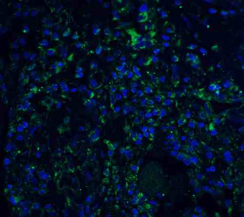 |
| Immunofluorescence Validation of STAT1 alpha in Human Colon Tissue Immunofluorescent analysis of 4% paraformaldehyde-fixed Human Colon Tissue labeling STAT1 alpha with 4141 at 20 µg/mL, followed by goat anti-rabbit IgG secondary antibody at 1/500 dilution (green) and DAPI staining (blue). |
 |
| Immunohistochemistry Validation of STAT1 alpha in Human Colon Tissue Immunohistochemical analysis of paraffin-embedded Human Colon Tissue using anti-STAT1 alpha antibody (4141) at 2.5 µg/mL. Tissue was fixed with formaldehyde and blocked with 10% serum for 1 h at RT; antigen retrieval was by heat mediation with a citrate buffer (pH6). Samples were incubated with primary antibody overnight at 4C. A goat anti-rabbit IgG H&L (HRP) at 1/250 was used as secondary. Counter stained with Hematoxylin. |
 |
| Immunocytochemistry Validation of STAT1 alpha in Human HeLa Cells Immunocytochemical analysis of HeLa cells using anti-STAT1 alpha antibody (4141) at 10 µg/mL. Cells was fixed with formaldehyde and blocked with 10% serum for 1 h at RT; antigen retrieval was by heat mediation with a citrate buffer (pH6). Samples were incubated with primary antibody overnight at 4C. A goat anti-rabbit IgG H&L (HRP) at 1/250 was used as secondary. Counter stained with Hematoxylin. |
FAQ & Publications
Frequently Asked Questions
What applications is the rabbit anti-STAT1 α polyclonal antibody 4141 validated for?
This antibody is suitable and tested for ELISA, Western Blot (WB), Immunocytochemistry/Immunofluorescence (ICC/IF), Immunoprecipitation (IP), and Immunohistochemistry on paraffin-embedded sections (IHC-P). Recommended dilutions include 1:1,000 for immunoblotting and 2-4 µg per sample for immunoprecipitation.
How should the rabbit anti-STAT1 α polyclonal antibody 4141 be stored to maintain stability?
The antibody should be stored at -20°C and is stable for at least one year under these conditions. It is important to avoid multiple freeze-thaw cycles to preserve antibody integrity.
What is the immunogen used to generate the rabbit anti-STAT1 α polyclonal antibody 4141?
The immunogen is a peptide corresponding to amino acids 712-750 of human STAT1 alpha.
Which secondary antibodies are compatible with the rabbit anti-STAT1 α polyclonal antibody 4141 for detection purposes?
Compatible secondary antibodies include goat anti-rabbit IgG, H&L chain specific polyclonal antibodies conjugated to peroxidase, biotin, or FITC, with some available as cross-absorbed versions to reduce cross-reactivity.
Publications
| pmid | title | authors | citation |
|---|---|---|---|
| We haven't added any publications to our database yet. | |||
Published literature highly relevant to the biological target of this product and referencing this antibody or clone are retrieved from the PubMed database provided by the United States National Library of Medicine at the National Institutes of Health.
Protocols
| relevant to this product |
|---|
| Western blot IHC ICC |
Documents
| Batch Number | QC File | SDS |
|---|---|---|
| To view batch-specific Safety Datasheets and Quality Certificates associated with your account, please Log In. | ||
Only logged in customers who have purchased this product may leave a review.


Reviews
There are no reviews yet.