| Weight | 1 lbs |
|---|---|
| Dimensions | 9 × 5 × 2 in |
| host | rabbit |
| isotype | IgG |
| clonality | polyclonal |
| concentration | 1 mg/mL |
| applications | ICC/IF, WB |
| reactivity | Smac (CT) |
| available sizes | 100 µg |


rabbit anti-Smac (CT) polyclonal antibody 4798
$445.00
Antibody summary
- Rabbit polyclonal to Smac (CT)
- Suitable for: ELISA,WB,IHC-P,IP,IF
- Isotype: IgG
- 100 µg
rabbit anti-Smac (CT) polyclonal antibody 4798
| antibody |
|---|
| Tested applications WB,IHC,IHC,ELISA,IP |
| Recommended dilutions Immunoblotting: use at 1ug/mL. Positive control: Human heart tissue lysate. Immunohistochemistry: use at 5ug/mL These are recommended concentrations. Enduser should determine optimal concentrations for their applications. |
| Immunogen Synthetic peptide corresponding to aa 225-239 of human Smac (accession no. AAF87716). |
| Size and concentration 100µg and lot specific |
| Form liquid |
| Storage Instructions This antibody is stable for at least one (1) year at -20°C. Avoid multiple freeze-thaw cycles. |
| Storage buffer PBS, pH 7.4. |
| Purity peptide affinity purification |
| Clonality polyclonal |
| Isotype IgG |
| Compatible secondaries goat anti-rabbit IgG, H&L chain specific, peroxidase conjugated, conjugated polyclonal antibody 9512 goat anti-rabbit IgG, H&L chain specific, biotin conjugated polyclonal antibody 2079 goat anti-rabbit IgG, H&L chain specific, FITC conjugated polyclonal antibody 7863 goat anti-rabbit IgG, H&L chain specific, Cross Absorbed polyclonal antibody 2371 goat anti-rabbit IgG, H&L chain specific, biotin conjugated polyclonal antibody, crossabsorbed 1715 goat anti-rabbit IgG, H&L chain specific, FITC conjugated polyclonal antibody, crossabsorbed 1720 |
| Isotype control Rabbit polyclonal - Isotype Control |
| target relevance |
|---|
| Homo sapiens DIABLO Diablo IAP-binding mitochondrial protein |
| Protein names Diablo IAP-binding mitochondrial protein |
| Alternative names Diablo homolog, mitochondrial, Direct IAP-binding protein with low pI, Second mitochondria-derived activator of caspases |
| Gene names DIABLO |
| Protein family Belongs to the Smac/DIABLO protein family |
| Function Promotes apoptosis by activating caspases in the cytochrome c/Apaf-1/caspase-9 pathway. Acts by opposing the inhibitory activity of inhibitor of apoptosis proteins (IAP). Inhibits the activity of BIRC6/BRUCE by inhibiting its binding to caspases (PubMed:15200957, PubMed:36758104, PubMed:36758105, PubMed:36758106) |
| Subcellular location Mitochondrion, Cytoplasm, cytosol |
| Structure Homodimer (PubMed:10972280, PubMed:36758104, PubMed:36758105, PubMed:36758106). Interacts with BIRC2/c-IAP1 (via BIR3 domain) (PubMed:19153467). Interacts with BIRC6/BRUCE; inhibits BIRC6 activity (PubMed:15200957, PubMed:36758104, PubMed:36758105, PubMed:36758106). Interacts with BIRC7/livin (PubMed:16729033). Interacts with XIAP/BIRC4 (via BIR3 domain) (PubMed:11140637, PubMed:21695558, PubMed:28288130). Interacts with the monomeric and dimeric form of BIRC5/survivin (PubMed:21536684). Interacts with AREL1 (via HECT domain); in the cytoplasm following induction of apoptosis (PubMed:23479728). Interacts with BEX3 (By similarity) |
| Post-translational modification Ubiquitinated by BIRC7/livin (PubMed:16729033). Ubiquitinated by BIRC6 (PubMed:36758104, PubMed:36758105, PubMed:36758106) The precursor form is proteolytically cleaved by mitochondrial processing peptidase MPP to remove the transit peptide and produce an intermediate form. This is then processed by PARL to produce the mature cleaved form which is released from mitochondria into the cytosol in apoptotic cells |
| Involvement in disease Deafness, autosomal dominant, 64 A form of non-syndromic sensorineural hearing loss. Sensorineural deafness results from damage to the neural receptors of the inner ear, the nerve pathways to the brain, or the area of the brain that receives sound information. |
| Keywords 3D-structure, Alternative splicing, Apoptosis, Cytoplasm, Deafness, Direct protein sequencing, Disease variant, Mitochondrion, Non-syndromic deafness, Proteomics identification, Reference proteome, Transit peptide, Ubl conjugation |
| Sequence MAALKSWLSRSVTSFFRYRQCLCVPVVANFKKRCFSELIRPWHKTVTIGFGVTLCAVPIA QKSEPHSLSSEALMRRAVSLVTDSTSTFLSQTTYALIEAITEYTKAVYTLTSLYRQYTSL LGKMNSEEEDEVWQVIIGARAEMTSKHQEYLKLETTWMTAVGLSEMAAEAAYQTGADQAS ITARNHIQLVKLQVEEVHQLSRKAETKLAEAQIEELRQKTQEEGEERAESEQEAYLRED |
| UniProt accession: Q9NR28 |
Data
 |
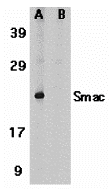
| Western Blot Validation in Human Heart Tissue Lysate Loading: 15 µg of lysates per lane. Antibodies: Smac 4798 (1 µg/mL), 1h incubation at RT in 5% NFDM/TBST.Secondary: Goat anti-rabbit IgG HRP conjugate at 1:10000 dilution.(A) Without blocking peptide(B) With blocking peptide |
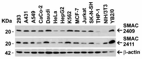 |
| Independent Antibody Validation (IAV) via Protein Expression Profile in Cell Lines Loading: 15 µg of lysates per lane. Antibodies: Smac 4798 (1 µg/mL), Smac 1411 (1 µg/mL), and beta-actin (1 µg/mL), 1h incubation at RT in 5% NFDM/TBST.Secondary: Goat anti-rabbit IgG HRP conjugate at 1:10000 dilution. |
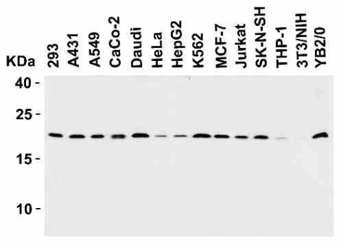 |
| Western Blot Validation in Human, Mouse and Rat Cell Lines Loading: 15 µg of lysates per lane. Antibodies: Smac 4798 (1 µg/mL), 1h incubation at RT in 5% NFM/TBST.Secondary: Goat anti-rabbit IgG HRP conjugate at 1:10000 dilution. |
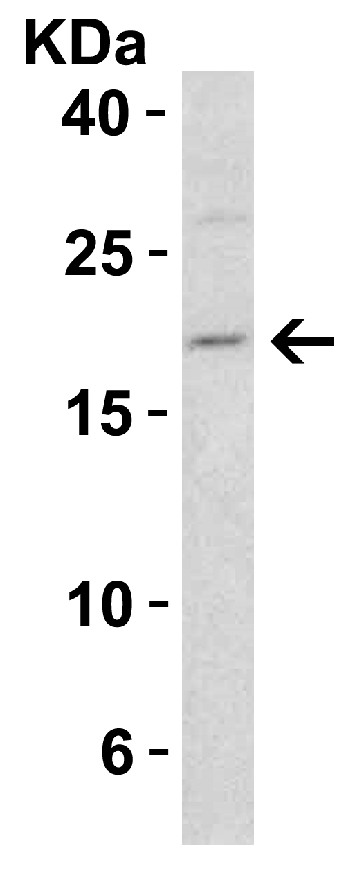 |
| Western Blot Validation in Mouse 3T3/NIH Cells Loading: 15 µg of lysate. Antibodies: Smac 4798 (1 µg/mL), 1h incubation at RT in 5% NFDM/TBST.Secondary: Goat anti-rabbit IgG HRP conjugate at 1:10000 dilution. |
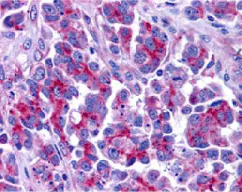 |
| Immunohistochemistry Validation of Smac in Human Ovary Tissue Immunohistochemical analysis of paraffin-embedded Human Ovary tissue using anti-Smac antibody (4798) at 5 µg/mL. Tissue was fixed with formaldehyde and blocked with 10% serum for 1 h at RT; antigen retrieval was by heat mediation with a citrate buffer (pH6). Samples were incubated with primary antibody overnight at 4C. A goat anti-rabbit IgG H&L (HRP) at 1/250 was used as secondary. Counter stained with Hematoxylin. |
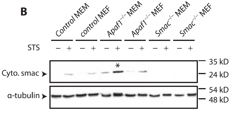 |
| KO Validation in Mouse Fibroblasts and Myoblasts (Ho et al., 2416) The indicated MEFs or MEMs were exposed to 2 uM STS for 4 h and analyzed by Western blot. Accumulation of Smac/Diablo in mitochondrion-depleted cytosol fractions fromSTS-treated Apaf-1 KO cells were detected by anti-smac antibodies. Smac expression was not detected in smac KO mice. |
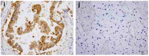 |
| Immunohistochemistry Validation of Smac in Human gastric carcinoma (Kim et al., 2011) Smac was highly expressed in gastric mucosa of patients with gastric carcinoma. |
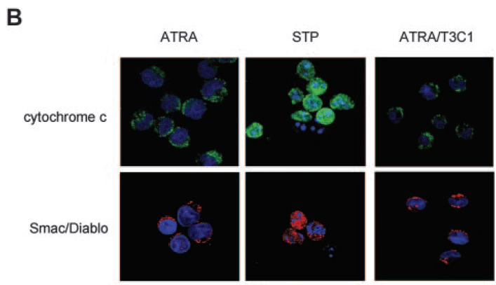 |
| Immunofluorescence Analysis of Smac in NB4-LR1 Cells (Saumet et al., 2005) NB4-LR1 cells were either treated with ATRA(1 µM) for 3 days without or with the T3C1 recombinant fragment (3 uM) or treated with staurosporine (STP; 5 uM ) for 3.5 hours. STP, but not ATRA or AYRA/T3C1 induced the release of smac. |
 |
| Induced Expression Validation in Rat Liver (Genestier et al., 2005) Mitochondria from rat liver were treated with increasing concentrations of rPVL (A), rLukS (B), or Bax alpha (C) for 1 hour at 30C. rPVL induces the release of the apoptogenic proteins cytochrome c and Smac/DIABLO from isolated mitochondria. |
FAQ & Publications
Frequently Asked Questions
What species reactivity does the rabbit anti-Smac (CT) polyclonal antibody 4798 exhibit?
This antibody reacts with the Smac (CT) protein and has been validated in human, mouse, and rat cell lines.
Which applications has the rabbit anti-Smac (CT) antibody 4798 been tested and recommended for?
The antibody is suitable and tested for Western blotting (WB), immunohistochemistry (IHC), ELISA, immunoprecipitation (IP), and immunofluorescence (IF/ICC). Recommended dilutions include 1 µg/mL for immunoblotting and 5 µg/mL for immunohistochemistry.
How should the rabbit anti-Smac (CT) polyclonal antibody 4798 be stored to maintain stability?
The antibody should be stored at -20°C and is stable for at least one year under these conditions. It is recommended to avoid multiple freeze-thaw cycles to preserve antibody integrity.
What is the immunogen used to generate the rabbit anti-Smac (CT) polyclonal antibody 4798?
The immunogen is a synthetic peptide corresponding to amino acids 225-239 of human Smac, based on accession number AAF87716.
What is the concentration and form of the rabbit anti-Smac (CT) polyclonal antibody 4798 supplied?
The antibody is supplied as a liquid at a concentration of 1 mg/mL, with a typical package size of 100 µg.
Publications
| pmid | title | authors | citation |
|---|---|---|---|
| We haven't added any publications to our database yet. | |||
Published literature highly relevant to the biological target of this product and referencing this antibody or clone are retrieved from the PubMed database provided by the United States National Library of Medicine at the National Institutes of Health.
Protocols
| relevant to this product |
|---|
| Western blot IHC ICC |
Documents
| Batch Number | QC File | SDS |
|---|---|---|
| To view batch-specific Safety Datasheets and Quality Certificates associated with your account, please Log In. | ||
Only logged in customers who have purchased this product may leave a review.


Reviews
There are no reviews yet.