| Weight | 1 lbs |
|---|---|
| Dimensions | 9 × 5 × 2 in |
| host | rabbit |
| isotype | IgG |
| clonality | polyclonal |
| concentration | 0.2 mg/mL |
| applications | IHC, IP, WB |
| reactivity | human, mouse |
| available sizes | 10 µL, 100 µL |

rabbit anti-PKc polyclonal antibody 1035
Price range: $100.00 through $2,600.00
Antibody summary
- Rabbit polyclonal to PKC (Protein kinase C) alpha
- Suitable for: WB, IHC, IP
- Reacts with: Hu, Ms presumed rat
- Isotype: IgG
- 100 µL, 10 µL
rabbit anti- pkc polyclonal antibody 1035
| target relevance |
|---|
| Homo sapiens PRKCA Protein kinase C alpha type |
| Protein names Protein kinase C alpha type |
| Gene names PRKCA |
| Protein family Belongs to the protein kinase superfamily. AGC Ser/Thr protein kinase family. PKC subfamily |
| Function Calcium-activated, phospholipid- and diacylglycerol (DAG)-dependent serine/threonine-protein kinase that is involved in positive and negative regulation of cell proliferation, apoptosis, differentiation, migration and adhesion, tumorigenesis, cardiac hypertrophy, angiogenesis, platelet function and inflammation, by directly phosphorylating targets such as RAF1, BCL2, CSPG4, TNNT2/CTNT, or activating signaling cascade involving MAPK1/3 (ERK1/2) and RAP1GAP. Involved in cell proliferation and cell growth arrest by positive and negative regulation of the cell cycle. Can promote cell growth by phosphorylating and activating RAF1, which mediates the activation of the MAPK/ERK signaling cascade, and/or by up-regulating CDKN1A, which facilitates active cyclin-dependent kinase (CDK) complex formation in glioma cells. In intestinal cells stimulated by the phorbol ester PMA, can trigger a cell cycle arrest program which is associated with the accumulation of the hyper-phosphorylated growth-suppressive form of RB1 and induction of the CDK inhibitors CDKN1A and CDKN1B. Exhibits anti-apoptotic function in glioma cells and protects them from apoptosis by suppressing the p53/TP53-mediated activation of IGFBP3, and in leukemia cells mediates anti-apoptotic action by phosphorylating BCL2. During macrophage differentiation induced by macrophage colony-stimulating factor (CSF1), is translocated to the nucleus and is associated with macrophage development. After wounding, translocates from focal contacts to lamellipodia and participates in the modulation of desmosomal adhesion. Plays a role in cell motility by phosphorylating CSPG4, which induces association of CSPG4 with extensive lamellipodia at the cell periphery and polarization of the cell accompanied by increases in cell motility. During chemokine-induced CD4(+) T cell migration, phosphorylates CDC42-guanine exchange factor DOCK8 resulting in its dissociation from LRCH1 and the activation of GTPase CDC42 (PubMed:28028151). Is highly expressed in a number of cancer cells where it can act as a tumor promoter and is implicated in malignant phenotypes of several tumors such as gliomas and breast cancers. Negatively regulates myocardial contractility and positively regulates angiogenesis, platelet aggregation and thrombus formation in arteries. Mediates hypertrophic growth of neonatal cardiomyocytes, in part through a MAPK1/3 (ERK1/2)-dependent signaling pathway, and upon PMA treatment, is required to induce cardiomyocyte hypertrophy up to heart failure and death, by increasing protein synthesis, protein-DNA ratio and cell surface area. Regulates cardiomyocyte function by phosphorylating cardiac troponin T (TNNT2/CTNT), which induces significant reduction in actomyosin ATPase activity, myofilament calcium sensitivity and myocardial contractility. In angiogenesis, is required for full endothelial cell migration, adhesion to vitronectin (VTN), and vascular endothelial growth factor A (VEGFA)-dependent regulation of kinase activation and vascular tube formation. Involved in the stabilization of VEGFA mRNA at post-transcriptional level and mediates VEGFA-induced cell proliferation. In the regulation of calcium-induced platelet aggregation, mediates signals from the CD36/GP4 receptor for granule release, and activates the integrin heterodimer ITGA2B-ITGB3 through the RAP1GAP pathway for adhesion. During response to lipopolysaccharides (LPS), may regulate selective LPS-induced macrophage functions involved in host defense and inflammation. But in some inflammatory responses, may negatively regulate NF-kappa-B-induced genes, through IL1A-dependent induction of NF-kappa-B inhibitor alpha (NFKBIA/IKBA). Upon stimulation with 12-O-tetradecanoylphorbol-13-acetate (TPA), phosphorylates EIF4G1, which modulates EIF4G1 binding to MKNK1 and may be involved in the regulation of EIF4E phosphorylation. Phosphorylates KIT, leading to inhibition of KIT activity. Phosphorylates ATF2 which promotes cooperation between ATF2 and JUN, activating transcription. Phosphorylates SOCS2 at 'Ser-52' facilitating its ubiquitination and proteasomal degradation (By similarity). Phosphorylates KLHL3 in response to angiotensin II signaling, decreasing the interaction between KLHL3 and WNK4 (PubMed:25313067). Phosphorylates and activates LRRK1, which phosphorylates RAB proteins involved in intracellular trafficking (PubMed:36040231) |
| Catalytic activity L-seryl-[protein] + ATP = O-phospho-L-seryl-[protein] + ADP + H(+) L-threonyl-[protein] + ATP = O-phospho-L-threonyl-[protein] + ADP + H(+) |
| Subcellular location Cytoplasm, Cell membrane, Mitochondrion membrane, Nucleus |
| Structure Recruited in a circadian manner into a nuclear complex which also includes BMAL1 and RACK1 (By similarity). Interacts with ADAP1/CENTA1 (PubMed:12893243). Interacts with CSPG4 (PubMed:15504744). Binds to CAVIN2 in the presence of phosphatidylserine (By similarity). Interacts with PRKCABP/PICK1 (via PDZ domain) (PubMed:15247289). Interacts with TRIM41 (PubMed:17893151). Interacts with PARD3 (PubMed:27925688). Interacts with SOCS2 (By similarity) |
| Post-translational modification In response to growth factors, phosphorylated at Thr-631 and Ser-657 by the mTORC2 complex, promoting autophosphorylation and activation of PRKCA |
| Keywords 3D-structure, Acetylation, Angiogenesis, Apoptosis, ATP-binding, Calcium, Cell adhesion, Cell membrane, Cytoplasm, Direct protein sequencing, Kinase, Membrane, Metal-binding, Mitochondrion, Nucleotide-binding, Nucleus, Phosphoprotein, Proteomics identification, Proto-oncogene, Reference proteome, Repeat, Serine/threonine-protein kinase, Transferase, Zinc, Zinc-finger |
| Sequence MADVFPGNDSTASQDVANRFARKGALRQKNVHEVKDHKFIARFFKQPTFCSHCTDFIWGF GKQGFQCQVCCFVVHKRCHEFVTFSCPGADKGPDTDDPRSKHKFKIHTYGSPTFCDHCGS LLYGLIHQGMKCDTCDMNVHKQCVINVPSLCGMDHTEKRGRIYLKAEVADEKLHVTVRDA KNLIPMDPNGLSDPYVKLKLIPDPKNESKQKTKTIRSTLNPQWNESFTFKLKPSDKDRRL SVEIWDWDRTTRNDFMGSLSFGVSELMKMPASGWYKLLNQEEGEYYNVPIPEGDEEGNME LRQKFEKAKLGPAGNKVISPSEDRKQPSNNLDRVKLTDFNFLMVLGKGSFGKVMLADRKG TEELYAIKILKKDVVIQDDDVECTMVEKRVLALLDKPPFLTQLHSCFQTVDRLYFVMEYV NGGDLMYHIQQVGKFKEPQAVFYAAEISIGLFFLHKRGIIYRDLKLDNVMLDSEGHIKIA DFGMCKEHMMDGVTTRTFCGTPDYIAPEIIAYQPYGKSVDWWAYGVLLYEMLAGQPPFDG EDEDELFQSIMEHNVSYPKSLSKEAVSVCKGLMTKHPAKRLGCGPEGERDVREHAFFRRI DWEKLENREIQPPFKPKVCGKGAENFDKFFTRGQPVLTPPDQLVIANIDQSDFEGFSYVN PQFVHPILQSAV |
| UniProt accession: P17252 |
Data
|
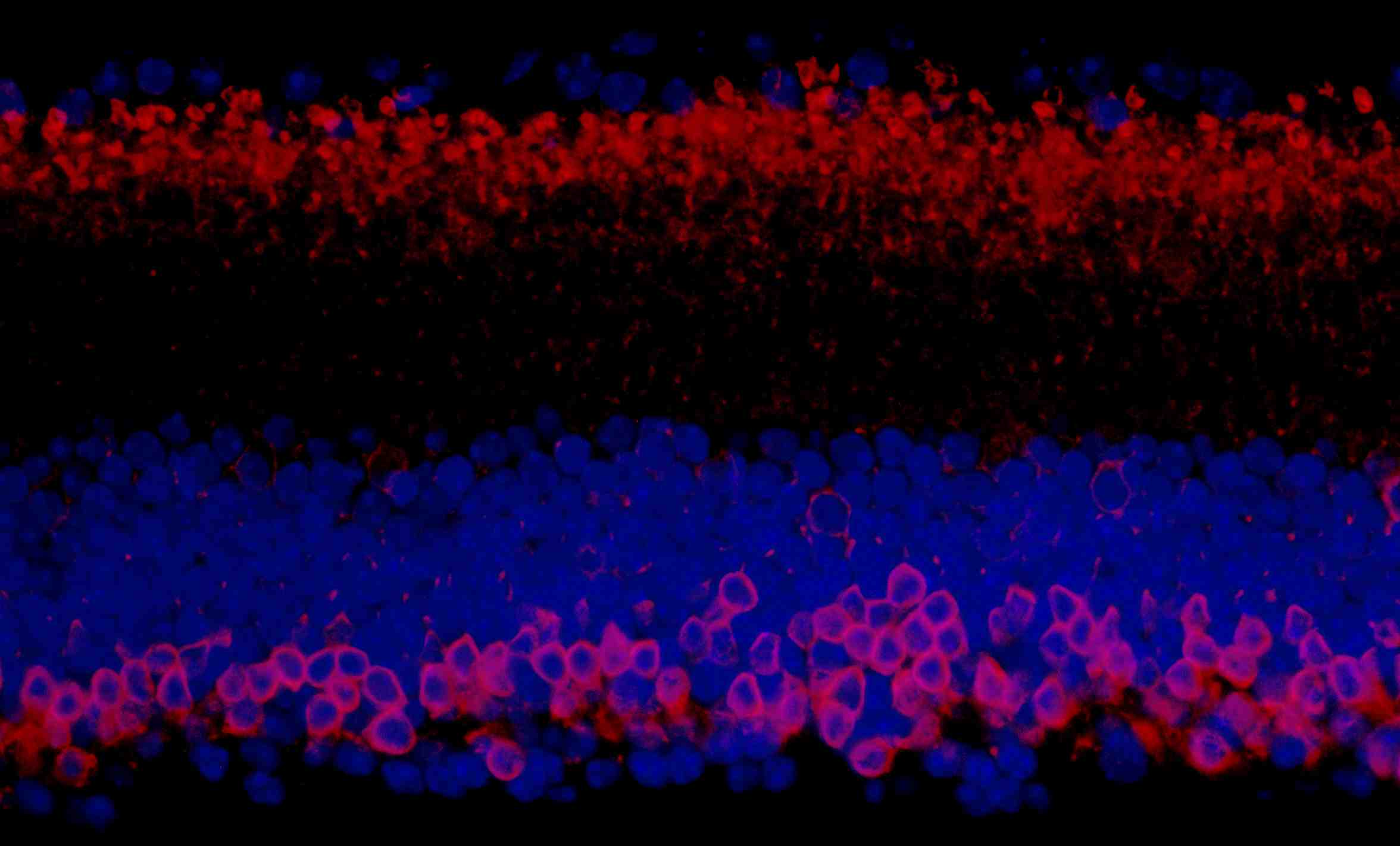
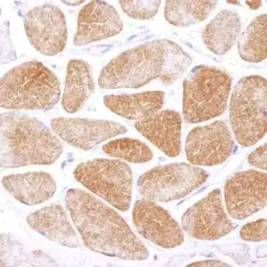
| Detection of mouse PKC-alpha by immunohistochemistry. Sample: FFPE section of mouse retina. Antibody: Affinity purified rabbit anti-PKC-alpha (#1035) used at a dilution of 1:100 (2µg/ml). Detection: Red-fluorescent goat anti-rabbit IgG-heavy and light chain cross-adsorbed Antibody DyLight-594 Conjugated used at a dilution of 1:100. Counterstain: DAPI (blue) |
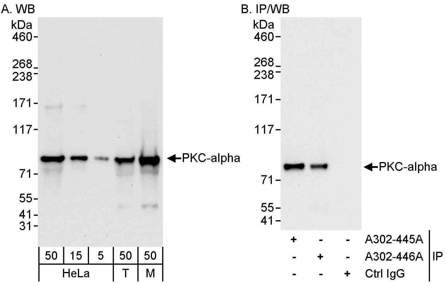 |
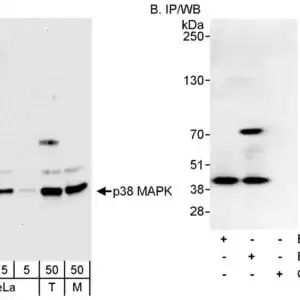
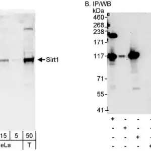
| Detection of human and mouse PKC-alpha by western blot (h & m) and immunoprecipitation (h). Samples: Whole cell lysate from HeLa (5, 15 and 50 µg for WB; 1 mg for IP, 20% of IP loaded), HEK293T (T; 50 µg), and mouse NIH 3T3 (M; 50 µg) cells. Antibodies: Affinity purified rabbit anti-PKC-alpha antibody #1035 used for WB at 0.04 µg/ml (A) and 1 µg/ml (B) and used for IP at 3 µg/mg lysate. PKC-alpha was also immunoprecipitated by rabbit anti-PKC-alpha antibody, which recognizes an upstream epitope. Detection: Chemiluminescence with exposure times of 10 seconds (A) and 3 seconds (B). |
FAQ & Publications
Frequently Asked Questions
What species does the rabbit anti-PKC polyclonal antibody 1035 react with and for which applications is it suitable?
This antibody reacts with human and mouse species and is suitable for Western blot (WB), immunohistochemistry (IHC), and immunoprecipitation (IP) applications.
How should the rabbit anti-PKC polyclonal antibody 1035 be stored to maintain its stability and what is its concentration?
The antibody should be stored at 2-8°C and is stable for 1 year from the date of receipt. It is supplied as a liquid at a concentration of 0.2 mg/mL in a Tris-buffered saline containing 0.1% BSA and 0.09% sodium azide.
Publications
| pmid | title | authors | citation |
|---|---|---|---|
| We haven't added any publications to our database yet. | |||
Published literature highly relevant to the biological target of this product and referencing this antibody or clone are retrieved from the PubMed database provided by the United States National Library of Medicine at the National Institutes of Health.
Protocols
| relevant to this product |
|---|
| Western blot IHC |
Documents
| Batch Number | QC File | SDS |
|---|---|---|
| To view batch-specific Safety Datasheets and Quality Certificates associated with your account, please Log In. | ||
Only logged in customers who have purchased this product may leave a review.


Reviews
There are no reviews yet.