| Weight | 1 lbs |
|---|---|
| Dimensions | 9 × 5 × 2 in |
| host | rabbit |
| isotype | IgG |
| clonality | polyclonal |
| concentration | 1 mg/mL |
| applications | ICC/IF, WB |
| reactivity | PHAP I (CT) |
| available sizes | 100 µg |


rabbit anti-PHAP I (CT) polyclonal antibody 6528
$445.00
Antibody summary
- Rabbit polyclonal to PHAP I (CT)
- Suitable for: ELISA,WB,IHC-P,IF
- Isotype: IgG
- 100 µg
rabbit anti-PHAP I (CT) polyclonal antibody 6528
| antibody |
|---|
| Tested applications WB,IHC,IHC,ICC/IF,ELISA |
| Recommended dilutions Immunoblotting: Use at 1ug/mL. Positive control: Raji cell lysate. Immunohistochemistry: use at 2ug/mL. These are recommended concentrations. Enduser should determine optimal concentrations for their applications. |
| Immunogen A synthetic peptide corresponding to amino acids at the carboxy terminus of human PHAP (accession no. P39687). |
| Size and concentration 100µg and lot specific |
| Form liquid |
| Storage Instructions This antibody is stable for at least one (1) year at -20°C. Avoid multiple freeze-thaw cycles. |
| Storage buffer PBS, pH 7.4. |
| Purity peptide affinity purification |
| Clonality polyclonal |
| Isotype IgG |
| Compatible secondaries goat anti-rabbit IgG, H&L chain specific, peroxidase conjugated, conjugated polyclonal antibody 9512 goat anti-rabbit IgG, H&L chain specific, biotin conjugated polyclonal antibody 2079 goat anti-rabbit IgG, H&L chain specific, FITC conjugated polyclonal antibody 7863 goat anti-rabbit IgG, H&L chain specific, Cross Absorbed polyclonal antibody 2371 goat anti-rabbit IgG, H&L chain specific, biotin conjugated polyclonal antibody, crossabsorbed 1715 goat anti-rabbit IgG, H&L chain specific, FITC conjugated polyclonal antibody, crossabsorbed 1720 |
| Isotype control Rabbit polyclonal - Isotype Control |
| target relevance |
|---|
| Homo sapiens ANP32A Acidic leucine-rich nuclear phosphoprotein 32 family member A |
| Protein names Acidic leucine-rich nuclear phosphoprotein 32 family member A |
| Alternative names Acidic nuclear phosphoprotein pp32, Leucine-rich acidic nuclear protein, Mapmodulin, Potent heat-stable protein phosphatase 2A inhibitor I1PP2A, Putative HLA-DR-associated protein I |
| Gene names ANP32A |
| Protein family Belongs to the ANP32 family |
| Function Multifunctional protein that is involved in the regulation of many processes including tumor suppression, apoptosis, cell cycle progression or transcription (PubMed:10400610, PubMed:11360199, PubMed:16341127, PubMed:18439902). Promotes apoptosis by favouring the activation of caspase-9/CASP9 and allowing apoptosome formation (PubMed:18439902). In addition, plays a role in the modulation of histone acetylation and transcription as part of the INHAT (inhibitor of histone acetyltransferases) complex. Inhibits the histone-acetyltranferase activity of EP300/CREBBP (CREB-binding protein) and EP300/CREBBP-associated factor by histone masking (PubMed:11830591). Preferentially binds to unmodified histone H3 and sterically inhibiting its acetylation and phosphorylation leading to cell growth inhibition (PubMed:16341127). Participates in other biochemical processes such as regulation of mRNA nuclear-to-cytoplasmic translocation and stability by its association with ELAVL1 (Hu-antigen R) (PubMed:18180367). Plays a role in E4F1-mediated transcriptional repression as well as inhibition of protein phosphatase 2A (PubMed:15642345, PubMed:17557114) |
| Subcellular location Nucleus, Cytoplasm, Endoplasmic reticulum |
| Structure (Microbial infection) Interacts (via C-terminus) with influenza virus C protein PB2; this interaction promotes viral replication by bridging viral replicase dimers together |
| Post-translational modification Phosphorylated on serine residues, at least in part by casein kinase 2/CK2 The N-terminus is blocked Some glutamate residues are glycylated by TTLL8. This modification occurs exclusively on glutamate residues and results in a glycine chain on the gamma-carboxyl group (By similarity) |
| Keywords 3D-structure, Cytoplasm, Direct protein sequencing, Endoplasmic reticulum, Host-virus interaction, Leucine-rich repeat, Nucleus, Phosphoprotein, Proteomics identification, Reference proteome, Repeat, Repressor, Transcription, Transcription regulation |
| Sequence MEMGRRIHLELRNRTPSDVKELVLDNSRSNEGKLEGLTDEFEELEFLSTINVGLTSIANL PKLNKLKKLELSDNRVSGGLEVLAEKCPNLTHLNLSGNKIKDLSTIEPLKKLENLKSLDL FNCEVTNLNDYRENVFKLLPQLTYLDGYDRDDKEAPDSDAEGYVEGLDDEEEDEDEEEYD EDAQVVEDEEDEDEEEEGEEEDVSGEEEEDEEGYNDGEVDDEEDEEELGEEERGQKRKRE PEDEGEDDD |
| UniProt accession: P39687 |
Data
 |
| Western Blot Validation in (A) Human Raji Cells , (B) Mouse testis tissue lysate and (C) Rat testis tissue lysate Loading: 15 µg of lysates per lane. Antibodies: PHAP I 6528 (1 µg/mL), 1h incubation at RT in 5% NFDM/TBST.Secondary: Goat anti-rabbit IgG HRP conjugate at 1:10000 dilution. |
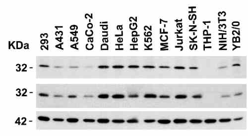 |
| Independent Antibody Validation (IAV) via Protein Expression Profile in Cell Lines Loading: 15 µg of lysates per lane. Antibodies: PHAP I 11082 (2 µg/mL), PHAP I 6528 (1 µg/mL), and beta-actin (1 µg/mL), 1h incubation at RT in 5% NFDM/TBST.Secondary: Goat anti-rabbit IgG HRP conjugate at 1:10000 dilution. |
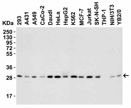 |
| Western Blot Validation in Human, Mouse and Rat Cell Lines Loading: 15 µg of lysates per lane. Antibodies: PHAP I 6528 (1 µg/mL), 1h incubation at RT in 5% NFDM/TBST.Secondary: Goat anti-rabbit IgG HRP conjugate at 1:10000 dilution. |
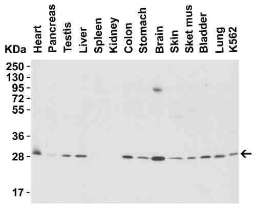 |
| Western Blot Validation in Mouse Tissues Loading: 15 µg of lysates per lane. Antibodies: PHAP I 6528 (1 µg/mL), 1h incubation at RT in 5% NFDM/TBST.Secondary: Goat anti-rabbit IgG HRP conjugate at 1:10000 dilution. |
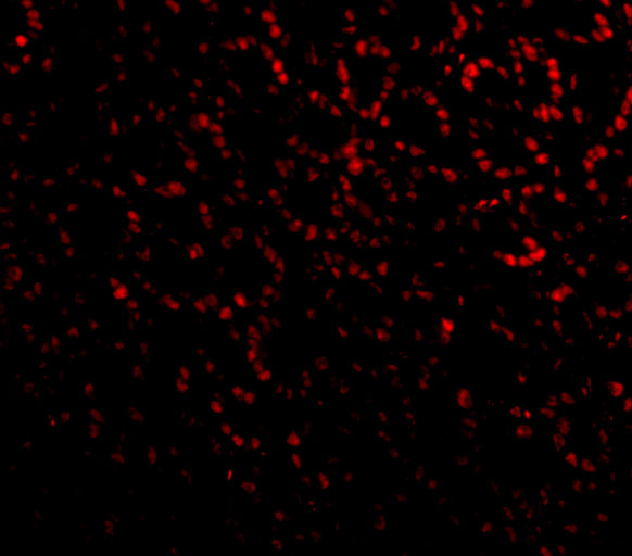 |
| Immunofluorescence Validation of PHAP I in Mouse Small Intestine cells Immunofluorescent analysis of 4% paraformaldehyde-fixed Mouse Small Intestine Cells labeling PHAP I with 6528 at 20 µg/mL, followed by goat anti-rabbit IgG secondary antibody at 1/500 dilution (red). |
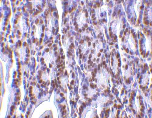 |
| Immunohistochemistry Validation of PHAP I in Mouse Small Intestine Tissue Immunohistochemical analysis of paraffin-embedded Mouse Small Intestine Tissue using anti-PHAP I antibody (6528) at 2 µg/mL. Tissue was fixed with formaldehyde and blocked with 10% serum for 1 h at RT; antigen retrieval was by heat mediation with a citrate buffer (pH6). Samples were incubated with primary antibody overnight at 4C. A goat anti-rabbit IgG H&L (HRP) at 1/250 was used as secondary. Counter stained with Hematoxylin. |
 |
| KD Validation of PHAPI in Human Breast Cancer Cells (Schafer et al., 2006) Human breast cancer cells (T47D cells) were transfected with control or PHAPI siRNA duplex. PHAPI was detected via Western Blot analysis by using the anti-PHAPI antibody. PHAPI expression was reduced after PHAPI siRNA knockdown. |
 |
| Increased Expression Validation of PHAPI in Patient Samples of BreastTumor Tissue (Schafer et al., 2006) PHAPI was overexpressed in all breast tumor samples of patients and human breast cancer cells (MDA-MB-453), but not in the normal breast tissue or human primary mammary epithelial cells (HMEC). |
 |
| Overexpression of PHAPI in Breast Cancer Cells (Schafer et al., 2006) Western blot analysis with anti-PHAPI antibodies was performed for PHAPI in human cell lines from breast, prostate and lung. PHAPI was overexpressed in breast cancer cells when compared with normal cells (HMEC) whereas there were no significant differences in PHAPI expression in normal and cancer cells of either prostate or lung origin. |
FAQ & Publications
Frequently Asked Questions
What applications is the rabbit anti-PHAP I (CT) polyclonal antibody 6528 suitable for?
This antibody is suitable for ELISA, Western blotting (WB), immunohistochemistry on paraffin sections (IHC-P), and immunofluorescence (IF/ICC) applications.
How should the rabbit anti-PHAP I (CT) polyclonal antibody be stored to maintain stability?
The antibody should be stored at -20°C and is stable for at least one year under these conditions. Avoid multiple freeze-thaw cycles to preserve antibody integrity.
What is the recommended dilution for using this antibody in immunoblotting and immunohistochemistry?
For immunoblotting (Western blot), use the antibody at 1 µg/mL. For immunohistochemistry, a concentration of 2 µg/mL is recommended. However, users should optimize concentrations for their specific applications.
What is the host species and clonality of the anti-PHAP I (CT) antibody 6528?
This antibody is a rabbit polyclonal IgG antibody raised against a synthetic peptide corresponding to the carboxy terminus of human PHAP.
Publications
| pmid | title | authors | citation |
|---|---|---|---|
| We haven't added any publications to our database yet. | |||
Published literature highly relevant to the biological target of this product and referencing this antibody or clone are retrieved from the PubMed database provided by the United States National Library of Medicine at the National Institutes of Health.
Protocols
| relevant to this product |
|---|
| Western blot IHC ICC |
Documents
| Batch Number | QC File | SDS |
|---|---|---|
| To view batch-specific Safety Datasheets and Quality Certificates associated with your account, please Log In. | ||
Only logged in customers who have purchased this product may leave a review.


Reviews
There are no reviews yet.