| Weight | 1 lbs |
|---|---|
| Dimensions | 9 × 5 × 2 in |
| host | mouse |
| isotype | IgG |
| clonality | monoclonal |
| concentration | concentrate, predilute |
| applications | IHC |
| reactivity | human |
| available size | 0.1 mL, 0.5 mL, 1 mL concentrated, 7 mL prediluted |

rabbit anti-PD-L1 monoclonal antibody (ZR3) 6328
Price range: $160.00 through $528.00
Antibody summary
- Rabbit monoclonal to PD-L1
- Suitable for: Immunohistochemistry (formalin-fixed, paraffin-embedded tissues)
- Reacts with: Human
- Isotype:IgG
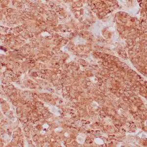
- Control: Placenta or lung adenocarcinoma
- Visualization: Membrane
- 0.1, 0.5, 1.0 mL concentrated, 7 mL prediluted
rabbit anti-PD-L1 monoclonal antibody ZR3 6328
| target relevance |
|---|
| Homo sapiens CD274 Programmed cell death 1 ligand 1 |
| Protein names Programmed cell death 1 ligand 1 |
| Alternative names B7 homolog 1 |
| Gene names CD274 |
| Protein family Belongs to the immunoglobulin superfamily. BTN/MOG family |
| Function Plays a critical role in induction and maintenance of immune tolerance to self (PubMed:11015443, PubMed:28813410, PubMed:28813417, PubMed:31399419). As a ligand for the inhibitory receptor PDCD1/PD-1, modulates the activation threshold of T-cells and limits T-cell effector response (PubMed:11015443, PubMed:28813410, PubMed:28813417, PubMed:36727298). Through a yet unknown activating receptor, may costimulate T-cell subsets that predominantly produce interleukin-10 (IL10) (PubMed:10581077). Can also act as a transcription coactivator: in response to hypoxia, translocates into the nucleus via its interaction with phosphorylated STAT3 and promotes transcription of GSDMC, leading to pyroptosis (PubMed:32929201) |
| Subcellular location Secreted |
| Structure May form homomultimers |
| Post-translational modification Ubiquitinated; STUB1 likely mediates polyubiquitination of PD-L1/CD274 triggering its degradation (PubMed:28813410). Ubiquitinated by MARCHF8; leading to degradation (PubMed:34183449). Deubiquitinated by USP22; leading to stabilization (PubMed:31399419) |
| Involvement in disease Autoimmune disease, multisystem, infantile-onset, 5 An autosomal recessive disorder characterized by autoimmune manifestations apparent in the first months or years of life. ADMIO5 patients have complete insulin deficiency and type 1 diabetes mellitus with neonatal onset. |
| Keywords 3D-structure, Adaptive immunity, Alternative splicing, Cell membrane, Diabetes mellitus, Disulfide bond, Endosome, Glycoprotein, Immunity, Immunoglobulin domain, Membrane, Nucleus, Proteomics identification, Receptor, Reference proteome, Repeat, Secreted, Signal, Transmembrane, Transmembrane helix, Ubl conjugation |
| Sequence MRIFAVFIFMTYWHLLNAFTVTVPKDLYVVEYGSNMTIECKFPVEKQLDLAALIVYWEME DKNIIQFVHGEEDLKVQHSSYRQRARLLKDQLSLGNAALQITDVKLQDAGVYRCMISYGG ADYKRITVKVNAPYNKINQRILVVDPVTSEHELTCQAEGYPKAEVIWTSSDHQVLSGKTT TTNSKREEKLFNVTSTLRINTTTNEIFYCTFRRLDPEENHTAELVIPELPLAHPPNERTH LVILGAILLCLGVALTFIFRLRKGRMMDVKKCGIQDTNSKKQSDTHLEET |
| UniProt accession: Q9NZQ7 |
Data
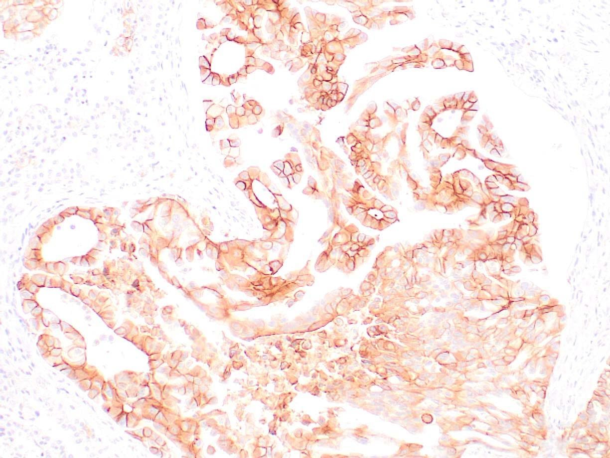 |
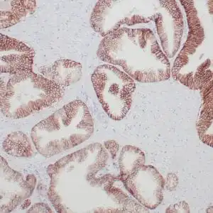
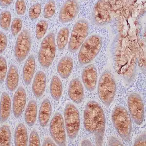
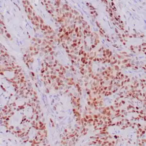
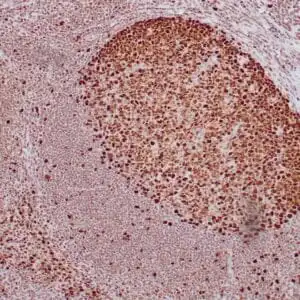
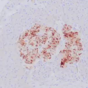
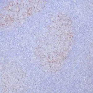
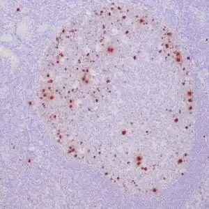
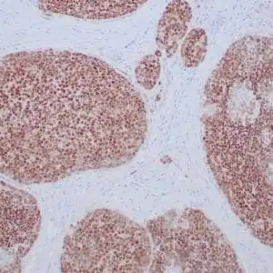
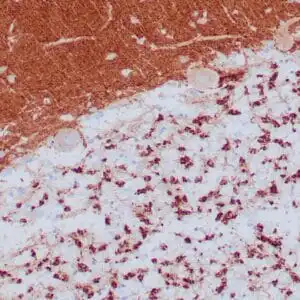
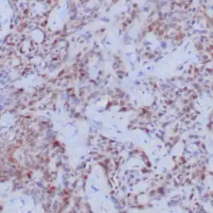
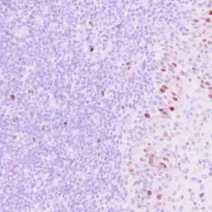
| Human lung adenocarcinoma stained with anti-PD-L1 (Clone ZR3) using peroxidase-conjugate and DAB chromogen. Note membranous staining of tumor cells. |
FAQ & Publications
Frequently Asked Questions
What species does the rabbit anti-PD-L1 monoclonal antibody (ZR3) specifically react with?
This antibody specifically reacts with human PD-L1 protein.
Which applications is this PD-L1 antibody validated for?
The rabbit anti-PD-L1 monoclonal antibody (ZR3) is validated for immunohistochemistry (IHC) on formalin-fixed, paraffin-embedded (FFPE) tissue samples.
What are the recommended storage conditions for the rabbit anti-PD-L1 monoclonal antibody (ZR3)?
For short-term storage, keep the antibody at 2-8°C; for long-term storage, it should be kept at -20°C. Avoid repeated freeze/thaw cycles to maintain antibody integrity.
What is the host species and clonality of the PD-L1 antibody (ZR3)?
The antibody is a rabbit monoclonal antibody, indicating it is derived from a single clone of rabbit B cells.
What positive controls are suggested for use with this anti-PD-L1 antibody in immunohistochemistry?
Placenta or lung adenocarcinoma tissues are recommended positive controls for validating staining with this anti-PD-L1 antibody in IHC assays.
Publications
| pmid | title | authors | citation |
|---|---|---|---|
| We haven't added any publications to our database yet. | |||
Published literature highly relevant to the biological target of this product and referencing this antibody or clone are retrieved from the PubMed database provided by the United States National Library of Medicine at the National Institutes of Health.
Protocols
| relevant to this product |
|---|
| IHC |
Documents
| Batch Number | QC File | SDS |
|---|---|---|
| To view batch-specific Safety Datasheets and Quality Certificates associated with your account, please Log In. | ||
Only logged in customers who have purchased this product may leave a review.

Reviews
There are no reviews yet.