| Weight | 1 lbs |
|---|---|
| Dimensions | 9 × 5 × 2 in |
| host | mouse |
| isotype | IgG |
| clonality | monoclonal |
| concentration | concentrate, predilute |
| applications | IHC |
| reactivity | human |
| available size | 0.1 mL, 0.5 mL, 1 mL concentrated, 7 mL prediluted |

rabbit anti-p120 monoclonal antibody (ZR316) 6302
Price range: $160.00 through $528.00
Antibody summary
- Rabbit monoclonal to p120
- Suitable for: Immunohistochemistry (formalin-fixed, paraffin-embedded tissues)
- Reacts with: Human
- Isotype:IgG
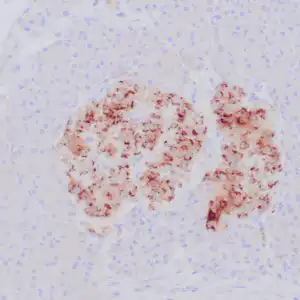
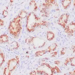
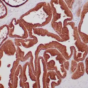
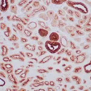
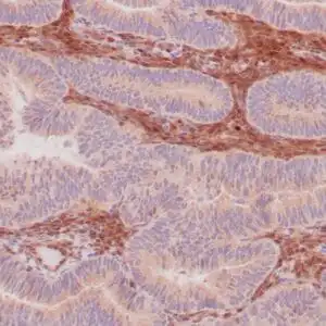
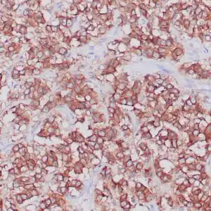
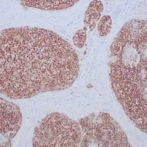
- Control: Breast lobular carcinoma, melanoma
- Visualization: Cytoplasmic and membranous
- 0.1, 0.5, 1.0 mL concentrated, 7 mL prediluted
rabbit anti-p120 monoclonal antibody ZR316 6302
| target relevance |
|---|
| Homo sapiens CTNND1 Catenin delta-1 |
| Protein names Catenin delta-1 |
| Alternative names Cadherin-associated Src substrate, p120 catenin, p120(cas) |
| Gene names CTNND1 |
| Protein family Belongs to the beta-catenin family |
| Function Key regulator of cell-cell adhesion that associates with and regulates the cell adhesion properties of both C-, E- and N-cadherins, being critical for their surface stability (PubMed:14610055, PubMed:20371349). Promotes localization and retention of DSG3 at cell-cell junctions, via its interaction with DSG3 (PubMed:18343367). Beside cell-cell adhesion, regulates gene transcription through several transcription factors including ZBTB33/Kaiso2 and GLIS2, and the activity of Rho family GTPases and downstream cytoskeletal dynamics (PubMed:10207085, PubMed:20371349). Implicated both in cell transformation by SRC and in ligand-induced receptor signaling through the EGF, PDGF, CSF-1 and ERBB2 receptors (PubMed:17344476) |
| Subcellular location Cytoplasm |
| Structure Belongs to a multiprotein cell-cell adhesion complex that also contains E-cadherin/CDH1, alpha-catenin/CTNNA1, beta-catenin/CTNNB1, and gamma-catenin/JUP (PubMed:15240885, PubMed:20371349). Component of a cadherin:catenin adhesion complex composed of at least of CDH26, beta-catenin/CTNNB1, alpha-catenin/CTNNA1 and p120 catenin/CTNND1 (PubMed:28051089). Binds to the C-terminal fragment of PSEN1 and mutually competes for CDH1. Interacts with ZBTB33 (PubMed:10207085). Interacts with GLIS2 (PubMed:17344476). Interacts with FER (PubMed:7623846). Interacts with NANOS1 (via N-terminal region) (PubMed:17047063). Interacts (via N-terminus) with GNA12; the interaction regulates CDH1-mediated cell-cell adhesion (PubMed:15240885). Interacts with GNA13 (PubMed:15240885). Interacts with CCDC85B (PubMed:25009281). Interacts with PLPP3; negatively regulates the PLPP3-mediated stabilization of CTNNB1 (PubMed:20123964). Interacts with DSG3; the interaction facilitates DSG3 localization and retention at cell-cell junctions (PubMed:18343367). Interacts with CTNND1/p120-catenin; the interaction controls CADH5 endocytosis (By similarity) |
| Post-translational modification Phosphorylated by FER and other protein-tyrosine kinases. Phosphorylated at Ser-288 by PAK5. Dephosphorylated by PTPRJ |
| Involvement in disease Blepharocheilodontic syndrome 2 A form of blepharocheilodontic syndrome, a rare autosomal dominant disorder. It is characterized by lower eyelid ectropion, upper eyelid distichiasis, euryblepharon, bilateral cleft lip and palate, and features of ectodermal dysplasia, including hair anomalies, conical teeth and tooth agenesis. An additional rare manifestation is imperforate anus. There is considerable phenotypic variability among affected individuals. |
| Keywords 3D-structure, Acetylation, Alternative initiation, Alternative splicing, Cell adhesion, Cell junction, Cell membrane, Coiled coil, Cytoplasm, Disease variant, Ectodermal dysplasia, Isopeptide bond, Membrane, Nucleus, Phosphoprotein, Proteomics identification, Reference proteome, Repeat, Transcription, Transcription regulation, Ubl conjugation, Wnt signaling pathway |
| Sequence MDDSEVESTASILASVKEQEAQFEKLTRALEEERRHVSAQLERVRVSPQDANPLMANGTL TRRHQNGRFVGDADLERQKFSDLKLNGPQDHSHLLYSTIPRMQEPGQIVETYTEEDPEGA MSVVSVETSDDGTTRRTETTVKKVVKTVTTRTVQPVAMGPDGLPVDASSVSNNYIQTLGR DFRKNGNGGPGPYVGQAGTATLPRNFHYPPDGYSRHYEDGYPGGSDNYGSLSRVTRIEER YRPSMEGYRAPSRQDVYGPQPQVRVGGSSVDLHRFHPEPYGLEDDQRSMGYDDLDYGMMS DYGTARRTGTPSDPRRRLRSYEDMIGEEVPSDQYYWAPLAQHERGSLASLDSLRKGGPPP PNWRQPELPEVIAMLGFRLDAVKSNAAAYLQHLCYRNDKVKTDVRKLKGIPVLVGLLDHP KKEVHLGACGALKNISFGRDQDNKIAIKNCDGVPALVRLLRKARDMDLTEVITGTLWNLS SHDSIKMEIVDHALHALTDEVIIPHSGWEREPNEDCKPRHIEWESVLTNTAGCLRNVSSE RSEARRKLRECDGLVDALIFIVQAEIGQKDSDSKLVENCVCLLRNLSYQVHREIPQAERY QEAAPNVANNTGPHAASCFGAKKGKDEWFSRGKKPIEDPANDTVDFPKRTSPARGYELLF QPEVVRIYISLLKESKTPAILEASAGAIQNLCAGRWTYGRYIRSALRQEKALSAIADLLT NEHERVVKAASGALRNLAVDARNKELIGKHAIPNLVKNLPGGQQNSSWNFSEDTVISILN TINEVIAENLEAAKKLRETQGIEKLVLINKSGNRSEKEVRAAALVLQTIWGYKELRKPLE KEGWKKSDFQVNLNNASRSQSSHSYDDSTLPLIDRNQKSDKKPDREEIQMSNMGSNTKSL DNNYSTPNERGDHNRTLDRSGDLGDMEPLKGTTPLMQDEGQESLEEELDVLVLDDEGGQV SYPSMQKI |
| UniProt accession: O60716 |
Data
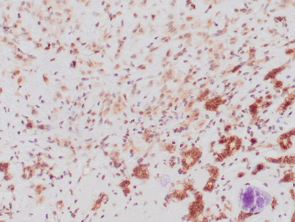 |
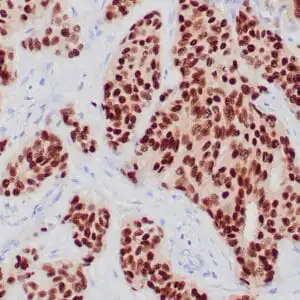
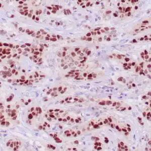
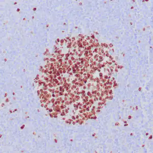
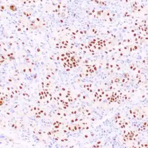
| Human breast lobular carcinoma stained with anti-P120 antibody using peroxidase-conjugate and DAB chromogen. Note: the cytoplasmic staining of lobular carcinoma cells and membranous staining of ductal epithelium (lower field). |
FAQ & Publications
Frequently Asked Questions
What is the recommended application and dilution range for the rabbit anti-p120 monoclonal antibody (ZR316)?
This antibody is suitable for immunohistochemistry (IHC) on formalin-fixed, paraffin-embedded human tissue samples. The recommended dilution for the concentrated antibody is between 1:100 and 1:200.
How should the rabbit anti-p120 monoclonal antibody (ZR316) be stored to maintain its stability?
For short-term storage, keep the antibody at 2-8°C. For longer-term storage, it should be kept at -20°C. It is important to avoid freeze/thaw cycles to preserve antibody integrity.
What species does the rabbit anti-p120 monoclonal antibody (ZR316) react with, and what controls are recommended for validation?
This antibody reacts specifically with human p120 protein. Positive controls recommended for immunohistochemistry validation include breast lobular carcinoma and melanoma tissue samples.
Publications
| pmid | title | authors | citation |
|---|---|---|---|
| We haven't added any publications to our database yet. | |||
Published literature highly relevant to the biological target of this product and referencing this antibody or clone are retrieved from the PubMed database provided by the United States National Library of Medicine at the National Institutes of Health.
Protocols
| relevant to this product |
|---|
| IHC |
Documents
| Batch Number | QC File | SDS |
|---|---|---|
| To view batch-specific Safety Datasheets and Quality Certificates associated with your account, please Log In. | ||
Only logged in customers who have purchased this product may leave a review.

Reviews
There are no reviews yet.