| Weight | 1 lbs |
|---|---|
| Dimensions | 9 × 5 × 2 in |
| host | mouse |
| isotype | IgG |
| clonality | monoclonal |
| concentration | concentrate, predilute |
| applications | IHC |
| reactivity | human |
| available size | 0.1 mL, 0.5 mL, 1 mL concentrated, 7 mL prediluted |

rabbit anti-Neurofilament monoclonal antibody (ZR216) 6287
Price range: $160.00 through $528.00
Antibody summary
- Rabbit monoclonal to Neurofilament
- Suitable for: Immunohistochemistry (formalin-fixed, paraffin-embedded tissues)
- Reacts with: Human
- Isotype:IgG
- Control: Brain
- Visualization: Cytoplasmic
- 0.1, 0.5, 1.0 mL concentrated, 7 mL prediluted
rabbit anti-Neurofilament monoclonal antibody ZR216 6287
| target relevance |
|---|
| Homo sapiens NEFL Neurofilament light polypeptide |
| Protein names Neurofilament light polypeptide |
| Alternative names 68 kDa neurofilament protein, Neurofilament triplet L protein |
| Gene names NEFL |
| Protein family Belongs to the intermediate filament family |
| Function Neurofilaments usually contain three intermediate filament proteins: NEFL, NEFM, and NEFH which are involved in the maintenance of neuronal caliber. May additionally cooperate with the neuronal intermediate filament proteins PRPH and INA to form neuronal filamentous networks (By similarity) |
| Subcellular location Cell projection, axon, Cytoplasm, cytoskeleton |
| Structure Forms homodimers (in vitro) (By similarity). Forms heterodimers with NEFH or NEFM; which can further hetero-oligomerize (in vitro) (By similarity). Forms heterodimers with INA (in vitro) (By similarity). Interacts with ARHGEF28. Interacts with TRIM2 |
| Post-translational modification O-glycosylated Phosphorylated in the head and rod regions by the PKC kinase PKN1, leading to the inhibition of polymerization Ubiquitinated in the presence of TRIM2 and UBE2D1 |
| Involvement in disease Charcot-Marie-Tooth disease, demyelinating, type 1F A dominant demyelinating form of Charcot-Marie-Tooth disease, a disorder of the peripheral nervous system, characterized by progressive weakness and atrophy, initially of the peroneal muscles and later of the distal muscles of the arms. Charcot-Marie-Tooth disease is classified in two main groups on the basis of electrophysiologic properties and histopathology: primary peripheral demyelinating neuropathies (designated CMT1 when they are dominantly inherited) and primary peripheral axonal neuropathies (CMT2). Demyelinating neuropathies are characterized by severely reduced nerve conduction velocities (less than 38 m/sec), segmental demyelination and remyelination with onion bulb formations on nerve biopsy, slowly progressive distal muscle atrophy and weakness, absent deep tendon reflexes, and hollow feet. CMT1F is characterized by onset in infancy or childhood (range 1 to 13 years). Charcot-Marie-Tooth disease, axonal, type 2E A dominant axonal form of Charcot-Marie-Tooth disease, a disorder of the peripheral nervous system, characterized by progressive weakness and atrophy, initially of the peroneal muscles and later of the distal muscles of the arms. Charcot-Marie-Tooth disease is classified in two main groups on the basis of electrophysiologic properties and histopathology: primary peripheral demyelinating neuropathies (designated CMT1 when they are dominantly inherited) and primary peripheral axonal neuropathies (CMT2). Neuropathies of the CMT2 group are characterized by signs of axonal degeneration in the absence of obvious myelin alterations, normal or slightly reduced nerve conduction velocities, and progressive distal muscle weakness and atrophy. Charcot-Marie-Tooth disease, dominant intermediate G An autosomal dominant, intermediate form of Charcot-Marie-Tooth disease, a disorder of the peripheral nervous system, characterized by progressive weakness and atrophy, initially of the peroneal muscles and later of the distal muscles of the arms. Dominant intermediate forms are characterized by clinical and pathologic features intermediate between demyelinating and axonal peripheral neuropathies, and motor median nerve conduction velocities ranging from 25 to 45 m/sec. CMTDIG is phenotypically variable. Most affected individuals have onset in the first or second decades of slowly progressive distal motor weakness and atrophy, resulting in gait instability and distal upper limb impairment, as well as distal sensory impairment. |
| Keywords Acetylation, Cell projection, Charcot-Marie-Tooth disease, Coiled coil, Cytoplasm, Cytoskeleton, Direct protein sequencing, Disease variant, Glycoprotein, Intermediate filament, Methylation, Neurodegeneration, Neuropathy, Phosphoprotein, Proteomics identification, Reference proteome, Ubl conjugation |
| Sequence MSSFSYEPYYSTSYKRRYVETPRVHISSVRSGYSTARSAYSSYSAPVSSSLSVRRSYSSS SGSLMPSLENLDLSQVAAISNDLKSIRTQEKAQLQDLNDRFASFIERVHELEQQNKVLEA ELLVLRQKHSEPSRFRALYEQEIRDLRLAAEDATNEKQALQGEREGLEETLRNLQARYEE EVLSREDAEGRLMEARKGADEAALARAELEKRIDSLMDEISFLKKVHEEEIAELQAQIQY AQISVEMDVTKPDLSAALKDIRAQYEKLAAKNMQNAEEWFKSRFTVLTESAAKNTDAVRA AKDEVSESRRLLKAKTLEIEACRGMNEALEKQLQELEDKQNADISAMQDTINKLENELRT TKSEMARYLKEYQDLLNVKMALDIEIAAYRKLLEGEETRLSFTSVGSITSGYSQSSQVFG RSAYGGLQTSSYLMSTRSFPSYYTSHVQEEQIEVEETIEAAKAEEAKDEPPSEGEAEEEE KDKEEAEEEEAAEEEEAAKEESEEAKEEEEGGEGEEGEETKEAEEEEKKVEGAGEEQAAK KKD |
| UniProt accession: P07196 |
Data
 |
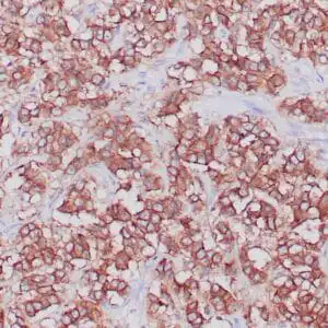
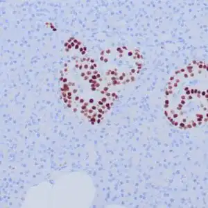
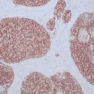
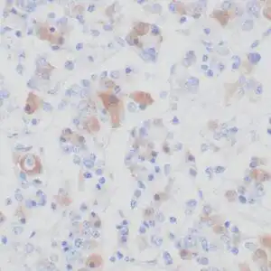
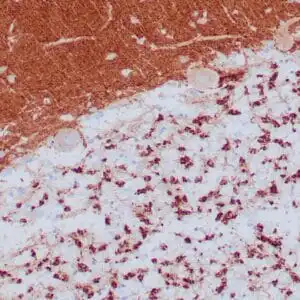
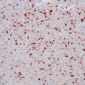
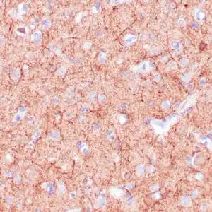
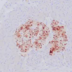
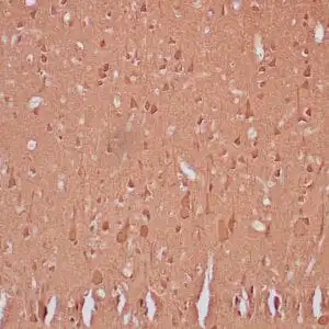
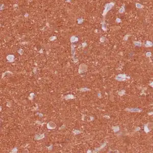
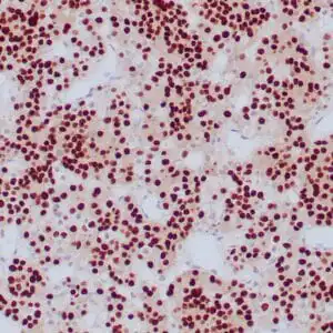
| Formalin-fixed, paraffin-embedded human cerebellum stained with anti-neurofilament antibody using peroxidase-conjugate and DAB chromogen. Note the cytoplasmic staining of neurons and neuronal processes |
FAQ & Publications
Frequently Asked Questions
What species reactivity does the rabbit anti-Neurofilament monoclonal antibody (ZR216) demonstrate?
This antibody is reactive with human species, specifically human tissues.
For which application has this antibody been validated and what is the recommended dilution?
The antibody is validated for immunohistochemistry (IHC) on formalin-fixed, paraffin-embedded tissues, with a recommended dilution range of 1:100 to 1:200 for the concentrated form.
How should the rabbit anti-Neurofilament monoclonal antibody be stored to maintain stability?
For short-term storage, keep the antibody at 2-8°C. For long-term preservation, store at -20°C and avoid repeated freeze/thaw cycles.
What is the host species and clonality of this Neurofilament antibody?
The antibody is a rabbit monoclonal antibody, meaning it is derived from a single clone produced in rabbits.
Which secondary antibodies are compatible with the rabbit anti-Neurofilament monoclonal antibody for detection?
Compatible secondary antibodies include goat anti-rabbit IgG (H&L chain specific) conjugated to peroxidase, biotin, or FITC, including cross-absorbed versions for enhanced specificity.
Publications
| pmid | title | authors | citation |
|---|---|---|---|
| We haven't added any publications to our database yet. | |||
Published literature highly relevant to the biological target of this product and referencing this antibody or clone are retrieved from the PubMed database provided by the United States National Library of Medicine at the National Institutes of Health.
Protocols
| relevant to this product |
|---|
| IHC |
Documents
| Batch Number | QC File | SDS |
|---|---|---|
| To view batch-specific Safety Datasheets and Quality Certificates associated with your account, please Log In. | ||
Only logged in customers who have purchased this product may leave a review.


Reviews
There are no reviews yet.