| Weight | 1 lbs |
|---|---|
| Dimensions | 9 × 5 × 2 in |
| host | mouse |
| isotype | IgG |
| clonality | monoclonal |
| concentration | concentrate, predilute |
| applications | IHC |
| reactivity | human |
| available size | 0.1 mL, 0.5 mL, 1 mL concentrated, 7 mL prediluted |

rabbit anti-Ki-67 monoclonal antibody (ZR433) 6236
Price range: $160.00 through $528.00
Antibody summary
- Rabbit monoclonal to Ki-67
- Suitable for: Immunohistochemistry (formalin-fixed, paraffin-embedded tissues)
- Reacts with: Human
- Isotype:IgG
- Control: Spleen, tonsil, or breast carcinomas
- Visualization: Nuclear
- 0.1, 0.5, 1.0 mL concentrated, 7 mL prediluted
rabbit anti-Ki-67 monoclonal antibody ZR433 6236
| target relevance |
|---|
| Protein names Proliferation marker protein Ki-67 (Antigen identified by monoclonal antibody Ki-67) (Antigen KI-67) (Antigen Ki67) |
| Gene names MKI67,MKI67 |
| Mass 358694Da |
| Function FUNCTION: Protein that associates with the surface of mitotic chromosomes and acts both as a chromosome repellent during early mitosis and chromosome attractant during late mitosis (PubMed:27362226, PubMed:32879492, PubMed:35513709, PubMed:39153474). Required to maintain individual mitotic chromosomes dispersed in the cytoplasm following nuclear envelope disassembly (PubMed:27362226). During early mitosis, relocalizes from nucleoli to the chromosome surface where it forms extended brush structures that cover a substantial fraction of the chromosome surface (PubMed:27362226). The MKI67 brush structure prevents chromosomes from collapsing into a single chromatin mass by forming a steric and electrostatic charge barrier: the protein has a high net electrical charge and acts as a surfactant, dispersing chromosomes and enabling independent chromosome motility (PubMed:27362226). During mitotic anaphase, the MKI67 brush structure collapses and MKI67 switches from a chromosome repellent to a chromosome attractant to promote chromosome clustering and facilitate the exclusion of large cytoplasmic particles from the future nuclear space (PubMed:32879492, PubMed:39153474). Mechanistically, dephosphorylation during mitotic exit and simultaneous exposure of a conserved basic patch induce the RNA-dependent formation of a liquid-like condensed phase on the chromosome surface, promoting coalescence of neighboring chromosome surfaces and clustering of chromosomes (PubMed:39153474). Binds premature ribosomal RNAs during anaphase; promoting liquid-liquid phase separation (PubMed:28935370, PubMed:39153474). Binds DNA, with a preference for supercoiled DNA and AT-rich DNA (PubMed:10878551). Does not contribute to the internal structure of mitotic chromosomes (By similarity). May play a role in chromatin organization; it is however unclear whether it plays a direct role in chromatin organization or whether it is an indirect consequence of its function in mitotic chromosome (PubMed:24867636). {ECO:0000250|UniProtKB:E9PVX6, ECO:0000269|PubMed:10878551, ECO:0000269|PubMed:24867636, ECO:0000269|PubMed:27362226, ECO:0000269|PubMed:28935370, ECO:0000269|PubMed:32879492, ECO:0000269|PubMed:35513709, ECO:0000269|PubMed:39153474}. |
| Subellular location SUBCELLULAR LOCATION: Chromosome {ECO:0000269|PubMed:15896774, ECO:0000269|PubMed:22002106, ECO:0000269|PubMed:27362226, ECO:0000269|PubMed:32879492, ECO:0000269|PubMed:35513709, ECO:0000269|PubMed:39153474, ECO:0000269|PubMed:9510506}. Nucleus {ECO:0000269|PubMed:10878551, ECO:0000269|PubMed:22002106}. Nucleus, nucleolus {ECO:0000269|PubMed:10878551, ECO:0000269|PubMed:22002106, ECO:0000269|PubMed:2674163, ECO:0000269|PubMed:8799815}. Note=During early mitosis, relocalizes from nucleoli to the surface of the mitotic chromosome, the perichromosomal layer, and covers a substantial fraction of the mitotic chromosome surface (PubMed:27362226). Associates with satellite DNA in G1 phase (PubMed:9510506). Binds tightly to chromatin in interphase, chromatin-binding decreases in mitosis when it associates with the surface of the condensed chromosomes (PubMed:15896774, PubMed:22002106). Predominantly localized in the G1 phase in the perinucleolar region, in the later phases it is also detected throughout the nuclear interior, being predominantly localized in the nuclear matrix (PubMed:22002106). {ECO:0000269|PubMed:15896774, ECO:0000269|PubMed:22002106, ECO:0000269|PubMed:27362226}. |
| Structure SUBUNIT: Interacts with KIF15 (PubMed:10878014). Interacts (via the FHA domain) with NIFK (PubMed:11342549, PubMed:14659764, PubMed:16244663). Interacts with PPP1CC (PubMed:24867636, PubMed:25012651). Component of a complex at least composed of ZNF335, HCFC1, CCAR2, EMSY, MKI67, RBBP5, ASH2L and WDR5; the complex is formed as a result of interactions between components of a nuclear receptor-mediated transcription complex and a histone methylation complex (PubMed:19131338). Interacts with ZNF335 (PubMed:19131338, PubMed:23178126). {ECO:0000269|PubMed:10878014, ECO:0000269|PubMed:11342549, ECO:0000269|PubMed:14659764, ECO:0000269|PubMed:16244663, ECO:0000269|PubMed:19131338, ECO:0000269|PubMed:23178126, ECO:0000269|PubMed:24867636, ECO:0000269|PubMed:25012651}. |
| Post-translational modification PTM: Hyperphosphorylated by CDK1 in mitosis; hyperphosphorylatiom prevents undergoing liquid-liquid phase separation (PubMed:10502411, PubMed:10653604, PubMed:25012651, PubMed:39153474). Dephosphorylated by PPP1CC at the onset of anaphase (PubMed:25012651). Dephosphorylated by protein phosphatase 2A (PP2A) at the onset of anaphase (By similarity). Dephosphorylation by protein phosphatase 2A (PP2A) and simultaneous exposure of the positively charged patch (CP) during mitotic exit induce the RNA-dependent formation of a liquid-like condensed phase on the chromosome surface (PubMed:39153474). {ECO:0000250|UniProtKB:E9PVX6, ECO:0000269|PubMed:10502411, ECO:0000269|PubMed:10653604, ECO:0000269|PubMed:25012651, ECO:0000269|PubMed:39153474}.; PTM: Ubiquitinated by the APC/C complex after neuronal progenitors exit mitosis during brain development, leading to clearance from constitutive heterochromatin. {ECO:0000269|PubMed:34942119}. |
| Domain DOMAIN: The positively charged patch (CP) region mediates liquid-liquid phase separation. {ECO:0000269|PubMed:39153474}. |
| Biotechnology BIOTECHNOLOGY: Widely used as a marker to assess cell proliferation, as it is detected in the nucleus of proliferating cells only (PubMed:21960707, PubMed:6339421). In cancer research field for example, MKI67 is the most widely used marker for comparing proliferation between tumor samples (PubMed:21960707, PubMed:26680267). {ECO:0000269|PubMed:6339421, ECO:0000303|PubMed:21960707, ECO:0000303|PubMed:26680267}. |
| Target Relevance information above includes information from UniProt accession: P46013 |
| The UniProt Consortium |
Data
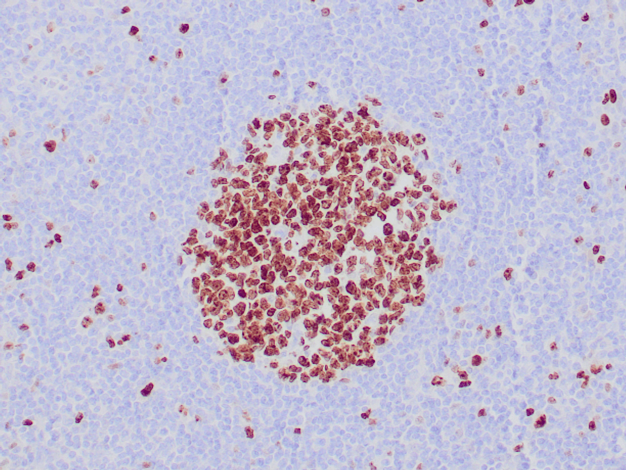 |
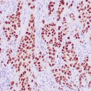
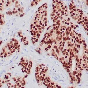
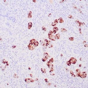
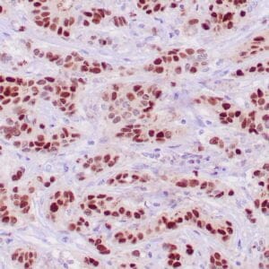
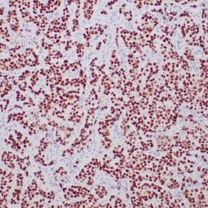
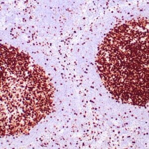
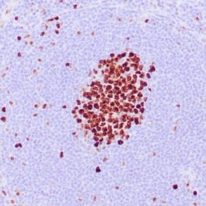
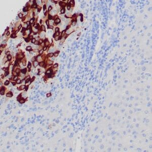
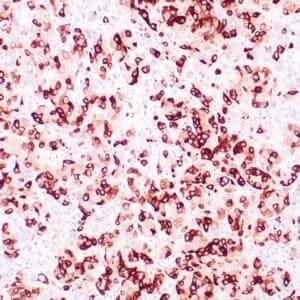
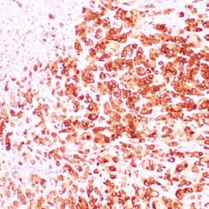
| Human tonsil stained with anti-Ki-67 antibody using peroxidase-conjugate and DAB chromogen. Note the nuclear staining of lymphocytes in follicles.. |
Publications
| pmid | title | authors | citation |
|---|---|---|---|
| We haven't added any publications to our database yet. | |||
Protocols
| relevant to this product |
|---|
| IHC |
Documents
| # | SDS | Certificate | |
|---|---|---|---|
| Please enter your product and batch number here to retrieve product datasheet, SDS, and QC information. | |||
Only logged in customers who have purchased this product may leave a review.
Reviews
There are no reviews yet.