| Weight | 1 lbs |
|---|---|
| Dimensions | 9 × 5 × 2 in |
| host | mouse |
| isotype | IgG |
| clonality | monoclonal |
| concentration | concentrate, predilute |
| applications | IHC |
| reactivity | human |
| available size | 0.1 mL, 0.5 mL, 1 mL concentrated, 7 mL prediluted |

rabbit anti-Her-2/neu monoclonal antibody (ZR218) 6212
Price range: $160.00 through $528.00
Antibody summary
- Rabbit monoclonal to Her-2/neu
- Suitable for: Immunohistochemistry (formalin-fixed, paraffin-embedded tissues)
- Reacts with: Human
- Isotype:IgG
- Control: Breast carcinomas
- Visualization: Cell membrane
- 0.1, 0.5, 1.0 mL concentrated, 7 mL prediluted
rabbit anti-Her-2/neu monoclonal antibody ZR218 6212
| target relevance |
|---|
| Homo sapiens ERBB2 Receptor tyrosine-protein kinase erbB-2 |
| Protein names Receptor tyrosine-protein kinase erbB-2 |
| Alternative names Metastatic lymph node gene 19 protein, Proto-oncogene Neu, Proto-oncogene c-ErbB-2, Tyrosine kinase-type cell surface receptor HER2, p185erbB2 |
| Gene names ERBB2 |
| Protein family Belongs to the protein kinase superfamily. Tyr protein kinase family. EGF receptor subfamily |
| Function Protein tyrosine kinase that is part of several cell surface receptor complexes, but that apparently needs a coreceptor for ligand binding. Essential component of a neuregulin-receptor complex, although neuregulins do not interact with it alone. GP30 is a potential ligand for this receptor. Regulates outgrowth and stabilization of peripheral microtubules (MTs). Upon ERBB2 activation, the MEMO1-RHOA-DIAPH1 signaling pathway elicits the phosphorylation and thus the inhibition of GSK3B at cell membrane. This prevents the phosphorylation of APC and CLASP2, allowing its association with the cell membrane. In turn, membrane-bound APC allows the localization of MACF1 to the cell membrane, which is required for microtubule capture and stabilization |
| Catalytic activity L-tyrosyl-[protein] + ATP = O-phospho-L-tyrosyl-[protein] + ADP + H(+) |
| Subcellular location Cytoplasm, Nucleus |
| Structure Homodimer (PubMed:21454582). Heterodimer with EGFR, ERBB3 and ERBB4 (PubMed:10358079, PubMed:15093539, PubMed:16978839, PubMed:21190959). Part of a complex with EGFR and either PIK3C2A or PIK3C2B. May interact with PIK3C2B when phosphorylated on Tyr-1196 (PubMed:10805725). Interacts with PLXNB1 (PubMed:15210733). Interacts (when phosphorylated on Tyr-1248) with MEMO1 (PubMed:15156151). Interacts with MUC1; the interaction is enhanced by heregulin (HRG) (PubMed:12939402). Interacts (when phosphorylated on Tyr-1139) with GRB7 (via SH2 domain) (PubMed:12975581). Interacts (when phosphorylated on Tyr-1248) with ERBIN (PubMed:12444095). Interacts with KPNB1, RANBP2, EEA1, CRM1 and CLTC (PubMed:16314522). Interacts with PTK6 (PubMed:18719096). Interacts with RPA194 and ACTB (PubMed:21555369). Interacts with PRKCABP, SRC and MYOC (By similarity). Interacts (preferentially with the tyrosine phosphorylated form) with CPNE3; this interaction occurs at the cell membrane and is increased in a growth factor heregulin-dependent manner (PubMed:20010870). Interacts with HSP90AA1 and HSP90AB1 in an ATP-dependent manner; the interaction suppresses ERBB2 kinase activity (PubMed:26517842). Interacts with SORL1; this interaction regulates ERBB2 subcellular distribution by promoting its recycling after internalization from endosomes back to the plasma membrane, hence stimulates ERBB2-mediated signaling (PubMed:31138794). Interacts with SH3BGRL (PubMed:32381043). Interacts with ROR1 (PubMed:36949068) |
| Post-translational modification Autophosphorylated. Autophosphorylation occurs in trans, i.e. one subunit of the dimeric receptor phosphorylates tyrosine residues on the other subunit (Probable). Ligand-binding increases phosphorylation on tyrosine residues (PubMed:27134172, PubMed:33497358). Signaling via SEMA4C promotes phosphorylation at Tyr-1248 (PubMed:17554007). Dephosphorylated by PTPN12 (PubMed:27134172) |
| Involvement in disease Glioma Gliomas are benign or malignant central nervous system neoplasms derived from glial cells. They comprise astrocytomas and glioblastoma multiforme that are derived from astrocytes, oligodendrogliomas derived from oligodendrocytes and ependymomas derived from ependymocytes. Ovarian cancer The term ovarian cancer defines malignancies originating from ovarian tissue. Although many histologic types of ovarian tumors have been described, epithelial ovarian carcinoma is the most common form. Ovarian cancers are often asymptomatic and the recognized signs and symptoms, even of late-stage disease, are vague. Consequently, most patients are diagnosed with advanced disease. Lung cancer A common malignancy affecting tissues of the lung. The most common form of lung cancer is non-small cell lung cancer (NSCLC) that can be divided into 3 major histologic subtypes: squamous cell carcinoma, adenocarcinoma, and large cell lung cancer. NSCLC is often diagnosed at an advanced stage and has a poor prognosis. Gastric cancer A malignant disease which starts in the stomach, can spread to the esophagus or the small intestine, and can extend through the stomach wall to nearby lymph nodes and organs. It also can metastasize to other parts of the body. The term gastric cancer or gastric carcinoma refers to adenocarcinoma of the stomach that accounts for most of all gastric malignant tumors. Two main histologic types are recognized, diffuse type and intestinal type carcinomas. Diffuse tumors are poorly differentiated infiltrating lesions, resulting in thickening of the stomach. In contrast, intestinal tumors are usually exophytic, often ulcerating, and associated with intestinal metaplasia of the stomach, most often observed in sporadic disease. Visceral neuropathy, familial, 2, autosomal recessive An autosomal recessive disorder characterized by intestinal dysmotility due to aganglionosis (Hirschsprung disease), hypoganglionosis, and/or chronic intestinal pseudoobstruction. Patients also show peripheral axonal neuropathy, hypotonia, mild developmental delay, unilateral ptosis, and sensorineural hearing loss. |
| Keywords 3D-structure, Activator, Alternative initiation, Alternative splicing, ATP-binding, Cell membrane, Cell projection, Chromosomal rearrangement, Cytoplasm, Disease variant, Disulfide bond, Endosome, Glycoprotein, Kinase, Membrane, Nucleotide-binding, Nucleus, Phosphoprotein, Proteomics identification, Receptor, Reference proteome, Signal, Transcription, Transcription regulation, Transferase, Transmembrane, Transmembrane helix, Tyrosine-protein kinase |
| Sequence MELAALCRWGLLLALLPPGAASTQVCTGTDMKLRLPASPETHLDMLRHLYQGCQVVQGNL ELTYLPTNASLSFLQDIQEVQGYVLIAHNQVRQVPLQRLRIVRGTQLFEDNYALAVLDNG DPLNNTTPVTGASPGGLRELQLRSLTEILKGGVLIQRNPQLCYQDTILWKDIFHKNNQLA LTLIDTNRSRACHPCSPMCKGSRCWGESSEDCQSLTRTVCAGGCARCKGPLPTDCCHEQC AAGCTGPKHSDCLACLHFNHSGICELHCPALVTYNTDTFESMPNPEGRYTFGASCVTACP YNYLSTDVGSCTLVCPLHNQEVTAEDGTQRCEKCSKPCARVCYGLGMEHLREVRAVTSAN IQEFAGCKKIFGSLAFLPESFDGDPASNTAPLQPEQLQVFETLEEITGYLYISAWPDSLP DLSVFQNLQVIRGRILHNGAYSLTLQGLGISWLGLRSLRELGSGLALIHHNTHLCFVHTV PWDQLFRNPHQALLHTANRPEDECVGEGLACHQLCARGHCWGPGPTQCVNCSQFLRGQEC VEECRVLQGLPREYVNARHCLPCHPECQPQNGSVTCFGPEADQCVACAHYKDPPFCVARC PSGVKPDLSYMPIWKFPDEEGACQPCPINCTHSCVDLDDKGCPAEQRASPLTSIISAVVG ILLVVVLGVVFGILIKRRQQKIRKYTMRRLLQETELVEPLTPSGAMPNQAQMRILKETEL RKVKVLGSGAFGTVYKGIWIPDGENVKIPVAIKVLRENTSPKANKEILDEAYVMAGVGSP YVSRLLGICLTSTVQLVTQLMPYGCLLDHVRENRGRLGSQDLLNWCMQIAKGMSYLEDVR LVHRDLAARNVLVKSPNHVKITDFGLARLLDIDETEYHADGGKVPIKWMALESILRRRFT HQSDVWSYGVTVWELMTFGAKPYDGIPAREIPDLLEKGERLPQPPICTIDVYMIMVKCWM IDSECRPRFRELVSEFSRMARDPQRFVVIQNEDLGPASPLDSTFYRSLLEDDDMGDLVDA EEYLVPQQGFFCPDPAPGAGGMVHHRHRSSSTRSGGGDLTLGLEPSEEEAPRSPLAPSEG AGSDVFDGDLGMGAAKGLQSLPTHDPSPLQRYSEDPTVPLPSETDGYVAPLTCSPQPEYV NQPDVRPQPPSPREGPLPAARPAGATLERPKTLSPGKNGVVKDVFAFGGAVENPEYLTPQ GGAAPQPHPPPAFSPAFDNLYYWDQDPPERGAPPSTFKGTPTAENPEYLGLDVPV |
| UniProt accession: P04626 |
Data
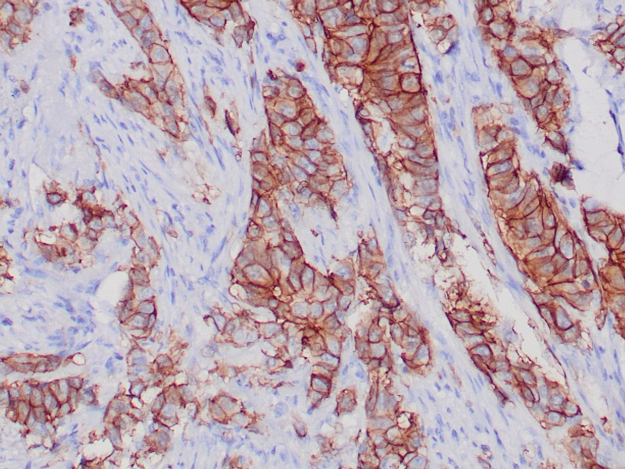 |
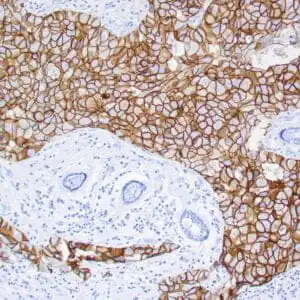
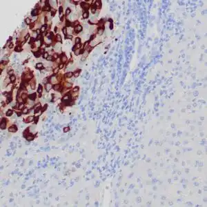
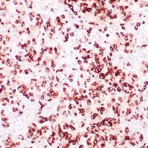
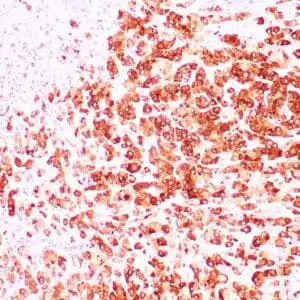
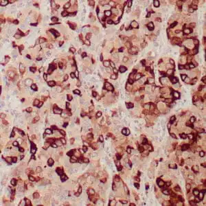
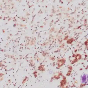
| Human breast infiltrating ductal carcinoma stained with anti-Her-2/neu antibody using peroxidase-conjugate and DAB chromogen. Note the strong membrane staining (3+) of carcinoma cells. |
FAQ & Publications
Frequently Asked Questions
What species reactivity does the rabbit anti-Her-2/neu monoclonal antibody (ZR218) exhibit?
This antibody specifically reacts with human tissues.
For which applications is the rabbit anti-Her-2/neu monoclonal antibody (ZR218) validated?
It is suitable for immunohistochemistry (IHC) on formalin-fixed, paraffin-embedded tissues.
What are the recommended storage conditions for the rabbit anti-Her-2/neu monoclonal antibody (ZR218)?
Store the antibody at 2-8°C for short term use and at -20°C for long term storage, avoiding freeze/thaw cycles.
What immunogen was used to generate the rabbit anti-Her-2/neu monoclonal antibody (ZR218)?
The immunogen is a recombinant protein encoding the extracellular domain of human c-erbB2.
Publications
| pmid | title | authors | citation |
|---|---|---|---|
| We haven't added any publications to our database yet. | |||
Published literature highly relevant to the biological target of this product and referencing this antibody or clone are retrieved from the PubMed database provided by the United States National Library of Medicine at the National Institutes of Health.
Protocols
| relevant to this product |
|---|
| IHC |
Documents
| Batch Number | QC File | SDS |
|---|---|---|
| To view batch-specific Safety Datasheets and Quality Certificates associated with your account, please Log In. | ||
Only logged in customers who have purchased this product may leave a review.

Reviews
There are no reviews yet.