| Weight | 1 lbs |
|---|---|
| Dimensions | 9 × 5 × 2 in |
| host | rabbit |
| isotype | IgG |
| clonality | polyclonal |
| concentration | 1 mg/mL |
| applications | ICC/IF, WB |
| reactivity | IRAK (CT) |
| available sizes | 100 µg |


rabbit anti-IRAK (CT) polyclonal antibody 5960
$445.00
Antibody summary
- Rabbit polyclonal to IRAK (CT)
- Suitable for: ELISA,WB,ICC,IP,IF
- Isotype: IgG
- 100 µg
rabbit anti-IRAK (CT) polyclonal antibody 5960
| antibody |
|---|
| Tested applications WB,IHC,IHC,ICC/IF,ELISA,IP |
| Recommended dilutions Immunoblotting: use at 1:1,000-1:2,000 dilution. Immunoprecipitation: use 2-4 ug antibody per sample. Positive control: Whole cell lysate from HeLa cells or THP-1 cells. |
| Immunogen Peptide corresponding the C- terminus of human IRAK. |
| Size and concentration 100µg and lot specific |
| Form liquid |
| Storage Instructions This antibody is stable for at least one (1) year at -20°C. Avoid multiple freeze-thaw cycles. |
| Storage buffer PBS, pH 7.4. |
| Purity peptide affinity purification |
| Clonality polyclonal |
| Isotype IgG |
| Compatible secondaries goat anti-rabbit IgG, H&L chain specific, peroxidase conjugated, conjugated polyclonal antibody 9512 goat anti-rabbit IgG, H&L chain specific, biotin conjugated polyclonal antibody 2079 goat anti-rabbit IgG, H&L chain specific, FITC conjugated polyclonal antibody 7863 goat anti-rabbit IgG, H&L chain specific, Cross Absorbed polyclonal antibody 2371 goat anti-rabbit IgG, H&L chain specific, biotin conjugated polyclonal antibody, crossabsorbed 1715 goat anti-rabbit IgG, H&L chain specific, FITC conjugated polyclonal antibody, crossabsorbed 1720 |
| Isotype control Rabbit polyclonal - Isotype Control |
| target relevance |
|---|
| Homo sapiens IRAK1 Interleukin-1 receptor-associated kinase 1 |
| Protein names Interleukin-1 receptor-associated kinase 1 |
| Gene names IRAK1 |
| Protein family Belongs to the protein kinase superfamily. TKL Ser/Thr protein kinase family. Pelle subfamily |
| Function Serine/threonine-protein kinase that plays a critical role in initiating innate immune response against foreign pathogens. Involved in Toll-like receptor (TLR) and IL-1R signaling pathways. Is rapidly recruited by MYD88 to the receptor-signaling complex upon TLR activation. Association with MYD88 leads to IRAK1 phosphorylation by IRAK4 and subsequent autophosphorylation and kinase activation. Phosphorylates E3 ubiquitin ligases Pellino proteins (PELI1, PELI2 and PELI3) to promote pellino-mediated polyubiquitination of IRAK1. Then, the ubiquitin-binding domain of IKBKG/NEMO binds to polyubiquitinated IRAK1 bringing together the IRAK1-MAP3K7/TAK1-TRAF6 complex and the NEMO-IKKA-IKKB complex. In turn, MAP3K7/TAK1 activates IKKs (CHUK/IKKA and IKBKB/IKKB) leading to NF-kappa-B nuclear translocation and activation. Alternatively, phosphorylates TIRAP to promote its ubiquitination and subsequent degradation. Phosphorylates the interferon regulatory factor 7 (IRF7) to induce its activation and translocation to the nucleus, resulting in transcriptional activation of type I IFN genes, which drive the cell in an antiviral state. When sumoylated, translocates to the nucleus and phosphorylates STAT3 |
| Catalytic activity L-seryl-[protein] + ATP = O-phospho-L-seryl-[protein] + ADP + H(+) L-threonyl-[protein] + ATP = O-phospho-L-threonyl-[protein] + ADP + H(+) |
| Subcellular location Cytoplasm, Nucleus, Lipid droplet |
| Structure (Microbial infection) Interacts with alphaviruses SINV, CHIKV, RRV, VEEV and EEEV capsid proteins; the interactions lead to inhibition of IRAK1-dependent signaling |
| Post-translational modification Following recruitment on the activated receptor complex, phosphorylated on Thr-209, probably by IRAK4, resulting in a conformational change of the kinase domain, allowing further phosphorylations to take place. Thr-387 phosphorylation in the activation loop is required to achieve full enzymatic activity Polyubiquitinated by TRAF6 after cell stimulation with IL-1-beta by PELI1, PELI2 and PELI3. Polyubiquitination occurs with polyubiquitin chains linked through 'Lys-63'. Ubiquitination promotes interaction with NEMO/IKBKG. Also sumoylated; leading to nuclear translocation |
| Keywords 3D-structure, Alternative splicing, ATP-binding, Cytoplasm, Direct protein sequencing, Host-virus interaction, Immunity, Innate immunity, Isopeptide bond, Kinase, Lipid droplet, Magnesium, Nucleotide-binding, Nucleus, Phosphoprotein, Proteomics identification, Reference proteome, Serine/threonine-protein kinase, Transferase, Ubl conjugation |
| Sequence MAGGPGPGEPAAPGAQHFLYEVPPWVMCRFYKVMDALEPADWCQFAALIVRDQTELRLCE RSGQRTASVLWPWINRNARVADLVHILTHLQLLRARDIITAWHPPAPLPSPGTTAPRPSS IPAPAEAEAWSPRKLPSSASTFLSPAFPGSQTHSGPELGLVPSPASLWPPPPSPAPSSTK PGPESSVSLLQGARPFPFCWPLCEISRGTHNFSEELKIGEGGFGCVYRAVMRNTVYAVKR LKENADLEWTAVKQSFLTEVEQLSRFRHPNIVDFAGYCAQNGFYCLVYGFLPNGSLEDRL HCQTQACPPLSWPQRLDILLGTARAIQFLHQDSPSLIHGDIKSSNVLLDERLTPKLGDFG LARFSRFAGSSPSQSSMVARTQTVRGTLAYLPEEYIKTGRLAVDTDTFSFGVVVLETLAG QRAVKTHGARTKYLKDLVEEEAEEAGVALRSTQSTLQAGLAADAWAAPIAMQIYKKHLDP RPGPCPPELGLGLGQLACCCLHRRAKRRPPMTQVYERLEKLQAVVAGVPGHSEAASCIPP SPQENSYVSSTGRAHSGAAPWQPLAAPSGASAQAAEQLQRGPNQPVESDESLGGLSAALR SWHLTPSCPLDPAPLREAGCPQGDTAGESSWGSGPGSRPTAVEGLALGSSASSSSEPPQI IINPARQKMVQKLALYEDGALDSLQLLSSSSLPGLGLEQDRQGPEESDEFQS |
| UniProt accession: P51617 |
Data
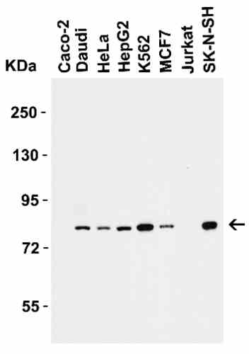 |
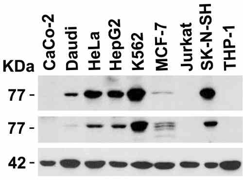
| Western Blot Validation in Human Cell Lines Loading: 15 µg of lysates per lane. Antibodies: IRAK 5960 (1 µg/mL), 1h incubation at RT in 5% NFDM/TBST.Secondary: Goat anti-rabbit IgG HRP conjugate at 1:10000 dilution. |
 |
| Independent Antibody Validation (IAV) via Protein Expression Profile in Cell Lines Loading: 15 µg of lysates per lane. Antibodies: IRAK 5960 (1 µg/mL), IRAK 64-231 (2 µg/mL), beta-actin (1 µg/mL), 1h incubation at RT in 5% NFDM/TBST.Secondary: Goat anti-rabbit IgG HRP conjugate at 1:10000 dilution. |
 |
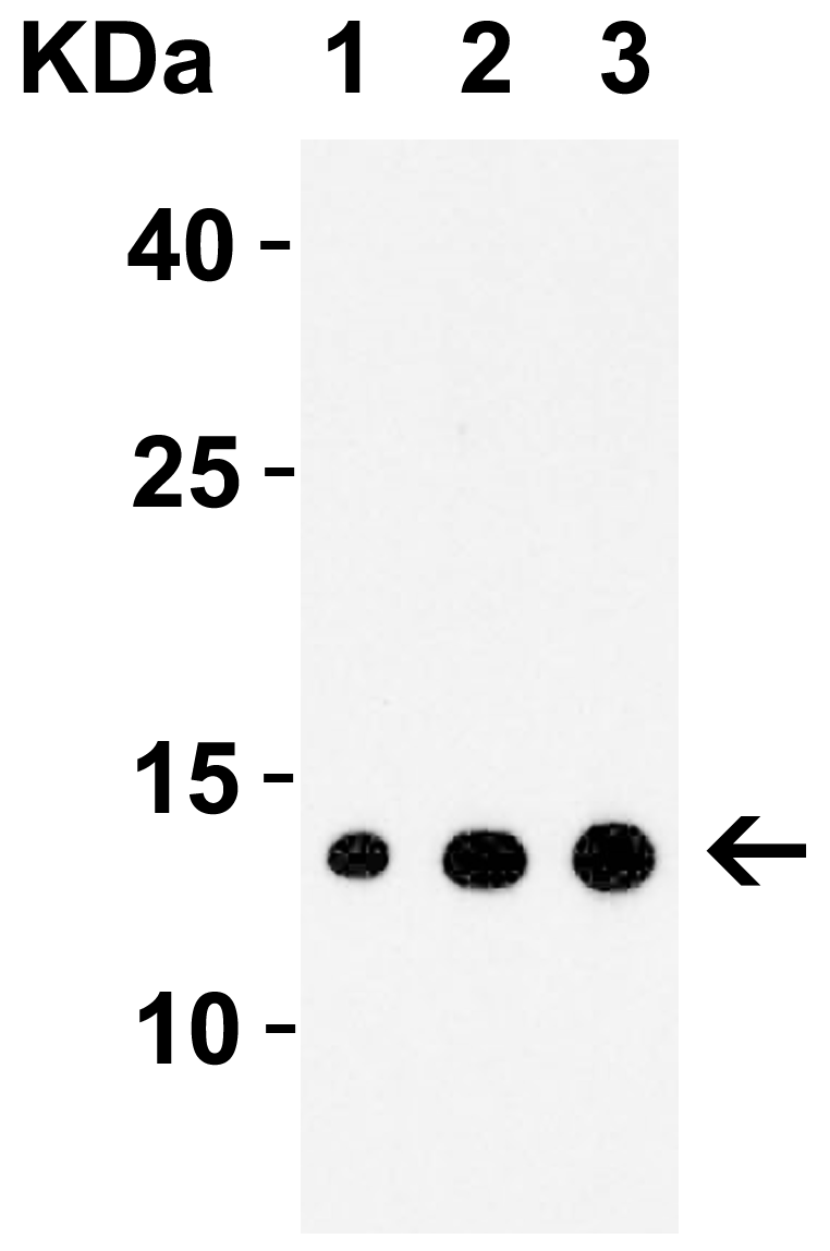
| Western Blot Validation with Recombinant Protein Loading: 30 ng of human IRAK recombinant protein per lane. Antibodies: IRAK 5960 (1: 1 µg/mL, 2: 2 µg/mL and 3: 4 µg/mL), 1h incubation at RT in 5% NFDM/TBST.Secondary: Goat anti-rabbit IgG HRP conjugate at 1:10000 dilution. |
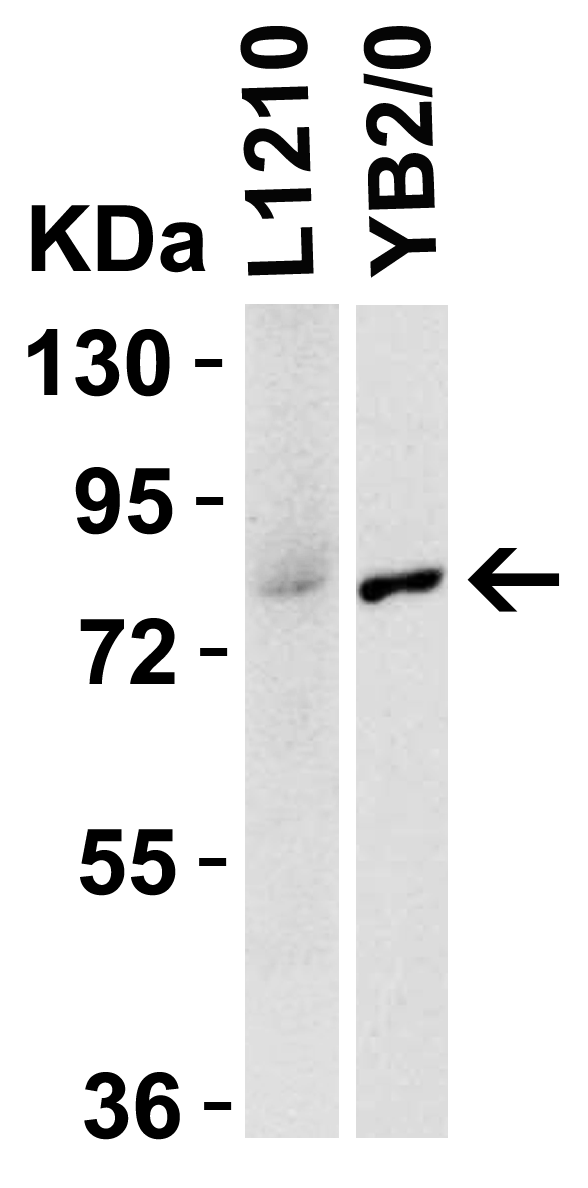 |
| Species Activity in Mouse and Rat Cell Lines Loading: 15 µg of lysates per lane. Antibodies: IRAK 5960 (1 µg /mL,), 1h incubation at RT in 5% NFDM/TBST.Secondary: Goat anti-rabbit IgG HRP conjugate at 1:10000 dilution. |
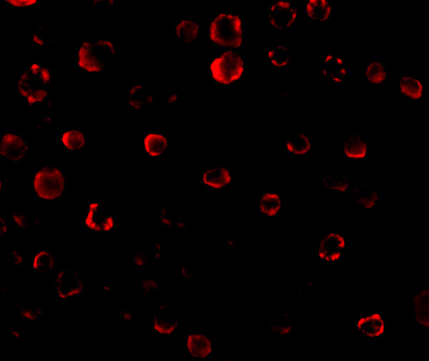 |
| Immunofluorescence Validation of IRAK in Human HeLa Cells Immunofluorescent analysis of 4% paraformaldehyde-fixed HeLa Cells labeling IRAK with 5960 at 20 µg/mL, followed by goat anti-rabbit IgG secondary antibody at 1/500 dilution (red). |
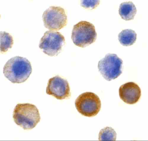 |
| Immunocytochemistry Validation of IRAK in Human HeLa Cells Immunocytochemical analysis of HeLa cells using anti-IRAK antibody (5960) at 10 µg/mL. Cells was fixed with formaldehyde and blocked with 10% serum for 1 h at RT; antigen retrieval was by heat mediation with a citrate buffer (pH6). Samples were incubated with primary antibody overnight at 4 C. A goat anti-rabbit IgG H&L (HRP) at 1/250 was used as secondary. Counter stained with Hematoxylin. |
 |
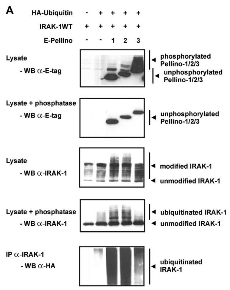
| Immunoprecipitation and Overexpression Validation in HEK293T Cells(Schauvliege et al., 2006) Co-expression of Pellino proteins and IRAK-1 leads to Pellino phosphorylation and IRAK-1 polyubiquitination. (-) E-tagged Pellino proteins were co-expressed with IRAK-1WT and HA ubiquitin in HEK293T cells. For assessment of IRAK-1 polyubiquitination, the same cellextracts, untreated or treated with phosphatase as described above, were analysed for slower migrating forms of IRAK-1 by Western blotting withanti-IRAK-1 (5960). Ubiquitination was specifically detected by IRAK-1 immunoprecipitation followed by Western blotting with anti-HA antibodies. |
 |
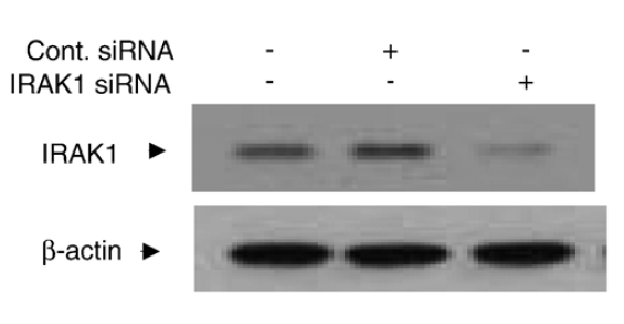
| KD Validation in Human Chondrocytes (Ahmad et al., 2416) Chondrocytes were transfected with 250 nM of IRAK1 or control siRNA for 48 h and lysates were analyzed for IRAK1 or beta-actin expression levels by immunoblotting. IRAK1 signal was disrupted in IRAK1 KD lysate. |
FAQ & Publications
Frequently Asked Questions
What applications has the rabbit anti-IRAK (CT) polyclonal antibody 5960 been validated for?
This antibody has been tested and validated for use in Western blotting (WB), immunohistochemistry (IHC), immunocytochemistry/immunofluorescence (ICC/IF), enzyme-linked immunosorbent assay (ELISA), and immunoprecipitation (IP).
What is the recommended dilution range for immunoblotting using this IRAK antibody?
For immunoblotting, it is recommended to use the antibody at a dilution between 1:1,000 and 1:2,000.
How should the rabbit anti-IRAK (CT) polyclonal antibody 5960 be stored to maintain stability?
The antibody should be stored at -20°C and is stable for at least one year under these conditions. It is important to avoid multiple freeze-thaw cycles to preserve antibody integrity.
Is this antibody suitable for detecting IRAK in species other than human?
Yes, the antibody has been validated for reactivity in mouse and rat cell lines, indicating cross-species activity.
What is the immunogen used to generate the rabbit anti-IRAK (CT) polyclonal antibody 5960?
The immunogen is a peptide corresponding to the C-terminus of the human IRAK protein.
Publications
| pmid | title | authors | citation |
|---|---|---|---|
| We haven't added any publications to our database yet. | |||
Published literature highly relevant to the biological target of this product and referencing this antibody or clone are retrieved from the PubMed database provided by the United States National Library of Medicine at the National Institutes of Health.
Protocols
| relevant to this product |
|---|
| Western blot IHC ICC |
Documents
| Batch Number | QC File | SDS |
|---|---|---|
| To view batch-specific Safety Datasheets and Quality Certificates associated with your account, please Log In. | ||
Only logged in customers who have purchased this product may leave a review.


Reviews
There are no reviews yet.