| Weight | 1 lbs |
|---|---|
| Dimensions | 9 × 5 × 2 in |
| host | rabbit |
| isotype | IgG |
| clonality | polyclonal |
| concentration | 1 mg/mL |
| applications | ICC/IF, WB |
| reactivity | IL-1RAcP |
| available sizes | 100 µg |


rabbit anti-IL-1RAcP (CT) polyclonal antibody 2928
$445.00
Antibody summary
- Rabbit polyclonal to IL-1RAcP (CT)
- Suitable for: ELISA,WB,ICC,IF
- Isotype: IgG
- 100 µg
rabbit anti-IL-1RAcP (CT) polyclonal antibody 2928
| antibody |
|---|
| Tested applications WB,ICC/IF,ELISA |
| Recommended dilutions Immunoblotting: use at 1:1,000-1:2,000 dilution. Positive control: Whole cell lysate from HeLa cells. |
| Immunogen Peptide corresponding to aa 525-540 of human IL-1RAcP. The sequence is identical to that of mouse and rat IL-1RAcP. |
| Size and concentration 100µg and lot specific |
| Form liquid |
| Storage Instructions This antibody is stable for at least one (1) year at -20°C. Avoid multiple freeze- thaw cycles. |
| Storage buffer PBS, pH 7.4. |
| Purity peptide affinity purification |
| Clonality polyclonal |
| Isotype IgG |
| Compatible secondaries goat anti-rabbit IgG, H&L chain specific, peroxidase conjugated, conjugated polyclonal antibody 9512 goat anti-rabbit IgG, H&L chain specific, biotin conjugated polyclonal antibody 2079 goat anti-rabbit IgG, H&L chain specific, FITC conjugated polyclonal antibody 7863 goat anti-rabbit IgG, H&L chain specific, Cross Absorbed polyclonal antibody 2371 goat anti-rabbit IgG, H&L chain specific, biotin conjugated polyclonal antibody, crossabsorbed 1715 goat anti-rabbit IgG, H&L chain specific, FITC conjugated polyclonal antibody, crossabsorbed 1720 |
| Isotype control Rabbit polyclonal - Isotype Control |
| target relevance |
|---|
| Homo sapiens IL1RAP Interleukin-1 receptor accessory protein |
| Protein names Interleukin-1 receptor accessory protein |
| Alternative names Interleukin-1 receptor 3 |
| Gene names IL1RAP |
| Protein family Belongs to the interleukin-1 receptor family |
| Function Coreceptor for IL1RL2 in the IL-36 signaling system (By similarity). Coreceptor with IL1R1 in the IL-1 signaling system. Associates with IL1R1 bound to IL1B to form the high affinity interleukin-1 receptor complex which mediates interleukin-1-dependent activation of NF-kappa-B and other pathways. Signaling involves the recruitment of adapter molecules such as TOLLIP, MYD88, and IRAK1 or IRAK2 via the respective TIR domains of the receptor/coreceptor subunits. Recruits TOLLIP to the signaling complex. Does not bind to interleukin-1 alone; binding of IL1RN to IL1R1, prevents its association with IL1R1 to form a signaling complex. The cellular response is modulated through a non-signaling association with the membrane IL1R2 decoy receptor. Coreceptor for IL1RL1 in the IL-33 signaling system. Can bidirectionally induce pre- and postsynaptic differentiation of neurons by trans-synaptically binding to PTPRD (By similarity). May play a role in IL1B-mediated costimulation of IFNG production from T-helper 1 (Th1) cells (Probable) |
| Catalytic activity NAD(+) + H2O = ADP-D-ribose + nicotinamide + H(+) |
| Subcellular location Secreted |
| Structure The interleukin-36 receptor complex is a heterodimer of IL1RL2 and IL1RAP; the association is inhibited by IL36RN (By similarity). The interleukin-1 receptor complex is a heterodimer of IL1R1 and IL1RAP. Associates with IL1R2 to form a non-signaling interleukin-1 receptor complex. Isoform 4 interacts with IL1R1 in an interleukin-1-dependent manner. Interacts with IL-33-bound IL1RL1 to form the minimal interleukin-33 signaling complex with a 1:1:1 stoichiometry. Interacts with KIT (independently of stimulation with KITLG/SCF). A mast cell-specific KITLG/SCF-induced interleukin-33 signaling complex contains IL1RL1, IL1RAP, KIT and MYD88 (By similarity). Interacts (via the first immunoglobilin domain) with PTPRD (via the third immunoglobilin domain); induces pre- and postsynaptic differentiation of neurons (By similarity) |
| Keywords 3D-structure, Alternative splicing, Cell membrane, Disulfide bond, Glycoprotein, Hydrolase, Immunity, Immunoglobulin domain, Inflammatory response, Innate immunity, Membrane, NAD, Phosphoprotein, Proteomics identification, Receptor, Reference proteome, Repeat, Secreted, Signal, Transmembrane, Transmembrane helix |
| Sequence MTLLWCVVSLYFYGILQSDASERCDDWGLDTMRQIQVFEDEPARIKCPLFEHFLKFNYST AHSAGLTLIWYWTRQDRDLEEPINFRLPENRISKEKDVLWFRPTLLNDTGNYTCMLRNTT YCSKVAFPLEVVQKDSCFNSPMKLPVHKLYIEYGIQRITCPNVDGYFPSSVKPTITWYMG CYKIQNFNNVIPEGMNLSFLIALISNNGNYTCVVTYPENGRTFHLTRTLTVKVVGSPKNA VPPVIHSPNDHVVYEKEPGEELLIPCTVYFSFLMDSRNEVWWTIDGKKPDDITIDVTINE SISHSRTEDETRTQILSIKKVTSEDLKRSYVCHARSAKGEVAKAAKVKQKVPAPRYTVEL ACGFGATVLLVVILIVVYHVYWLEMVLFYRAHFGTDETILDGKEYDIYVSYARNAEEEEF VLLTLRGVLENEFGYKLCIFDRDSLPGGIVTDETLSFIQKSRRLLVVLSPNYVLQGTQAL LELKAGLENMASRGNINVILVQYKAVKETKVKELKRAKTVLTVIKWKGEKSKYPQGRFWK QLQVAMPVKKSPRRSSSDEQGLSYSSLKNV |
| UniProt accession: Q9NPH3 |
Data
 |
| Western blot analysis of IL-1RAcP in HeLa whole cell lysate with IL-1RAcP antibody at 1 µg/mL. |
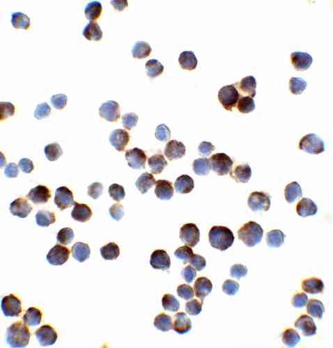 |
| Immunocytochemistry of IL-1RAcP in HeLa cells with IL-1RAcP antibody at 2 µg/mL. |
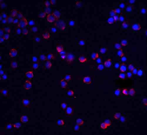 |
| Immunofluorescence of IL-1RAcP in HeLa cells with IL-1RAcP antibody at 5 µg/mL.
Red: IL-1RAcP Antibody (2928) Blue: DAPI staining |
FAQ & Publications
Frequently Asked Questions
What applications has the rabbit anti-IL-1RAcP (CT) polyclonal antibody 2928 been validated for?
This antibody is suitable and has been tested for ELISA, Western blot (WB), Immunocytochemistry (ICC), and Immunofluorescence (IF) applications.
How should the rabbit anti-IL-1RAcP (CT) antibody be stored to maintain stability?
The antibody should be stored at -20°C and is stable for at least one year under these conditions. It is recommended to avoid multiple freeze-thaw cycles to preserve antibody integrity.
What is the immunogen used to generate the rabbit anti-IL-1RAcP (CT) polyclonal antibody?
The immunogen is a peptide corresponding to amino acids 525-540 of human IL-1RAcP, which is identical in mouse and rat IL-1RAcP.
What is the host species and clonality of this IL-1RAcP antibody?
The antibody is a rabbit polyclonal IgG.
Which secondary antibodies are compatible with this rabbit anti-IL-1RAcP (CT) polyclonal antibody?
Compatible secondary antibodies include goat anti-rabbit IgG, H&L chain specific, conjugated to peroxidase, biotin, or FITC, including cross-absorbed versions for reduced cross-reactivity.
Publications
| pmid | title | authors | citation |
|---|---|---|---|
| We haven't added any publications to our database yet. | |||
Published literature highly relevant to the biological target of this product and referencing this antibody or clone are retrieved from the PubMed database provided by the United States National Library of Medicine at the National Institutes of Health.
Protocols
| relevant to this product |
|---|
| Western blot IHC ICC |
Documents
| Batch Number | QC File | SDS |
|---|---|---|
| To view batch-specific Safety Datasheets and Quality Certificates associated with your account, please Log In. | ||
Only logged in customers who have purchased this product may leave a review.


Reviews
There are no reviews yet.