| Weight | 1 lbs |
|---|---|
| Dimensions | 9 × 5 × 2 in |
| host | mouse |
| isotype | IgG |
| clonality | monoclonal |
| concentration | concentrate, predilute |
| applications | IHC |
| reactivity | human |
| available size | 0.1 mL, 0.5 mL, 1 mL concentrated, 7 mL prediluted |

rabbit anti-EMA monoclonal antibody (ZR133) 6168
Price range: $160.00 through $528.00
Antibody summary
- Rabbit monoclonal to EMA
- Suitable for: Immunohistochemistry (formalin-fixed, paraffin-embedded tissues)
- Reacts with: Human
- Isotype:IgG
- Control: Breast or colon carcinoma
- Visualization: Cytoplasmic and membranous
- 0.1, 0.5, 1.0 mL concentrated, 7 mL prediluted
rabbit anti-EMA monoclonal antibody ZR133 6168
| target relevance |
|---|
| Protein names Mucin-1 (MUC-1) (Breast carcinoma-associated antigen DF3) (Cancer antigen 15-3) (CA 15-3) (Carcinoma-associated mucin) (Episialin) (H23AG) (Krebs von den Lungen-6) (KL-6) (PEMT) (Peanut-reactive urinary mucin) (PUM) (Polymorphic epithelial mucin) (PEM) (Tumor-associated epithelial membrane antigen) (EMA) (Tumor-associated mucin) (CD antigen CD227) [Cleaved into: Mucin-1 subunit alpha (MUC1-NT) (MUC1-alpha); Mucin-1 subunit beta (MUC1-beta) (MUC1-CT)] |
| Gene names MUC1,MUC1 PUM |
| Mass 122102Da |
| Function FUNCTION: The alpha subunit has cell adhesive properties. Can act both as an adhesion and an anti-adhesion protein. May provide a protective layer on epithelial cells against bacterial and enzyme attack.; FUNCTION: The beta subunit contains a C-terminal domain which is involved in cell signaling, through phosphorylations and protein-protein interactions. Modulates signaling in ERK, SRC and NF-kappa-B pathways. In activated T-cells, influences directly or indirectly the Ras/MAPK pathway. Promotes tumor progression. Regulates TP53-mediated transcription and determines cell fate in the genotoxic stress response. Binds, together with KLF4, the PE21 promoter element of TP53 and represses TP53 activity. |
| Subellular location SUBCELLULAR LOCATION: Apical cell membrane {ECO:0000269|PubMed:11118479, ECO:0000269|PubMed:12832415, ECO:0000269|PubMed:12939402, ECO:0000269|PubMed:15471854, ECO:0000269|PubMed:15972891, ECO:0000269|PubMed:16507569, ECO:0000269|PubMed:17524503, ECO:0000269|PubMed:17545600}; Single-pass type I membrane protein {ECO:0000269|PubMed:11118479, ECO:0000269|PubMed:12832415, ECO:0000269|PubMed:12939402, ECO:0000269|PubMed:15471854, ECO:0000269|PubMed:15972891, ECO:0000269|PubMed:16507569, ECO:0000269|PubMed:17524503, ECO:0000269|PubMed:17545600}. Note=Exclusively located in the apical domain of the plasma membrane of highly polarized epithelial cells. After endocytosis, internalized and recycled to the cell membrane. Located to microvilli and to the tips of long filopodial protusions.; SUBCELLULAR LOCATION: [Isoform 5]: Secreted.; SUBCELLULAR LOCATION: [Isoform Y]: Secreted.; SUBCELLULAR LOCATION: [Isoform 9]: Secreted.; SUBCELLULAR LOCATION: [Mucin-1 subunit beta]: Cell membrane. Cytoplasm. Nucleus. Note=On EGF and PDGFRB stimulation, transported to the nucleus through interaction with CTNNB1, a process which is stimulated by phosphorylation. On HRG stimulation, colocalizes with JUP/gamma-catenin at the nucleus. |
| Tissues TISSUE SPECIFICITY: Expressed on the apical surface of epithelial cells, especially of airway passages, breast and uterus. Also expressed in activated and unactivated T-cells. Overexpressed in epithelial tumors, such as breast or ovarian cancer and also in non-epithelial tumor cells. Isoform Y is expressed in tumor cells only. {ECO:0000269|PubMed:15513966, ECO:0000269|PubMed:9212228}. |
| Structure SUBUNIT: The alpha subunit forms a tight, non-covalent heterodimeric complex with the proteolytically-released beta-subunit. Interaction, via the tandem repeat region, with domain 1 of ICAM1 is implicated in cell migration and metastases. Isoform 1 binds directly the SH2 domain of GRB2, and forms a MUC1/GRB2/SOS1 complex involved in RAS signaling. The cytoplasmic tail (MUC1CT) interacts with several proteins such as SRC, CTNNB1 and ERBs. Interaction with the SH2 domain of CSK decreases interaction with GSK3B. Interacts with CTNNB1/beta-catenin and JUP/gamma-catenin and promotes cell adhesion. Interaction with JUP/gamma-catenin is induced by heregulin. Binds PRKCD, ERBB2, ERBB3 and ERBB4. Heregulin (HRG) stimulates the interaction with ERBB2 and, to a much lesser extent, the interaction with ERBB3 and ERBB4. Interacts with P53 in response to DNA damage. Interacts with KLF4. Interacts with estrogen receptor alpha/ESR1, through its DNA-binding domain, and stimulates its transcription activity. Binds ADAM17. Isoform ZD forms disulfide-linked oligomers. {ECO:0000269|PubMed:11152665, ECO:0000269|PubMed:11173916, ECO:0000269|PubMed:11483589, ECO:0000269|PubMed:11877440, ECO:0000269|PubMed:12441351, ECO:0000269|PubMed:12750561, ECO:0000269|PubMed:12832415, ECO:0000269|PubMed:12939402, ECO:0000269|PubMed:14688481, ECO:0000269|PubMed:15471854, ECO:0000269|PubMed:15513966, ECO:0000269|PubMed:15710329, ECO:0000269|PubMed:16288032, ECO:0000269|PubMed:16427018, ECO:0000269|PubMed:16507569, ECO:0000269|PubMed:16983337, ECO:0000269|PubMed:17308127, ECO:0000269|PubMed:17524503, ECO:0000269|PubMed:7664271, ECO:0000269|PubMed:9139698, ECO:0000269|PubMed:9819408}. |
| Post-translational modification PTM: Highly glycosylated (N- and O-linked carbohydrates and sialic acid). O-glycosylated to a varying degree on serine and threonine residues within each tandem repeat, ranging from mono- to penta-glycosylation. The average density ranges from about 50% in human milk to over 90% in T47D breast cancer cells. Further sialylation occurs during recycling. Membrane-shed glycoproteins from kidney and breast cancer cells have preferentially sialyated core 1 structures, while secreted forms from the same tissues display mainly core 2 structures. The O-glycosylated content is overlapping in both these tissues with terminal fucose and galactose, 2- and 3-linked galactose, 3- and 3,6-linked GalNAc-ol and 4-linked GlcNAc predominating. Differentially O-glycosylated in breast carcinomas with 3,4-linked GlcNAc. N-glycosylation consists of high-mannose, acidic complex-type and hybrid glycans in the secreted form MUC1/SEC, and neutral complex-type in the transmembrane form, MUC1/TM. {ECO:0000269|PubMed:10373415, ECO:0000269|PubMed:7744025, ECO:0000269|PubMed:9312074, ECO:0000269|PubMed:9597769}.; PTM: Proteolytic cleavage in the SEA domain occurs in the endoplasmic reticulum by an autoproteolytic mechanism and requires the full-length SEA domain as well as requiring a Ser, Thr or Cys residue at the P + 1 site. Cleavage at this site also occurs on isoform MUC1/X but not on isoform MUC1/Y. Ectodomain shedding is mediated by ADAM17. {ECO:0000269|PubMed:11341784, ECO:0000269|PubMed:12441351, ECO:0000269|PubMed:15987679}.; PTM: Dual palmitoylation on cysteine residues in the CQC motif is required for recycling from endosomes back to the plasma membrane. {ECO:0000269|PubMed:16507569}.; PTM: Phosphorylated on tyrosines and serine residues in the C-terminal. Phosphorylation on tyrosines in the C-terminal increases the nuclear location of MUC1 and beta-catenin. Phosphorylation by PKC delta induces binding of MUC1 to beta-catenin/CTNNB1 and thus decreases the formation of the beta-catenin/E-cadherin complex. Src-mediated phosphorylation inhibits interaction with GSK3B. Src- and EGFR-mediated phosphorylation on Tyr-1229 increases binding to beta-catenin/CTNNB1. GSK3B-mediated phosphorylation on Ser-1227 decreases this interaction but restores the formation of the beta-cadherin/E-cadherin complex. On T-cell receptor activation, phosphorylated by LCK. PDGFR-mediated phosphorylation increases nuclear colocalization of MUC1CT and CTNNB1. {ECO:0000269|PubMed:11152665, ECO:0000269|PubMed:11483589, ECO:0000269|PubMed:11877440, ECO:0000269|PubMed:12750561, ECO:0000269|PubMed:14521915, ECO:0000269|PubMed:15513966, ECO:0000269|PubMed:16288032, ECO:0000269|PubMed:17545600, ECO:0000269|PubMed:7664271, ECO:0000269|PubMed:7988707, ECO:0000269|PubMed:9819408}.; PTM: The N-terminal sequence has been shown to begin at position 24 or 28. {ECO:0000269|PubMed:11341784}. |
| Involvement in disease DISEASE: Note=MUC1/CA 15-3 is used as a serological clinical marker of breast cancer to monitor response to breast cancer treatment and disease recurrence (PubMed:20816948). Decreased levels over time may be indicative of a positive response to treatment. Conversely, increased levels may indicate disease progression. At an early stage disease, only 21% of patients exhibit high MUC1/CA 15-3 levels, that is why CA 15-3 is not a useful screening test. Most antibodies target the highly immunodominant core peptide domain of 20 amino acid (APDTRPAPGSTAPPAHGVTS) tandem repeats. Some antibodies recognize glycosylated epitopes. {ECO:0000269|PubMed:20816948}.; DISEASE: Tubulointerstitial kidney disease, autosomal dominant, 2 (ADTKD2) [MIM:174000]: A form of autosomal dominant tubulointerstitial kidney disease, a genetically heterogeneous disorder characterized by slowly progressive loss of kidney function, bland urinary sediment, hyperuricemia, absent or mildly increased albuminuria, lack of severe hypertension during the early stages, and normal or small kidneys on ultrasound. Renal histology shows variable abnormalities including interstitial fibrosis with tubular atrophy, microcystic dilatation of the tubules, thickening of tubular basement membranes, medullary cysts, and secondary glomerulosclerotic or glomerulocystic changes with abnormal glomerular tufting. There is significant variability, as well as incomplete penetrance. {ECO:0000269|PubMed:23396133}. Note=The disease is caused by variants affecting the gene represented in this entry. |
| Target Relevance information above includes information from UniProt accession: P15941 |
| The UniProt Consortium |
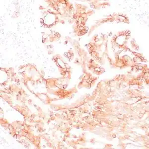
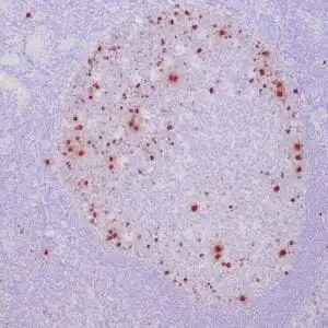
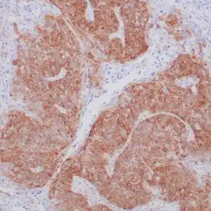
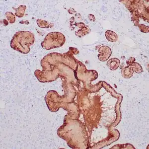
Data
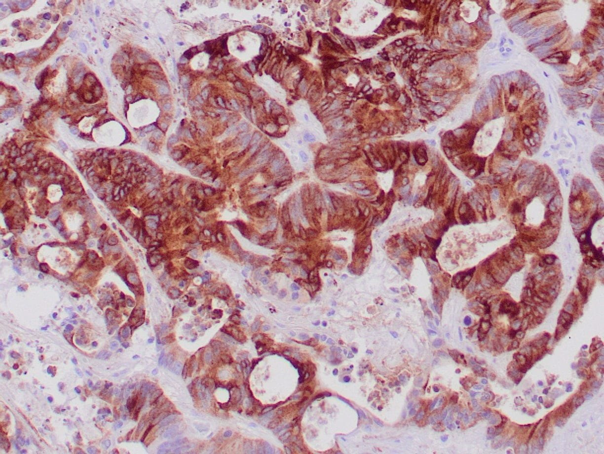 |
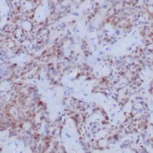
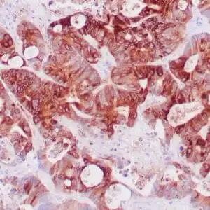
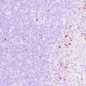
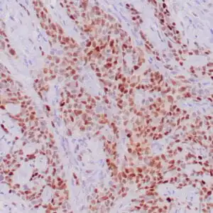
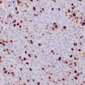
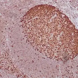
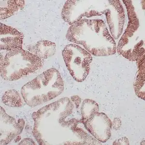
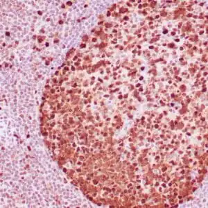
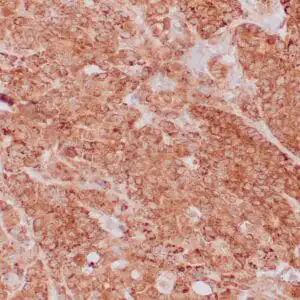
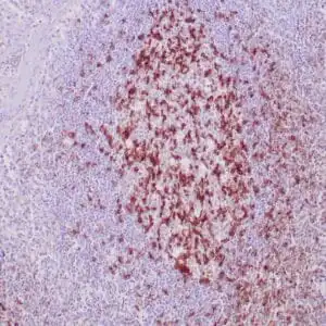
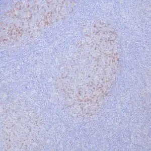
| Formalin-fixed, paraffin-embedded human colon cancer stained with anti-EMA antibody using peroxidase-conjugate and DAB chromogen. Note the membrane and cytoplasmic staining of tumor cells |
FAQ & Publications
Frequently Asked Questions
What applications is the rabbit anti-EMA monoclonal antibody (ZR133) suitable for?
This antibody is suitable for immunohistochemistry (IHC) on formalin-fixed, paraffin-embedded human tissue samples.
What species does the rabbit anti-EMA monoclonal antibody (ZR133) react with?
The antibody specifically reacts with human tissues.
How should the rabbit anti-EMA monoclonal antibody (ZR133) be stored to maintain stability?
Store the antibody at 2-8°C for short term use, and for longer-term storage, keep it at -20°C while avoiding freeze/thaw cycles.
What is the recommended dilution range when using the concentrated form of this antibody for IHC?
The concentrated antibody should be diluted between 1:100 and 1:200 for optimal immunohistochemical staining.
What is the immunogen used to generate the rabbit anti-EMA monoclonal antibody (ZR133)?
The antibody was raised against recombinant full-length human MUC1 protein.
Publications
| pmid | title | authors | citation |
|---|---|---|---|
| We haven't added any publications to our database yet. | |||
Published literature highly relevant to the biological target of this product and referencing this antibody or clone are retrieved from the PubMed database provided by the United States National Library of Medicine at the National Institutes of Health.
Protocols
| relevant to this product |
|---|
| IHC |
Documents
| Batch Number | QC File | SDS |
|---|---|---|
| To view batch-specific Safety Datasheets and Quality Certificates associated with your account, please Log In. | ||
Only logged in customers who have purchased this product may leave a review.
Reviews
There are no reviews yet.