| Weight | 1 lbs |
|---|---|
| Dimensions | 9 × 5 × 2 in |
| host | mouse |
| isotype | IgG |
| clonality | monoclonal |
| concentration | concentrate, predilute |
| applications | IHC |
| reactivity | human |
| available size | 0.1 mL, 0.5 mL, 1 mL concentrated, 7 mL prediluted |

rabbit anti-E-Cadherin monoclonal antibody (ZR375) 6164
Price range: $160.00 through $528.00
Antibody summary
- Rabbit monoclonal to E-Cadherin
- Suitable for: Immunohistochemistry (formalin-fixed, paraffin-embedded tissues)
- Reacts with: Human
- Isotype:IgG
- Control: Skin or breast carcinoma
- Visualization: Membrane
- 0.1, 0.5, 1.0 mL concentrated, 7 mL prediluted
rabbit anti-E-Cadherin monoclonal antibody ZR375 6164
| target relevance |
|---|
| Homo sapiens CDH1 Cadherin-1 |
| Protein names Cadherin-1 |
| Alternative names CAM 120/80, Epithelial cadherin, Uvomorulin |
| Gene names CDH1 |
| Function Cadherins are calcium-dependent cell adhesion proteins (PubMed:11976333). They preferentially interact with themselves in a homophilic manner in connecting cells; cadherins may thus contribute to the sorting of heterogeneous cell types. CDH1 is involved in mechanisms regulating cell-cell adhesions, mobility and proliferation of epithelial cells (PubMed:11976333). Promotes organization of radial actin fiber structure and cellular response to contractile forces, via its interaction with AMOTL2 which facilitates anchoring of radial actin fibers to CDH1 junction complexes at the cell membrane (By similarity). Plays a role in the early stages of desmosome cell-cell junction formation via facilitating the recruitment of DSG2 and DSP to desmosome plaques (PubMed:29999492). Has a potent invasive suppressor role. It is a ligand for integrin alpha-E/beta-7 |
| Subcellular location Cell junction, adherens junction, Cell membrane, Endosome, Golgi apparatus, trans-Golgi network, Cytoplasm, Cell junction, desmosome |
| Structure (Microbial infection) Interacts with L.monocytogenes InlA (PubMed:12526809, PubMed:17540170, PubMed:17715295). The formation of the complex between InlA and cadherin-1 is calcium-dependent (PubMed:12526809) |
| Post-translational modification During apoptosis or with calcium influx, cleaved by a membrane-bound metalloproteinase (ADAM10), PS1/gamma-secretase and caspase-3 (PubMed:10597309, PubMed:11076937, PubMed:11953314). Processing by the metalloproteinase, induced by calcium influx, causes disruption of cell-cell adhesion and the subsequent release of beta-catenin into the cytoplasm (PubMed:10597309). The residual membrane-tethered cleavage product is rapidly degraded via an intracellular proteolytic pathway (PubMed:10597309). Cleavage by caspase-3 releases the cytoplasmic tail resulting in disintegration of the actin microfilament system (PubMed:11076937). The gamma-secretase-mediated cleavage promotes disassembly of adherens junctions (PubMed:11953314). During development of the cochlear organ of Corti, cleavage by ADAM10 at adherens junctions promotes pillar cell separation (By similarity) N-glycosylation at Asn-637 is essential for expression, folding and trafficking. Addition of bisecting N-acetylglucosamine by MGAT3 modulates its cell membrane location (PubMed:19403558) Ubiquitinated by a SCF complex containing SKP2, which requires prior phosphorylation by CK1/CSNK1A1. Ubiquitinated by CBLL1/HAKAI, requires prior phosphorylation at Tyr-754 O-glycosylated. O-manosylated by TMTC1, TMTC2, TMTC3 or TMTC4. Thr-285 and Thr-509 are O-mannosylated by TMTC2 or TMTC4 but not TMTC1 or TMTC3 (Microbial infection) Cleaved by S.pyogenes SpeB protease; leading to its degradation (PubMed:23532847). Degradation by SpeB promotes bacterial translocation across the host epithelial barrier (PubMed:23532847) |
| Involvement in disease Diffuse gastric and lobular breast cancer syndrome A cancer predisposition syndrome with increased susceptibility to diffuse gastric cancer. Diffuse gastric cancer is a malignant disease characterized by poorly differentiated infiltrating lesions resulting in thickening of the stomach. Malignant tumors start in the stomach, can spread to the esophagus or the small intestine, and can extend through the stomach wall to nearby lymph nodes and organs. It also can metastasize to other parts of the body. In addition to gastric cancer, most female mutation carriers develop lobular carcinoma of the breast. Endometrial cancer A malignancy of endometrium, the mucous lining of the uterus. Most endometrial cancers are adenocarcinomas, cancers that begin in cells that make and release mucus and other fluids. Ovarian cancer The term ovarian cancer defines malignancies originating from ovarian tissue. Although many histologic types of ovarian tumors have been described, epithelial ovarian carcinoma is the most common form. Ovarian cancers are often asymptomatic and the recognized signs and symptoms, even of late-stage disease, are vague. Consequently, most patients are diagnosed with advanced disease. Breast cancer, lobular A type of breast cancer that begins in the milk-producing glands (lobules) of the breast. Blepharocheilodontic syndrome 1 A form of blepharocheilodontic syndrome, a rare autosomal dominant disorder. It is characterized by lower eyelid ectropion, upper eyelid distichiasis, euryblepharon, bilateral cleft lip and palate, and features of ectodermal dysplasia, including hair anomalies, conical teeth and tooth agenesis. An additional rare manifestation is imperforate anus. There is considerable phenotypic variability among affected individuals. |
| Keywords 3D-structure, Alternative splicing, Calcium, Cell adhesion, Cell junction, Cell membrane, Cleavage on pair of basic residues, Cytoplasm, Direct protein sequencing, Disease variant, Disulfide bond, Ectodermal dysplasia, Endosome, Glycoprotein, Golgi apparatus, Membrane, Metal-binding, Phosphoprotein, Proteomics identification, Reference proteome, Repeat, Signal, Transmembrane, Transmembrane helix, Ubl conjugation |
| Sequence MGPWSRSLSALLLLLQVSSWLCQEPEPCHPGFDAESYTFTVPRRHLERGRVLGRVNFEDC TGRQRTAYFSLDTRFKVGTDGVITVKRPLRFHNPQIHFLVYAWDSTYRKFSTKVTLNTVG HHHRPPPHQASVSGIQAELLTFPNSSPGLRRQKRDWVIPPISCPENEKGPFPKNLVQIKS NKDKEGKVFYSITGQGADTPPVGVFIIERETGWLKVTEPLDRERIATYTLFSHAVSSNGN AVEDPMEILITVTDQNDNKPEFTQEVFKGSVMEGALPGTSVMEVTATDADDDVNTYNAAI AYTILSQDPELPDKNMFTINRNTGVISVVTTGLDRESFPTYTLVVQAADLQGEGLSTTAT AVITVTDTNDNPPIFNPTTYKGQVPENEANVVITTLKVTDADAPNTPAWEAVYTILNDDG GQFVVTTNPVNNDGILKTAKGLDFEAKQQYILHVAVTNVVPFEVSLTTSTATVTVDVLDV NEAPIFVPPEKRVEVSEDFGVGQEITSYTAQEPDTFMEQKITYRIWRDTANWLEINPDTG AISTRAELDREDFEHVKNSTYTALIIATDNGSPVATGTGTLLLILSDVNDNAPIPEPRTI FFCERNPKPQVINIIDADLPPNTSPFTAELTHGASANWTIQYNDPTQESIILKPKMALEV GDYKINLKLMDNQNKDQVTTLEVSVCDCEGAAGVCRKAQPVEAGLQIPAILGILGGILAL LILILLLLLFLRRRAVVKEPLLPPEDDTRDNVYYYDEEGGGEEDQDFDLSQLHRGLDARP EVTRNDVAPTLMSVPRYLPRPANPDEIGNFIDENLKAADTDPTAPPYDSLLVFDYEGSGS EAASLSSLNSSESDKDQDYDYLNEWGNRFKKLADMYGGGEDD |
| UniProt accession: P12830 |
Data
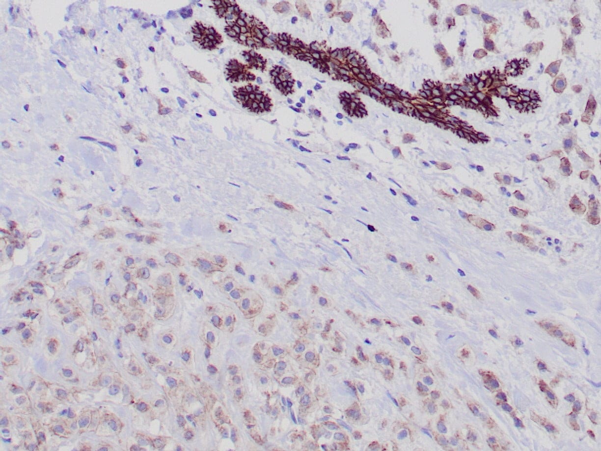 |
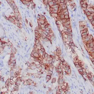
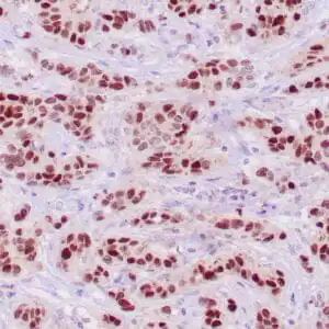
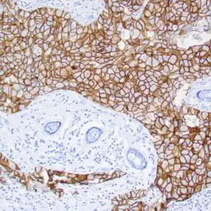
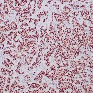
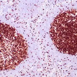
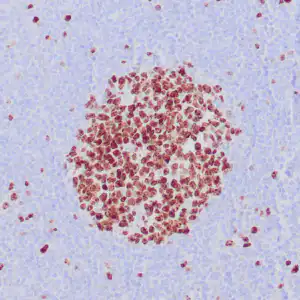
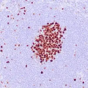
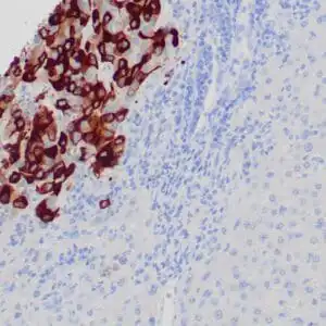
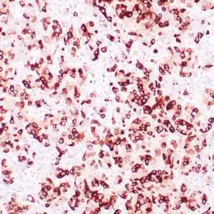
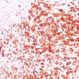
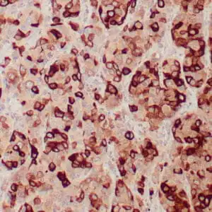
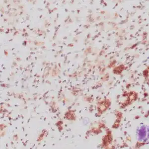
| Formalin-fixed paraffin-embedded human breast lobular anti-E-Cadherin antibody using peroxidase-conjugate and DAB chromogen. Note the strong membranous staining of ductal cells (top), whereas tumor cells show weak cytoplasmic staining. |
FAQ & Publications
Frequently Asked Questions
What is the recommended dilution for using the rabbit anti-E-Cadherin monoclonal antibody (ZR375) in immunohistochemistry?
For immunohistochemistry applications, the concentrated rabbit anti-E-Cadherin monoclonal antibody (ZR375) is recommended to be diluted between 1:100 and 1:200.
Which species does the rabbit anti-E-Cadherin monoclonal antibody (ZR375) specifically react with?
This antibody is reactive with human tissues and is suitable for detecting E-Cadherin in human samples.
What are the storage conditions for maintaining the stability of the rabbit anti-E-Cadherin monoclonal antibody (ZR375)?
For short-term storage, keep the antibody at 2-8°C. For long-term preservation, it should be stored at -20°C, avoiding repeated freeze-thaw cycles to maintain antibody integrity.
Publications
| pmid | title | authors | citation |
|---|---|---|---|
| We haven't added any publications to our database yet. | |||
Published literature highly relevant to the biological target of this product and referencing this antibody or clone are retrieved from the PubMed database provided by the United States National Library of Medicine at the National Institutes of Health.
Protocols
| relevant to this product |
|---|
| IHC |
Documents
| Batch Number | QC File | SDS |
|---|---|---|
| To view batch-specific Safety Datasheets and Quality Certificates associated with your account, please Log In. | ||
Only logged in customers who have purchased this product may leave a review.
Reviews
There are no reviews yet.