| Weight | 1 lbs |
|---|---|
| Dimensions | 9 × 5 × 2 in |
| host | rabbit |
| isotype | IgG |
| clonality | polyclonal |
| concentration | 1 mg/mL |
| applications | ICC/IF, WB |
| reactivity | Chk2 (NT) |
| available sizes | 100 µg |


rabbit anti-Chk2 (NT) polyclonal antibody 7313
$469.00
Antibody summary
- Rabbit polyclonal to Chk2 (NT)
- Suitable for: ELISA,WB,ICC,IF
- Isotype: IgG
- 100 µg
rabbit anti-Chk2 (NT) polyclonal antibody 7313
| antibody |
|---|
| Tested applications WB,ICC/IF,ELISA |
| Recommended dilutions Immunoblotting: use at 0.5-1 ug/mL. In immunoblots, a band of 60 kD is detected. Positive control: Jurkat cell lysate. |
| Immunogen Peptide corresponding to aa 2-18 of human Chk2. |
| Size and concentration 100µg and lot specific |
| Form liquid |
| Storage Instructions This antibody is stable for at least one (1) year at -20°C. Avoid multiple freeze- thaw cycles. |
| Storage buffer PBS, pH 7.4. |
| Purity peptide affinity purification |
| Clonality polyclonal |
| Isotype IgG |
| Compatible secondaries goat anti-rabbit IgG, H&L chain specific, peroxidase conjugated, conjugated polyclonal antibody 9512 goat anti-rabbit IgG, H&L chain specific, biotin conjugated polyclonal antibody 2079 goat anti-rabbit IgG, H&L chain specific, FITC conjugated polyclonal antibody 7863 goat anti-rabbit IgG, H&L chain specific, Cross Absorbed polyclonal antibody 2371 goat anti-rabbit IgG, H&L chain specific, biotin conjugated polyclonal antibody, crossabsorbed 1715 goat anti-rabbit IgG, H&L chain specific, FITC conjugated polyclonal antibody, crossabsorbed 1720 |
| Isotype control Rabbit polyclonal - Isotype Control |
| target relevance |
|---|
| Homo sapiens CHEK2 Serine/threonine-protein kinase Chk2 |
| Protein names Serine/threonine-protein kinase Chk2 |
| Alternative names CHK2 checkpoint homolog, Cds1 homolog, Checkpoint kinase 2 |
| Gene names CHEK2 |
| Protein family Belongs to the protein kinase superfamily. CAMK Ser/Thr protein kinase family. CHK2 subfamily |
| Function Serine/threonine-protein kinase which is required for checkpoint-mediated cell cycle arrest, activation of DNA repair and apoptosis in response to the presence of DNA double-strand breaks. May also negatively regulate cell cycle progression during unperturbed cell cycles. Following activation, phosphorylates numerous effectors preferentially at the consensus sequence [L-X-R-X-X-S/T] (PubMed:37943659). Regulates cell cycle checkpoint arrest through phosphorylation of CDC25A, CDC25B and CDC25C, inhibiting their activity. Inhibition of CDC25 phosphatase activity leads to increased inhibitory tyrosine phosphorylation of CDK-cyclin complexes and blocks cell cycle progression. May also phosphorylate NEK6 which is involved in G2/M cell cycle arrest. Regulates DNA repair through phosphorylation of BRCA2, enhancing the association of RAD51 with chromatin which promotes DNA repair by homologous recombination. Also stimulates the transcription of genes involved in DNA repair (including BRCA2) through the phosphorylation and activation of the transcription factor FOXM1. Regulates apoptosis through the phosphorylation of p53/TP53, MDM4 and PML. Phosphorylation of p53/TP53 at 'Ser-20' by CHEK2 may alleviate inhibition by MDM2, leading to accumulation of active p53/TP53. Phosphorylation of MDM4 may also reduce degradation of p53/TP53. Also controls the transcription of pro-apoptotic genes through phosphorylation of the transcription factor E2F1. Tumor suppressor, it may also have a DNA damage-independent function in mitotic spindle assembly by phosphorylating BRCA1. Its absence may be a cause of the chromosomal instability observed in some cancer cells. Promotes the CCAR2-SIRT1 association and is required for CCAR2-mediated SIRT1 inhibition (PubMed:25361978). Under oxidative stress, promotes ATG7 ubiquitination by phosphorylating the E3 ubiquitin ligase TRIM32 at 'Ser-55' leading to positive regulation of the autophagosme assembly (PubMed:37943659) |
| Catalytic activity L-seryl-[protein] + ATP = O-phospho-L-seryl-[protein] + ADP + H(+) L-threonyl-[protein] + ATP = O-phospho-L-threonyl-[protein] + ADP + H(+) |
| Subcellular location Nucleus, PML body, Nucleus, nucleoplasm |
| Structure Homodimer. Homodimerization is part of the activation process but the dimer may dissociate following activation. Interacts with PML. Interacts with TP53. Interacts with RB1; phosphorylates RB1. Interacts with BRCA1. Interacts (phosphorylated at Thr-68) with MDC1; requires ATM-mediated phosphorylation of CHEK2. Interacts with TP53BP1; modulates CHEK2 phosphorylation at Thr-68 in response to ionizing radiation. Interacts with CDC25A; phosphorylates CDC25A and mediates its degradation in response to ionizing radiation. Interacts with CUL1; mediates CHEK2 ubiquitination and regulation. Interacts with CDKN2AIP. Interacts (via protein kinase domain) with CCAR2 (via N-terminus). Interacts with SIRT1 |
| Post-translational modification Phosphorylated. Phosphorylated at Ser-73 by PLK3 in response to DNA damage, promoting phosphorylation at Thr-68 by ATM and the G2/M transition checkpoint. Phosphorylation at Thr-68 induces homodimerization. Autophosphorylates at Thr-383 and Thr-387 in the T-loop/activation segment upon dimerization to become fully active and phosphorylate its substrates like for instance CDC25C. DNA damage-induced autophosphorylation at Ser-379 induces CUL1-mediated ubiquitination and regulates the pro-apoptotic function. Phosphorylation at Ser-456 also regulates ubiquitination. Phosphorylated by PLK4 Ubiquitinated. CUL1-mediated ubiquitination regulates the pro-apoptotic function. Ubiquitination may also regulate protein stability. Ubiquitinated by RNF8 via 'Lys-48'-linked ubiquitination |
| Involvement in disease Tumor predisposition syndrome 4 A disorder characterized by an increased risk for developing various types of benign and/or malignant neoplasms that arise at an accelerated rate and in different organs. Prostate cancer A malignancy originating in tissues of the prostate. Most prostate cancers are adenocarcinomas that develop in the acini of the prostatic ducts. Other rare histopathologic types of prostate cancer that occur in approximately 5% of patients include small cell carcinoma, mucinous carcinoma, prostatic ductal carcinoma, transitional cell carcinoma, squamous cell carcinoma, basal cell carcinoma, adenoid cystic carcinoma (basaloid), signet-ring cell carcinoma and neuroendocrine carcinoma. Osteogenic sarcoma A sarcoma originating in bone-forming cells, affecting the ends of long bones. Breast cancer A common malignancy originating from breast epithelial tissue. Breast neoplasms can be distinguished by their histologic pattern. Invasive ductal carcinoma is by far the most common type. Breast cancer is etiologically and genetically heterogeneous. Important genetic factors have been indicated by familial occurrence and bilateral involvement. Mutations at more than one locus can be involved in different families or even in the same case. |
| Keywords 3D-structure, Alternative splicing, Apoptosis, ATP-binding, Cell cycle, Cell division, Disease variant, DNA damage, DNA repair, Host-virus interaction, Kinase, Li-Fraumeni syndrome, Magnesium, Metal-binding, Mitosis, Nucleotide-binding, Nucleus, Phosphoprotein, Proteomics identification, Reference proteome, Serine/threonine-protein kinase, Transcription, Transcription regulation, Transferase, Tumor suppressor, Ubl conjugation |
| Sequence MSRESDVEAQQSHGSSACSQPHGSVTQSQGSSSQSQGISSSSTSTMPNSSQSSHSSSGTL SSLETVSTQELYSIPEDQEPEDQEPEEPTPAPWARLWALQDGFANLECVNDNYWFGRDKS CEYCFDEPLLKRTDKYRTYSKKHFRIFREVGPKNSYIAYIEDHSGNGTFVNTELVGKGKR RPLNNNSEIALSLSRNKVFVFFDLTVDDQSVYPKALRDEYIMSKTLGSGACGEVKLAFER KTCKKVAIKIISKRKFAIGSAREADPALNVETEIEILKKLNHPCIIKIKNFFDAEDYYIV LELMEGGELFDKVVGNKRLKEATCKLYFYQMLLAVQYLHENGIIHRDLKPENVLLSSQEE DCLIKITDFGHSKILGETSLMRTLCGTPTYLAPEVLVSVGTAGYNRAVDCWSLGVILFIC LSGYPPFSEHRTQVSLKDQITSGKYNFIPEVWAEVSEKALDLVKKLLVVDPKARFTTEEA LRHPWLQDEDMKRKFQDLLSEENESTALPQVLAQPSTSRKRPREGEAEGAETTKRPAVCA AVL |
| UniProt accession: O96017 |
Data
 |
| Western blot analysis of Chk2 expression in (A) K562, (B) Jurkat, and (C) HL-60 whole cell lysates with Chk2 antibody at 1 µg/mL. |
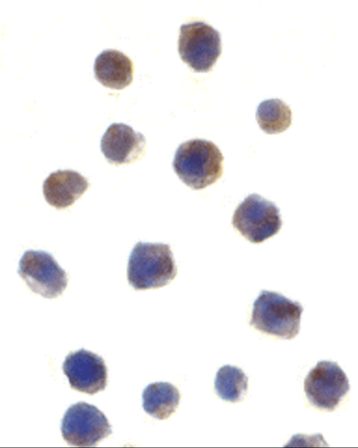 |
| Immunocytochemistry of Chk2 in Jurkat cells with Chk2 antibody at 1 µg/mL. |
 |
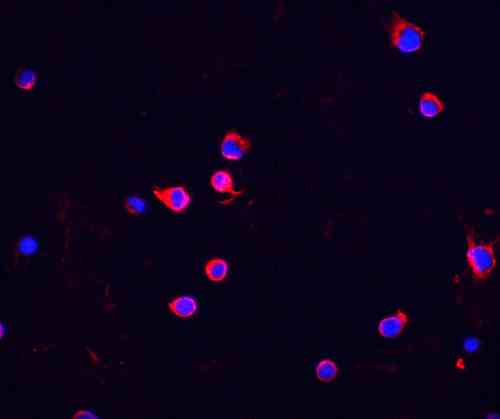
| Immunofluorescence of Chk2 in Jurkat cells with Chk2 antibody at 5 µg/mL
Red: Chk2 Antibody (7313) Blue: DAPI staining |
FAQ & Publications
Frequently Asked Questions
What applications has the rabbit anti-Chk2 (NT) polyclonal antibody 7313 been validated for?
This antibody is validated for use in ELISA, Western blotting (WB), Immunocytochemistry (ICC), and Immunofluorescence (IF) applications.
How should the rabbit anti-Chk2 (NT) polyclonal antibody 7313 be stored to maintain its stability?
The antibody should be stored at -20°C and is stable for at least one year under these conditions. It is important to avoid multiple freeze-thaw cycles to preserve antibody integrity.
What is the immunogen used to generate the rabbit anti-Chk2 (NT) polyclonal antibody 7313?
The immunogen is a peptide corresponding to amino acids 2-18 of the human Chk2 protein.
Which secondary antibodies are compatible with the rabbit anti-Chk2 (NT) polyclonal antibody 7313 for detection?
Compatible secondary antibodies include goat anti-rabbit IgG, H&L chain specific antibodies conjugated to peroxidase, biotin, or FITC, including cross-absorbed versions for reduced background.
Publications
| pmid | title | authors | citation |
|---|---|---|---|
| We haven't added any publications to our database yet. | |||
Published literature highly relevant to the biological target of this product and referencing this antibody or clone are retrieved from the PubMed database provided by the United States National Library of Medicine at the National Institutes of Health.
Protocols
| relevant to this product |
|---|
| Western blot IHC ICC |
Documents
| Batch Number | QC File | SDS |
|---|---|---|
| To view batch-specific Safety Datasheets and Quality Certificates associated with your account, please Log In. | ||
Only logged in customers who have purchased this product may leave a review.


Reviews
There are no reviews yet.