| Weight | 1 lbs |
|---|---|
| Dimensions | 9 × 5 × 2 in |
| host | rabbit |
| isotype | IgG |
| clonality | polyclonal |
| concentration | 1 mg/mL |
| applications | ICC/IF, WB |
| reactivity | BACE (CT) |
| available sizes | 100 µg |


rabbit anti-BACE (CT) polyclonal antibody 7999
$445.00
Antibody summary
- Rabbit polyclonal to BACE (CT)
- Suitable for: ELISA,WB,ICC,IHC-P,IF
- Isotype: IgG
- 100 µg
rabbit anti-BACE (CT) polyclonal antibody 7999
| antibody |
|---|
| Tested applications WB,IHC,IHC,ICC/IF,ELISA |
| Recommended dilutions Immunoblotting: use at 1:500-1:1,000 dilution. Positive control: Human brain tissue lysate. |
| Immunogen Peptide corresponding to aa 485-501 of human BACE. This sequence differs from those of mouse and rat by one amino acid. |
| Size and concentration 100µg and lot specific |
| Form liquid |
| Storage Instructions This antibody is stable for at least one (1) year at -20°C. Avoid multiple freeze-thaw cycles. |
| Storage buffer PBS, pH 7.4. |
| Purity immunogen affinity purification |
| Clonality polyclonal |
| Isotype IgG |
| Compatible secondaries goat anti-rabbit IgG, H&L chain specific, peroxidase conjugated, conjugated polyclonal antibody 9512 goat anti-rabbit IgG, H&L chain specific, biotin conjugated polyclonal antibody 2079 goat anti-rabbit IgG, H&L chain specific, FITC conjugated polyclonal antibody 7863 goat anti-rabbit IgG, H&L chain specific, Cross Absorbed polyclonal antibody 2371 goat anti-rabbit IgG, H&L chain specific, biotin conjugated polyclonal antibody, crossabsorbed 1715 goat anti-rabbit IgG, H&L chain specific, FITC conjugated polyclonal antibody, crossabsorbed 1720 |
| Isotype control Rabbit polyclonal - Isotype Control |
| target relevance |
|---|
| Homo sapiens BACE1 Beta-secretase 1 |
| Protein names Beta-secretase 1 |
| Alternative names Aspartyl protease 2, Beta-site amyloid precursor protein cleaving enzyme 1, Memapsin-2, Membrane-associated aspartic protease 2 |
| Gene names BACE1 |
| Protein family Belongs to the peptidase A1 family |
| Function Responsible for the proteolytic processing of the amyloid precursor protein (APP). Cleaves at the N-terminus of the A-beta peptide sequence, between residues 671 and 672 of APP, leads to the generation and extracellular release of beta-cleaved soluble APP, and a corresponding cell-associated C-terminal fragment which is later released by gamma-secretase (PubMed:10656250, PubMed:10677483, PubMed:20354142). Cleaves CHL1 (By similarity) |
| Catalytic activity Broad endopeptidase specificity. Cleaves Glu-Val-Asn-Leu-|-Asp-Ala-Glu-Phe in the Swedish variant of Alzheimer's amyloid precursor protein. |
| Subcellular location Cell membrane, Golgi apparatus, trans-Golgi network, Endoplasmic reticulum, Endosome, Cell surface, Cytoplasmic vesicle membrane, Membrane raft, Lysosome, Late endosome, Early endosome, Recycling endosome, Cell projection, axon, Cell projection, dendrite |
| Structure Monomer. Interacts (via DXXLL motif) with GGA1, GGA2 and GGA3 (via their VHS domain); the interaction highly increases when BACE1 is phosphorylated at Ser-498 (PubMed:14567678, PubMed:15886016). Interacts with RTN1; RTN2; RTN3 and RTN4; the interaction leads to inhibition of amyloid precursor protein processing (PubMed:15286784, PubMed:16965550, PubMed:16979658). Interacts with SNX6 (PubMed:20354142). Interacts with PCSK9 (PubMed:18660751). Interacts with NAT8 and NAT8B (PubMed:19011241). Interacts with BIN1 (PubMed:27179792). Interacts (via extracellular domain) with ADAM10 (via extracellular domain) (By similarity). Interacts with SORL1; this interaction may affect binding with APP and hence reduce APP cleavage (PubMed:16407538). Interacts with NRDC AND NRG1 (By similarity) |
| Post-translational modification N-Glycosylated (PubMed:11083922, PubMed:17425515). Addition of a bisecting N-acetylglucosamine by MGAT3 blocks lysosomal targeting, further degradation and is required for maintaining stability under stress conditions (By similarity) Acetylated in the endoplasmic reticulum at Lys-126, Lys-275, Lys-279, Lys-285, Lys-299, Lys-300 and Lys-307. Acetylation by NAT8 and NAT8B is transient and deacetylation probably occurs in the Golgi. Acetylation regulates the maturation, the transport to the plasma membrane, the stability and the expression of the protein Palmitoylation mediates lipid raft localization Ubiquitinated at Lys-501, ubiquitination leads to lysosomal degradation (PubMed:16033761, PubMed:20484053, PubMed:23109336, PubMed:27302062). Monoubiquitinated and 'Lys-63'-linked polyubitinated (PubMed:20484053). Deubiquitnated by USP8; inhibits lysosomal degradation (PubMed:27302062) Phosphorylation at Ser-498 is required for interaction with GGA1 and retrograded transport from endosomal compartments to the trans-Golgi network. Non-phosphorylated BACE1 enters a direct recycling route to the cell surface |
| Keywords 3D-structure, Acetylation, Alternative splicing, Aspartyl protease, Cell membrane, Cell projection, Cytoplasmic vesicle, Direct protein sequencing, Disulfide bond, Endoplasmic reticulum, Endosome, Glycoprotein, Golgi apparatus, Hydrolase, Isopeptide bond, Lipoprotein, Lysosome, Membrane, Palmitate, Phosphoprotein, Protease, Proteomics identification, Reference proteome, Signal, Transmembrane, Transmembrane helix, Ubl conjugation, Zymogen |
| Sequence MAQALPWLLLWMGAGVLPAHGTQHGIRLPLRSGLGGAPLGLRLPRETDEEPEEPGRRGSF VEMVDNLRGKSGQGYYVEMTVGSPPQTLNILVDTGSSNFAVGAAPHPFLHRYYQRQLSST YRDLRKGVYVPYTQGKWEGELGTDLVSIPHGPNVTVRANIAAITESDKFFINGSNWEGIL GLAYAEIARPDDSLEPFFDSLVKQTHVPNLFSLQLCGAGFPLNQSEVLASVGGSMIIGGI DHSLYTGSLWYTPIRREWYYEVIIVRVEINGQDLKMDCKEYNYDKSIVDSGTTNLRLPKK VFEAAVKSIKAASSTEKFPDGFWLGEQLVCWQAGTTPWNIFPVISLYLMGEVTNQSFRIT ILPQQYLRPVEDVATSQDDCYKFAISQSSTGTVMGAVIMEGFYVVFDRARKRIGFAVSAC HVHDEFRTAAVEGPFVTLDMEDCGYNIPQTDESTLMTIAYVMAAICALFMLPLCLMVCQW RCLRCLRQQHDDFADDISLLK |
| UniProt accession: P56817 |
Data
 |
| Western Blot Validation of BACE Loading: 15 µg of lysates per lane. Antibodies: BACE (1 µg/mL), 1h incubation at RT in 5% NFDM/TBST.Secondary: Goat anti-rabbit IgG HRP conjugate at 1:10000 dilution.Lane A-C: human brain tissue lysate in the absence (A) or presence (B) of blocking peptide and mouse 3T3/NIH cell lysate (C). |
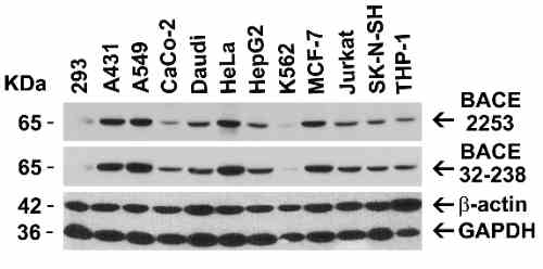 |
| Independent Antibody Validation (IAV) via Protein Expression Profile in Cell Lines Loading: 15 µg of lysates per lane. Antibodies: BACE 7999 (1 µg/mL), BACE 32-238 (1 µg/mL), beta-actin (1 µg/mL), and GAPDH (0.02 µg/mL), 1h incubation at RT in 5% NFDM/TBST.Secondary: Goat anti-rabbit IgG HRP conjugate at 1:10000 dilution. |
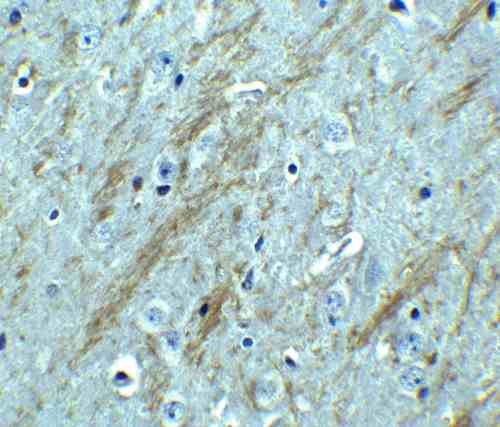 |
| Immunohistochemistry Validation of BACE in Mouse Brain Immunohistochemical analysis of paraffin-embedded mouse brain tissue using anti-BACE antibody (7999) at 2.5 µg/mL. Tissue was fixed with formaldehyde and blocked with 10% serum for 1 h at RT; antigen retrieval was by heat mediation with a citrate buffer (pH6). Samples were incubated with primary antibody overnight at 4C. A goat anti-rabbit IgG H&L (HRP) at 1/250 was used as secondary. Counter stained with Hematoxylin. |
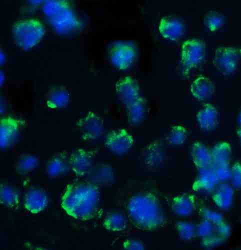 |
| Immunofluorescence Validation of BACE in 3T3/NIH Cells Immunofluorescent analysis of 4% paraformaldehyde-fixed mouse 3T3/NIH cells labeling BACE with 7999 at 20 µg/mL, followed by goat anti-rabbit IgG secondary antibody at 1/500 dilution (green) and DAPI staining (blue). Image showing both membrane and cytosol staining on 3T3/NIH cells. |
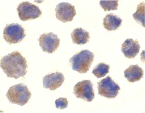 |
| Immunocytochemistry Validation of BACE in 3T3/NIH Cells Immunocytochemical analysis of 3T3/NIH cells using anti-BACE antibody (7999) at 10 µg/mL. Cells was fixed with formaldehyde and blocked with 10% serum for 1 h at RT; antigen retrieval was by heat mediation with a citrate buffer (pH6). Samples were incubated with primary antibody overnight at 4C. A goat anti-rabbit IgG H&L (HRP) at 1/250 was used as secondary. Counter stained with Hematoxylin. |
 |
| KO and Overexpression Validation of BACE in Human and Mouse Brain and 293 Cells. (Singer et al., 2005) Western blot analysis of the BACE1 (7999) antibody'sability to recognize human and murine BACE1. The BACE1 antibody recognized both the mouse and human forms of BACE1. Lanes 1-4 are frontal cortex homogenates from human and mouse brains. Lane 1 is from a neurologically unimpaired aged human control case, lane 2 from a BACE1-deficient mouse, lane 3 from a nontransgenic mouse and lane 4 from hBACE1 transgenic mouse. Lanes 5-7 are lysates from HEK293T cells transfected with a plasmid vector expressing eGFP, mBACE1 and hBACE1, respectively. |
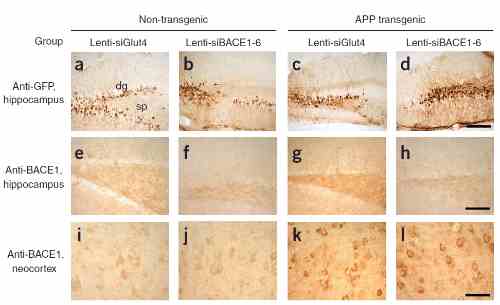 |
| KD Validation of BACE in Mouse Brain (Singer et al., 2005) Characterization of the effects of lenti-siBACE1-6 expression in the brains of APP transgenic mice. (a-d) Anti-eGFP immunoreactivity in the hippocampus (the injection site) shows comparable and consistent expression of lenti-siRNA constructs in the dentate gyrus (dg) and stratus polymorphus (sp). (-) Anti-BACE1 immunoreactivity in the hippocampus of nontransgenic mice treated with lenti-siGlut4. (-) Reduced BACE1 immunostaining in the hippocampus of nontransgenic mice treated with lenti-siBACE1-6 vector. (-) Intense BACE1 immunoreactivity in the hippocampus of APP transgenic mice treated with lenti-siGlut4. (-) Reduced BACE1 expression in APP transgenic mice treated with lenti-siBACE1-6 vector. (i,j) Anti-BACE1 reacted with pyramidal cell bodies in the neocortex, which was not injected, |
 |
| KD Validation of BACE in Mouse Brain (Singer et al., 2005) Immunolabeling patterns of BACE1 expression and the lenti-siRNA distribution. Sections from APP transgenic mice treated with the eGFPtagged lenti siRNA vectors (green) were co-immunolabeled with an antibody against BACE1 (red) and imaged with the LSCM. All sections are from the hippocampus of treated mice. (a°C) Lenti-siBACE1-6 treated mice. Areas within the hippocampus expressing the eGFP tagged vector have reduced BACE1 immunolabeling. (d-f) Mice treated with the eGFP-tagged control lenti-siGlut4 show unchanged expression of BACE1 in the hippocampus. (g-i) Mice treated with a saline vehicle show unchanged expression of BACE1 in the hippocampus. |
 |
| KO Validation of BACE in MEF Cells (Jo et al., 2010) Wildtype and BACE -/- MEFs were exposed to HNE (15_M) for 2 h. BACE1 levels were examined by Western blot with anti-BACE antibodies (7999). |
FAQ & Publications
Frequently Asked Questions
What applications is the rabbit anti-BACE (CT) polyclonal antibody 7999 validated for?
This antibody is suitable for ELISA, Western Blot (WB), Immunocytochemistry/Immunofluorescence (ICC/IF), Immunohistochemistry on paraffin-embedded sections (IHC-P), and Immunohistochemistry (IHC). Recommended dilutions for immunoblotting are 1:500 to 1:1,000, using human brain tissue lysate as a positive control.
How should the rabbit anti-BACE (CT) polyclonal antibody 7999 be stored to maintain stability?
The antibody should be stored at -20°C and is stable for at least one year under these conditions. It is provided in PBS buffer at pH 7.4 and users should avoid multiple freeze-thaw cycles to preserve antibody integrity.
Publications
| pmid | title | authors | citation |
|---|---|---|---|
| We haven't added any publications to our database yet. | |||
Published literature highly relevant to the biological target of this product and referencing this antibody or clone are retrieved from the PubMed database provided by the United States National Library of Medicine at the National Institutes of Health.
Protocols
| relevant to this product |
|---|
| Western blot IHC ICC |
Documents
| Batch Number | QC File | SDS |
|---|---|---|
| To view batch-specific Safety Datasheets and Quality Certificates associated with your account, please Log In. | ||
Only logged in customers who have purchased this product may leave a review.


Reviews
There are no reviews yet.