| Weight | 1 lbs |
|---|---|
| Dimensions | 9 × 5 × 2 in |
| host | rabbit |
| isotype | IgG |
| clonality | polyclonal |
| concentration | 1 mg/mL |
| applications | ICC/IF, WB |
| reactivity | ASC |
| available sizes | 100 µg |


rabbit anti-ASC polyclonal antibody 1972
$445.00
Antibody summary
- Rabbit polyclonal to ASC
- Suitable for: ELISA,WB,ICC,IHC-P
- Isotype: IgG
- 100 µg
rabbit anti-ASC polyclonal antibody 1972
| antibody |
|---|
| Tested applications WB,IHC,IHC,ICC/IF,ELISA |
| Recommended dilutions Immunoblotting: use at 1ug/mL. Immunocytochemistry: use at 5ug/mL. These are recommended concentrations. Enduser should determine optimal concentrations for their applications. Positive control: Whole cell lysate of HL60 cells. |
| Immunogen Peptide corresponding to aa 182-195 of human ASC. |
| Size and concentration 100µg and lot specific |
| Form liquid |
| Storage Instructions This antibody is stable for at least one (1) year at -20°C. Avoid multiple freeze-thaw cycles. |
| Storage buffer PBS, pH 7.4. |
| Purity peptide affinity purification |
| Clonality polyclonal |
| Isotype IgG |
| Compatible secondaries goat anti-rabbit IgG, H&L chain specific, peroxidase conjugated, conjugated polyclonal antibody 9512 goat anti-rabbit IgG, H&L chain specific, biotin conjugated polyclonal antibody 2079 goat anti-rabbit IgG, H&L chain specific, FITC conjugated polyclonal antibody 7863 goat anti-rabbit IgG, H&L chain specific, Cross Absorbed polyclonal antibody 2371 goat anti-rabbit IgG, H&L chain specific, biotin conjugated polyclonal antibody, crossabsorbed 1715 goat anti-rabbit IgG, H&L chain specific, FITC conjugated polyclonal antibody, crossabsorbed 1720 |
| Isotype control Rabbit polyclonal - Isotype Control |
| target relevance |
|---|
| Homo sapiens PYCARD Apoptosis-associated speck-like protein containing a CARD |
| Protein names Apoptosis-associated speck-like protein containing a CARD |
| Alternative names Caspase recruitment domain-containing protein 5, PYD and CARD domain-containing protein, Target of methylation-induced silencing 1 |
| Gene names PYCARD |
| Function Functions as a key mediator in apoptosis and inflammation (PubMed:11103777, PubMed:12646168, PubMed:15030775, PubMed:17349957, PubMed:17599095, PubMed:19158675, PubMed:19158676, PubMed:19234215, PubMed:19494289, PubMed:21487011, PubMed:24630722, PubMed:25847972, PubMed:30674671, PubMed:34678144, PubMed:36050480). Promotes caspase-mediated apoptosis involving predominantly caspase-8 and also caspase-9 in a probable cell type-specific manner (PubMed:11103777, PubMed:12646168). Involved in activation of the mitochondrial apoptotic pathway, promotes caspase-8-dependent proteolytic maturation of BID independently of FADD in certain cell types and also mediates mitochondrial translocation of BAX and activates BAX-dependent apoptosis coupled to activation of caspase-9, -2 and -3 (PubMed:14730312, PubMed:16964285). Involved in innate immune response by acting as an integral adapter in the assembly of various inflammasomes (NLRP1, NLRP2, NLRP3, NLRP6, AIM2 and probably IFI16) which recruit and activate caspase-1 leading to processing and secretion of pro-inflammatory cytokines (PubMed:15030775, PubMed:16982856, PubMed:17349957, PubMed:17599095, PubMed:19158675, PubMed:19158676, PubMed:19234215, PubMed:21487011, PubMed:23530044, PubMed:24630722, PubMed:25847972, PubMed:29440442, PubMed:30674671, PubMed:33980849, PubMed:34678144, PubMed:34706239). Caspase-1-dependent inflammation leads to macrophage pyroptosis, a form of cell death (PubMed:24630722). The function as activating adapter in different types of inflammasomes is mediated by the pyrin and CARD domains and their homotypic interactions (PubMed:14499617, PubMed:19234215, PubMed:24630722). Clustered PYCARD nucleates the formation of caspase-1 filaments through the interaction of their respective CARD domains, acting as a platform for of caspase-1 polymerization (PubMed:24630722). In the NLRP1 and NLRC4 inflammasomes seems not be required but facilitates the processing of procaspase-1 (PubMed:17349957). In cooperation with NOD2 involved in an inflammasome activated by bacterial muramyl dipeptide leading to caspase-1 activation (PubMed:16964285). May be involved in RIGI-triggered pro-inflammatory responses and inflammasome activation (PubMed:19915568). In collaboration with AIM2 which detects cytosolic double-stranded DNA may also be involved in a caspase-1-independent cell death that involves caspase-8 (PubMed:19158675, PubMed:19158676). In adaptive immunity may be involved in maturation of dendritic cells to stimulate T-cell immunity and in cytoskeletal rearrangements coupled to chemotaxis and antigen uptake may be involved in post-transcriptional regulation of the guanine nucleotide exchange factor DOCK2; the latter function is proposed to involve the nuclear form (PubMed:22732093). Also involved in transcriptional activation of cytokines and chemokines independent of the inflammasome; this function may involve AP-1, NF-kappa-B, MAPK and caspase-8 signaling pathways (PubMed:12486103, PubMed:16585594). For regulation of NF-kappa-B activating and inhibiting functions have been reported (PubMed:12486103). Modulates NF-kappa-B induction at the level of the IKK complex by inhibiting kinase activity of CHUK and IKBK (PubMed:12486103, PubMed:16585594). Proposed to compete with RIPK2 for association with CASP1 thereby down-regulating CASP1-mediated RIPK2-dependent NF-kappa-B activation and activating interleukin-1 beta processing (PubMed:16585594). Modulates host resistance to DNA virus infection, probably by inducing the cleavage of and inactivating CGAS in presence of cytoplasmic double-stranded DNA (PubMed:28314590) |
| Subcellular location Golgi apparatus membrane |
| Structure Self-associates; enforced oligomerization induces apoptosis, NF-kappa-B regulation and interleukin-1 beta secretion (PubMed:15641782, PubMed:17599095, PubMed:33420028, PubMed:33420033, PubMed:34706239). Homooligomers can form disk-like particles of approximately 12 nm diameter and approximately 1 nm height (PubMed:15641782, PubMed:17599095). Next to isoform 1, also isoform 2 and isoform 3 may be involved in oligomerization leading to functional regulation (Probable). Component of several inflammasomes containing one pattern recognition receptor/sensor, such as NLRP1, NLRP2, NLRP3, NLRP6, NLRC4, AIM2, MEFV or NOD2, and probably NLRC4, NLRP12 or IFI16 (PubMed:11374873, PubMed:12191486, PubMed:15030775, PubMed:15456791, PubMed:19158676, PubMed:23530044, PubMed:27432880, PubMed:29440442, PubMed:30674671, PubMed:33980849, PubMed:34678144, PubMed:35559676). Major component of the ASC pyroptosome, a 1-2 um supramolecular assembly (one per macrophage cell) which consists of oligomerized PYCARD dimers and CASP1 (PubMed:17599095). Interacts with CASP1 (precursor form); the interaction induces activation of CASP1 leading to the processing of interleukin-1 beta; PYCARD competes with RIPK2 for binding to CASP1 (PubMed:11967258, PubMed:14634131, PubMed:16585594, PubMed:17599095, PubMed:33420033). Interacts with NLRP3; the interaction requires the homooligomerization of NLRP3 (PubMed:11786556, PubMed:15020601, PubMed:15030775, PubMed:34341353, PubMed:35559676). Interacts with NLRP2, NLRC4, MEFV, CARD16, AIM2, IFI16, NOD2, RIGI, RIPK2, PYDC1, PYDC2, NLRP10, CASP8, CHUK, IKBKB and BAX (PubMed:11374873, PubMed:11498534, PubMed:12486103, PubMed:12646168, PubMed:12656673, PubMed:14730312, PubMed:15096476, PubMed:15456791, PubMed:17178784, PubMed:17339483, PubMed:18362139, PubMed:19158675, PubMed:19158676, PubMed:19915568, PubMed:21575908, PubMed:23530044, PubMed:29440442, PubMed:33980849). Component of the AIM2 PANoptosome complex, a multiprotein complex that drives inflammatory cell death (PANoptosis) (By similarity) |
| Post-translational modification Phosphorylated 'Lys-63'-linked polyubiquitination by TRAF3 is critical for speck formation and inflammasome activation (PubMed:25847972). 'Lys-63'-linked deubiquitinated by USP50; a crucial step for NLRP3-mediated inflammasome activation (PubMed:28094437). 'Lys-63'-linked polyubiquitination by PELI1 is also critical for speck formation and inflammasome activation (PubMed:34706239). Deubiquitinated by USP3 that cleaves 'Lys-48'-linked ubiquitin chains and strengthens its stability by blocking proteasomal degradation (PubMed:36050480) |
| Keywords 3D-structure, Alternative splicing, Apoptosis, Cytoplasm, Endoplasmic reticulum, Golgi apparatus, Immunity, Inflammasome, Inflammatory response, Innate immunity, Isopeptide bond, Membrane, Mitochondrion, Nucleus, Phosphoprotein, Proteomics identification, Reference proteome, Tumor suppressor, Ubl conjugation |
| Sequence MGRARDAILDALENLTAEELKKFKLKLLSVPLREGYGRIPRGALLSMDALDLTDKLVSFY LETYGAELTANVLRDMGLQEMAGQLQAATHQGSGAAPAGIQAPPQSAAKPGLHFIDQHRA ALIARVTNVEWLLDALYGKVLTDEQYQAVRAEPTNPSKMRKLFSFTPAWNWTCKDLLLQA LRESQSYLVEDLERS |
| UniProt accession: Q9ULZ3 |
Data
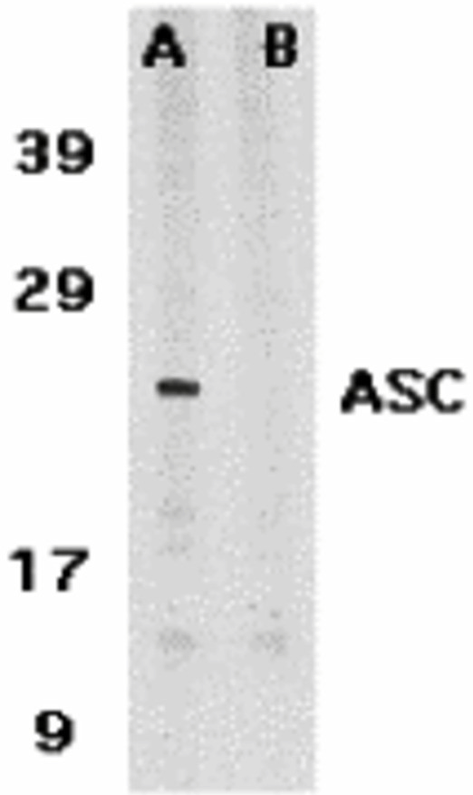 |
| Western Blot Validation in Human HL60 Cells Loading: 15 µg of lysates per lane. Antibodies: ASC 1972, (1 µg/mL) in the absence (A) or presence (B) of blocking peptide, 1h incubation at RT in 5% NFDM/TBST.Secondary: Goat anti-rabbit IgG HRP conjugate at 1:10000 dilution. |
 |
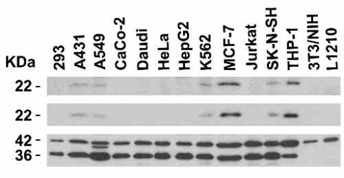
| Independent Antibody Validation (IAV) via Protein Expression Profile in Cell Lines Loading: 15 µg of lysates per lane. Antibodies: ASC 1972, (2 µg/mL), ASC 39-001, (2 µg/mL), beta-actin (1 µg/mL) and GAPDH (0.02 µg/mL), 1h incubation at RT in 5% NFDM/TBST.Secondary: Goat anti-rabbit or goat anti-mouse (for ASC 39001) IgG HRP conjugate at 1:10000 dilution. |
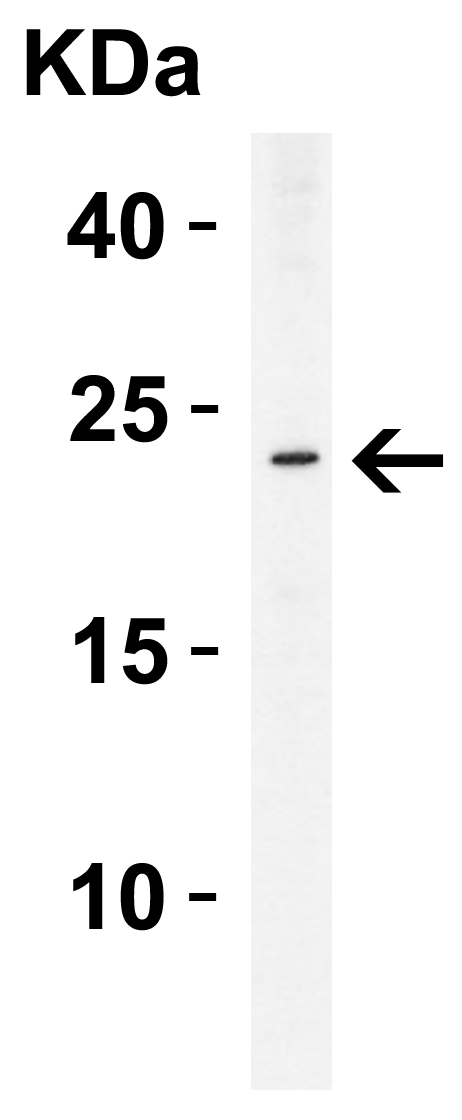 |
| Western Blot Validation in Human THP-1 Cells Loading: 15 µg of lysate per lane. Antibodies: ASC 1972, (2 µg/mL), 1h incubation at RT in 5% NFDM/TBST.Secondary: Goat anti-rabbit IgG HRP conjugate at 1:10000 dilution. |
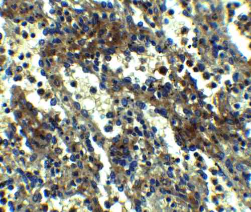 |
| Immunohistochemistry Validation of ASC in Human Spleen Tissue Immunohistochemical analysis of paraffin-embedded human spleen tissue using anti-ASC antibody (1972) at 2.5 µg/mL. Tissue was fixed with formaldehyde and blocked with 10% serum for 1 h at RT; antigen retrieval was by heat mediation with a citrate buffer (pH6). Samples were incubated with primary antibody overnight at 4C. A goat anti-rabbit IgG H&L (HRP) at 1/250 was used as secondary. Counter stained with Hematoxylin. |
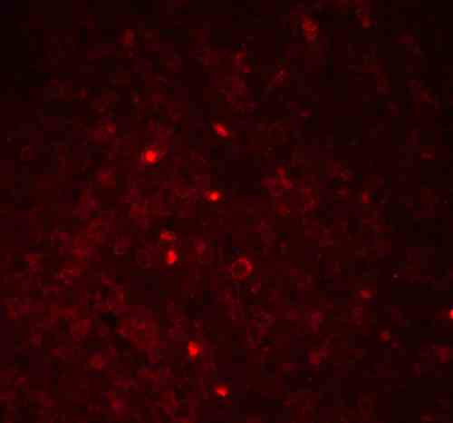 |
| Immunofluorescence Validation of ASC in Human Spleen Tissue Immunofluorescent analysis of 4% paraformaldehyde-fixed Human Spleen Tissue labeling ASC with 1972 at 20 µg/mL, followed by goat anti-rabbit IgG secondary antibody at 1/500 dilution (red). |
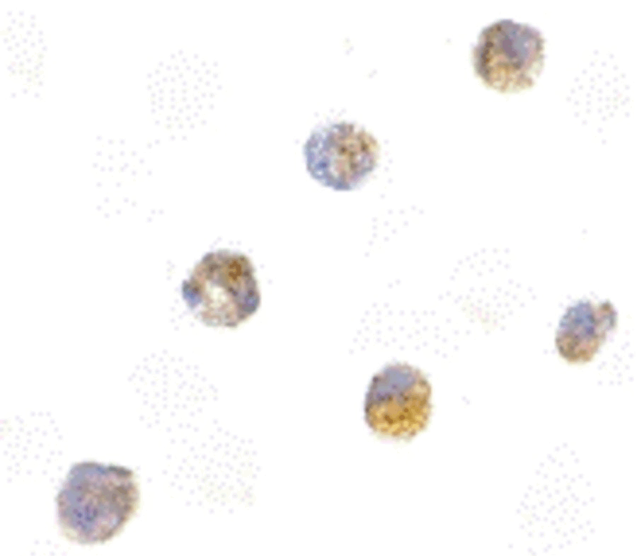 |
| Immunocytochemistry Validation of ASC in HL60 Cells Immunocytochemical analysis of HL60 cells using anti-ASC antibody (1972) at 5 µg/mL. Cells was fixed with formaldehyde and blocked with 10% serum for 1 h at RT; antigen retrieval was by heat mediation with a citrate buffer (pH6). Samples were incubated with primary antibody overnight at 4C. A goat anti-rabbit IgG H&L (HRP) at 1/250 was used as secondary. Counter stained with Hematoxylin. |
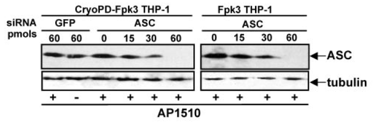 |
| KD Validation of ASC in THP-1 Cells (Dowds et al., 2004) Immunofluorescence analysis with anti-ASC antibodies (1972) was performed for BIM in 293 cells transfected with GFP siRNA or ASC siRNA. ASC expression was disrupted after ASC siRNA knockdown. |
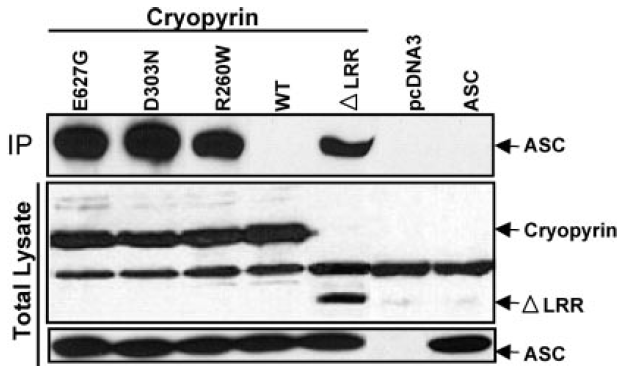 |
| Overexpression Validation of ASC in HEK293T Cells (Dowds et al., 2004) Western blot analysis with anti-ASC antibodies (1972) was performed for ASC in HEK293T cells transfected with pcDNA3-ASC. |
FAQ & Publications
Frequently Asked Questions
What applications has the rabbit anti-ASC polyclonal antibody 1972 been tested for and what are the recommended working concentrations?
The rabbit anti-ASC polyclonal antibody 1972 has been tested for Western Blot (WB), Immunohistochemistry (IHC), Immunocytochemistry/Immunofluorescence (ICC/IF), and ELISA. The recommended dilution for immunoblotting is 1 µg/mL and for immunocytochemistry is 5 µg/mL. Users should optimize concentrations for their specific applications.
How should the rabbit anti-ASC polyclonal antibody 1972 be stored to ensure stability?
This antibody should be stored at -20°C and is stable for at least one year under these conditions. It is important to avoid multiple freeze-thaw cycles to maintain antibody integrity. The antibody is supplied in PBS buffer at pH 7.4 in liquid form.
Publications
| pmid | title | authors | citation |
|---|---|---|---|
| We haven't added any publications to our database yet. | |||
Published literature highly relevant to the biological target of this product and referencing this antibody or clone are retrieved from the PubMed database provided by the United States National Library of Medicine at the National Institutes of Health.
Protocols
| relevant to this product |
|---|
| Western blot IHC ICC |
Documents
| Batch Number | QC File | SDS |
|---|---|---|
| To view batch-specific Safety Datasheets and Quality Certificates associated with your account, please Log In. | ||
Only logged in customers who have purchased this product may leave a review.


Reviews
There are no reviews yet.