| Weight | 1 lbs |
|---|---|
| Dimensions | 9 × 5 × 2 in |
| host | rabbit |
| isotype | IgG |
| clonality | monoclonal |
| concentration | 0.4 mg/mL |
| applications | Flow, WB |
| reactivity | human, mouse |
| available sizes | 100 µg, 100 µL, 20 µL |


rabbit anti-AKT1 polyclonal antibody BLR245L 1020
Price range: $127.00 through $794.00
Antibody summary
- Rabbit polyclonal to PI3K/Akt Pathway (Phosphoinositide 3-kinase/Akt)
- Suitable for: WB,ICC
- Reacts with: Hu, Ms
- Isotype: IgG
- 100 µg, 100 µL (10 blots), 20 µL
rabbit anti-akt polyclonal antibody BLR245L 1020
| antibody |
|---|
| Database link: human P31749 mouse P31750 |
| Tested applications WB,ICC |
| Recommended dilutions Flow Cytometry (Flow Cyt) Fixed in 4% formaldehyde and permeabilized with 90% methanol. 1 µl per 1 x 10^6 cells., Western Blot (WB) 1:1000 |
| Immunogen Between 100 and 150 |
| Size and concentration 100µL and 0.4 mg/mL |
| Form liquid |
| Storage Instructions Store at 2-8°C. Expires 1 year from date of receipt. |
| Storage buffer Tris-buffered Saline containing 0.1% BSA and 0.09% Sodium Azide |
| Purity affinity purified |
| Clonality monoclonal |
| Isotype IgG |
| Compatible secondaries goat anti-rabbit IgG, H&L chain specific, peroxidase conjugated, conjugated polyclonal antibody 9512 goat anti-rabbit IgG, H&L chain specific, biotin conjugated polyclonal antibody 2079 goat anti-rabbit IgG, H&L chain specific, FITC conjugated polyclonal antibody 7863 goat anti-rabbit IgG, H&L chain specific, Cross Absorbed polyclonal antibody 2371 goat anti-rabbit IgG, H&L chain specific, biotin conjugated polyclonal antibody, crossabsorbed 1715 goat anti-rabbit IgG, H&L chain specific, FITC conjugated polyclonal antibody, crossabsorbed 1720 |
| Isotype control Rabbit monoclonal - Isotype Control |
| target relevance |
|---|
| Protein names RAC-alpha serine/threonine-protein kinase (EC 2.7.11.1) (Protein kinase B) (PKB) (Protein kinase B alpha) (PKB alpha) (Proto-oncogene c-Akt) (RAC-PK-alpha) |
| Gene names AKT1,AKT1 PKB RAC |
| Protein family Protein kinase superfamily, AGC Ser/Thr protein kinase family, RAC subfamily |
| Mass 55686Da |
| Function FUNCTION: AKT1 is one of 3 closely related serine/threonine-protein kinases (AKT1, AKT2 and AKT3) called the AKT kinase, and which regulate many processes including metabolism, proliferation, cell survival, growth and angiogenesis (PubMed:11882383, PubMed:15526160, PubMed:15861136, PubMed:21432781, PubMed:21620960, PubMed:31204173). This is mediated through serine and/or threonine phosphorylation of a range of downstream substrates (PubMed:11882383, PubMed:15526160, PubMed:21432781, PubMed:21620960, PubMed:31204173). Over 100 substrate candidates have been reported so far, but for most of them, no isoform specificity has been reported (PubMed:11882383, PubMed:15526160, PubMed:21432781, PubMed:21620960). AKT is responsible of the regulation of glucose uptake by mediating insulin-induced translocation of the SLC2A4/GLUT4 glucose transporter to the cell surface (By similarity). Phosphorylation of PTPN1 at 'Ser-50' negatively modulates its phosphatase activity preventing dephosphorylation of the insulin receptor and the attenuation of insulin signaling (By similarity). Phosphorylation of TBC1D4 triggers the binding of this effector to inhibitory 14-3-3 proteins, which is required for insulin-stimulated glucose transport (PubMed:11994271). AKT also regulates the storage of glucose in the form of glycogen by phosphorylating GSK3A at 'Ser-21' and GSK3B at 'Ser-9', resulting in inhibition of its kinase activity (By similarity). Phosphorylation of GSK3 isoforms by AKT is also thought to be one mechanism by which cell proliferation is driven (By similarity). AKT also regulates cell survival via the phosphorylation of MAP3K5 (apoptosis signal-related kinase) (PubMed:11154276). Phosphorylation of 'Ser-83' decreases MAP3K5 kinase activity stimulated by oxidative stress and thereby prevents apoptosis (PubMed:11154276). AKT mediates insulin-stimulated protein synthesis by phosphorylating TSC2 at 'Ser-939' and 'Thr-1462', thereby activating the mTORC1 signaling pathway, and leading to both phosphorylation of 4E-BP1 and in activation of RPS6KB1 (PubMed:12150915, PubMed:12172553). Also regulates the mTORC1 signaling pathway by catalyzing phosphorylation of CASTOR1 and DEPDC5 (PubMed:31548394, PubMed:33594058). AKT plays a role as key modulator of the AKT-mTOR signaling pathway controlling the tempo of the process of newborn neurons integration during adult neurogenesis, including correct neuron positioning, dendritic development and synapse formation (By similarity). Part of a positive feedback loop of mTORC2 signaling by mediating phosphorylation of MAPKAP1/SIN1, promoting mTORC2 activation (By similarity). AKT is involved in the phosphorylation of members of the FOXO factors (Forkhead family of transcription factors), leading to binding of 14-3-3 proteins and cytoplasmic localization (PubMed:10358075). In particular, FOXO1 is phosphorylated at 'Thr-24', 'Ser-256' and 'Ser-319' (PubMed:10358075). FOXO3 and FOXO4 are phosphorylated on equivalent sites (PubMed:10358075). AKT has an important role in the regulation of NF-kappa-B-dependent gene transcription and positively regulates the activity of CREB1 (cyclic AMP (cAMP)-response element binding protein) (PubMed:9829964). The phosphorylation of CREB1 induces the binding of accessory proteins that are necessary for the transcription of pro-survival genes such as BCL2 and MCL1 (PubMed:9829964). AKT phosphorylates 'Ser-454' on ATP citrate lyase (ACLY), thereby potentially regulating ACLY activity and fatty acid synthesis (By similarity). Activates the 3B isoform of cyclic nucleotide phosphodiesterase (PDE3B) via phosphorylation of 'Ser-273', resulting in reduced cyclic AMP levels and inhibition of lipolysis (By similarity). Phosphorylates PIKFYVE on 'Ser-318', which results in increased PI(3)P-5 activity (By similarity). The Rho GTPase-activating protein DLC1 is another substrate and its phosphorylation is implicated in the regulation cell proliferation and cell growth (By similarity). Signals downstream of phosphatidylinositol 3-kinase (PI(3)K) to mediate the effects of various growth factors such as platelet-derived growth factor (PDGF), epidermal growth factor (EGF), insulin and insulin-like growth factor 1 (IGF1) (PubMed:12176338, PubMed:12964941). AKT mediates the antiapoptotic effects of IGF1 (By similarity). Essential for the SPATA13-mediated regulation of cell migration and adhesion assembly and disassembly (PubMed:19934221). May be involved in the regulation of the placental development (By similarity). Phosphorylates STK4/MST1 at 'Thr-120' and 'Thr-387' leading to inhibition of its: kinase activity, nuclear translocation, autophosphorylation and ability to phosphorylate FOXO3 (PubMed:17726016). Phosphorylates STK3/MST2 at 'Thr-117' and 'Thr-384' leading to inhibition of its: cleavage, kinase activity, autophosphorylation at Thr-180, binding to RASSF1 and nuclear translocation (PubMed:20086174). Phosphorylates SRPK2 and enhances its kinase activity towards SRSF2 and ACIN1 and promotes its nuclear translocation (PubMed:19592491). Phosphorylates RAF1 at 'Ser-259' and negatively regulates its activity (PubMed:10576742). Phosphorylation of BAD stimulates its pro-apoptotic activity (PubMed:10926925). Phosphorylates KAT6A at 'Thr-369' and this phosphorylation inhibits the interaction of KAT6A with PML and negatively regulates its acetylation activity towards p53/TP53 (PubMed:23431171). Phosphorylates palladin (PALLD), modulating cytoskeletal organization and cell motility (PubMed:20471940). Phosphorylates prohibitin (PHB), playing an important role in cell metabolism and proliferation (PubMed:18507042). Phosphorylates CDKN1A, for which phosphorylation at 'Thr-145' induces its release from CDK2 and cytoplasmic relocalization (PubMed:16982699). These recent findings indicate that the AKT1 isoform has a more specific role in cell motility and proliferation (PubMed:16139227). Phosphorylates CLK2 thereby controlling cell survival to ionizing radiation (PubMed:20682768). Phosphorylates PCK1 at 'Ser-90', reducing the binding affinity of PCK1 to oxaloacetate and changing PCK1 into an atypical protein kinase activity using GTP as donor (PubMed:32322062). Also acts as an activator of TMEM175 potassium channel activity in response to growth factors: forms the lysoK(GF) complex together with TMEM175 and acts by promoting TMEM175 channel activation, independently of its protein kinase activity (PubMed:32228865). Acts as a regulator of mitochondrial calcium uptake by mediating phosphorylation of MICU1 in the mitochondrial intermembrane space, impairing MICU1 maturation (PubMed:30504268). Acts as an inhibitor of tRNA methylation by mediating phosphorylation of the N-terminus of METTL1, thereby inhibiting METTL1 methyltransferase activity (PubMed:15861136). In response to LPAR1 receptor pathway activation, phosphorylates Rabin8/RAB3IP which alters its activity and phosphorylates WDR44 which induces WDR44 binding to Rab11, thereby switching Rab11 vesicular function from preciliary trafficking to endocytic recycling (PubMed:31204173). {ECO:0000250|UniProtKB:P31750, ECO:0000250|UniProtKB:P47196, ECO:0000269|PubMed:10358075, ECO:0000269|PubMed:10576742, ECO:0000269|PubMed:10926925, ECO:0000269|PubMed:11154276, ECO:0000269|PubMed:11994271, ECO:0000269|PubMed:12150915, ECO:0000269|PubMed:12172553, ECO:0000269|PubMed:12176338, ECO:0000269|PubMed:12964941, ECO:0000269|PubMed:15861136, ECO:0000269|PubMed:16139227, ECO:0000269|PubMed:16982699, ECO:0000269|PubMed:17726016, ECO:0000269|PubMed:18507042, ECO:0000269|PubMed:19592491, ECO:0000269|PubMed:19934221, ECO:0000269|PubMed:20086174, ECO:0000269|PubMed:20471940, ECO:0000269|PubMed:20682768, ECO:0000269|PubMed:23431171, ECO:0000269|PubMed:30504268, ECO:0000269|PubMed:31204173, ECO:0000269|PubMed:31548394, ECO:0000269|PubMed:32228865, ECO:0000269|PubMed:32322062, ECO:0000269|PubMed:33594058, ECO:0000269|PubMed:9829964, ECO:0000303|PubMed:11882383, ECO:0000303|PubMed:15526160, ECO:0000303|PubMed:21432781, ECO:0000303|PubMed:21620960}. |
| Catalytic activity CATALYTIC ACTIVITY: Reaction=L-seryl-[protein] + ATP = O-phospho-L-seryl-[protein] + ADP + H(+); Xref=Rhea:RHEA:17989, Rhea:RHEA-COMP:9863, Rhea:RHEA-COMP:11604, ChEBI:CHEBI:15378, ChEBI:CHEBI:29999, ChEBI:CHEBI:30616, ChEBI:CHEBI:83421, ChEBI:CHEBI:456216; EC=2.7.11.1; Evidence={ECO:0000269|PubMed:12172553, ECO:0000269|PubMed:15861136, ECO:0000269|PubMed:16139227, ECO:0000269|PubMed:1718748, ECO:0000269|PubMed:1851997, ECO:0000269|PubMed:26440888, ECO:0000269|PubMed:31548394, ECO:0000269|PubMed:32322062, ECO:0000269|PubMed:33594058}; CATALYTIC ACTIVITY: Reaction=L-threonyl-[protein] + ATP = O-phospho-L-threonyl-[protein] + ADP + H(+); Xref=Rhea:RHEA:46608, Rhea:RHEA-COMP:11060, Rhea:RHEA-COMP:11605, ChEBI:CHEBI:15378, ChEBI:CHEBI:30013, ChEBI:CHEBI:30616, ChEBI:CHEBI:61977, ChEBI:CHEBI:456216; EC=2.7.11.1; Evidence={ECO:0000269|PubMed:12172553, ECO:0000269|PubMed:16139227, ECO:0000269|PubMed:1718748, ECO:0000269|PubMed:1851997}; |
| Subellular location SUBCELLULAR LOCATION: Cytoplasm {ECO:0000250|UniProtKB:P31750}. Nucleus {ECO:0000269|PubMed:20333297}. Cell membrane {ECO:0000269|PubMed:20333297}. Mitochondrion intermembrane space {ECO:0000250|UniProtKB:P31750}. Note=Nucleus after activation by integrin-linked protein kinase 1 (ILK1). Nuclear translocation is enhanced by interaction with TCL1A. Phosphorylation on Tyr-176 by TNK2 results in its localization to the cell membrane where it is targeted for further phosphorylations on Thr-308 and Ser-473 leading to its activation and the activated form translocates to the nucleus. Colocalizes with WDFY2 in intracellular vesicles (PubMed:16792529). Also localizes to mitochondrial intermembrane space in response to rapamycin treatment (By similarity). {ECO:0000250|UniProtKB:P31750, ECO:0000269|PubMed:16792529}. |
| Tissues TISSUE SPECIFICITY: Expressed in prostate cancer and levels increase from the normal to the malignant state (at protein level). Expressed in all human cell types so far analyzed. The Tyr-176 phosphorylated form shows a significant increase in expression in breast cancers during the progressive stages i.e. normal to hyperplasia (ADH), ductal carcinoma in situ (DCIS), invasive ductal carcinoma (IDC) and lymph node metastatic (LNMM) stages. {ECO:0000269|PubMed:1718748, ECO:0000269|PubMed:17932490, ECO:0000269|PubMed:20333297}. |
| Structure SUBUNIT: Interacts with BTBD10 (By similarity). Interacts with KCTD20 (By similarity). Interacts (via the C-terminus) with CCDC88A (via its C-terminus). Interacts with GRB10; the interaction leads to GRB10 phosphorylation thus promoting YWHAE-binding (By similarity). Interacts with AGAP2 (isoform 2/PIKE-A); the interaction occurs in the presence of guanine nucleotides. Interacts with AKTIP. Interacts (via PH domain) with MTCP1, TCL1A and TCL1B. Interacts with CDKN1B; the interaction phosphorylates CDKN1B promoting 14-3-3 binding and cell-cycle progression. Interacts with MAP3K5 and TRAF6. Interacts with BAD, PPP2R5B, STK3 and STK4. Interacts (via PH domain) with SIRT1. Interacts with SRPK2 in a phosphorylation-dependent manner. Interacts with RAF1. Interacts with TRIM13; the interaction ubiquitinates AKT1 leading to its proteasomal degradation. Interacts with TNK2 and CLK2. Interacts (via the C-terminus) with THEM4 (via its C-terminus). Interacts with and phosphorylated by PDPK1. Interacts with PA2G4 (By similarity). Interacts with KIF14; the interaction is detected in the plasma membrane upon INS stimulation and promotes AKT1 phosphorylation (PubMed:24784001). Interacts with FAM83B; activates the PI3K/AKT signaling cascade (PubMed:23676467). Interacts with WDFY2 (via WD repeats 1-3) (PubMed:16792529). Forms a complex with WDFY2 and FOXO1 (By similarity). Interacts with FAM168A (PubMed:23251525). Interacts with SYAP1 (via phosphorylated form and BSD domain); this interaction is enhanced in a mTORC2-mediated manner in response to epidermal growth factor (EGF) stimulation and activates AKT1 (PubMed:23300339). Interacts with PKHM3 (By similarity). Interacts with FKBP5/FKBP51; promoting interaction between Akt/AKT1 and PHLPP1, thereby enhancing dephosphorylation and subsequent activation of Akt/AKT1 (PubMed:28147277). Interacts with TMEM175; leading to formation of the lysoK(GF) complex (PubMed:32228865). Acts as a negative regulator of the cGAS-STING pathway by mediating phosphorylation of CGAS during mitosis, leading to its inhibition (PubMed:26440888). {ECO:0000250|UniProtKB:P31750, ECO:0000250|UniProtKB:P47196, ECO:0000269|PubMed:10576742, ECO:0000269|PubMed:10716693, ECO:0000269|PubMed:10926925, ECO:0000269|PubMed:10983986, ECO:0000269|PubMed:11154276, ECO:0000269|PubMed:11598301, ECO:0000269|PubMed:11839817, ECO:0000269|PubMed:12042314, ECO:0000269|PubMed:12176338, ECO:0000269|PubMed:12244301, ECO:0000269|PubMed:12964941, ECO:0000269|PubMed:14749367, ECO:0000269|PubMed:14761976, ECO:0000269|PubMed:15118108, ECO:0000269|PubMed:16139227, ECO:0000269|PubMed:16417524, ECO:0000269|PubMed:16792529, ECO:0000269|PubMed:17726016, ECO:0000269|PubMed:17932490, ECO:0000269|PubMed:19592491, ECO:0000269|PubMed:19713527, ECO:0000269|PubMed:20086174, ECO:0000269|PubMed:20333297, ECO:0000269|PubMed:20682768, ECO:0000269|PubMed:21329884, ECO:0000269|PubMed:21333377, ECO:0000269|PubMed:21775285, ECO:0000269|PubMed:22629392, ECO:0000269|PubMed:23251525, ECO:0000269|PubMed:23300339, ECO:0000269|PubMed:23676467, ECO:0000269|PubMed:24784001, ECO:0000269|PubMed:26440888, ECO:0000269|PubMed:28147277, ECO:0000269|PubMed:32228865}. |
| Post-translational modification PTM: O-GlcNAcylation at Thr-305 and Thr-312 inhibits activating phosphorylation at Thr-308 via disrupting the interaction between AKT1 and PDPK1. O-GlcNAcylation at Ser-473 also probably interferes with phosphorylation at this site. {ECO:0000269|PubMed:14761976, ECO:0000269|PubMed:15047712, ECO:0000269|PubMed:15718470, ECO:0000269|PubMed:16266983, ECO:0000269|PubMed:17013611, ECO:0000269|PubMed:18456494, ECO:0000269|PubMed:20333297, ECO:0000269|PubMed:20481595, ECO:0000269|PubMed:20978158, ECO:0000269|PubMed:21464307, ECO:0000269|PubMed:23799035, ECO:0000269|PubMed:8978681, ECO:0000269|PubMed:9512493, ECO:0000269|PubMed:9736715}.; PTM: Phosphorylation on Thr-308, Ser-473 and Tyr-474 is required for full activity (PubMed:12149249, PubMed:15047712, PubMed:15262962, PubMed:16266983, PubMed:18456494, PubMed:20481595, PubMed:20978158, PubMed:8978681, PubMed:9512493, PubMed:9736715). Phosphorylation of the activation loop at Thr-308 by PDPK1/PDK1 is a prerequisite for full activation (PubMed:9512493). Phosphorylation by mTORC2 in response to growth factors plays a key role in AKT1 activation: mTORC2 phosphorylates different sites depending on the context, such as Thr-450, Ser-473, Ser-477 or Thr-479, thereby facilitating subsequent phosphorylation of the activation loop by PDPK1/PDK1 (PubMed:15718470, PubMed:24670654). Phosphorylation at Ser-473 by mTORC2 promotes ubiquitination and degradation by the proteasome (By similarity). Also phosphorylated at Ser-477 and Thr-479 by CDK2, facilitating subsequent phosphorylation of the activation loop by PDPK1/PDK1 (PubMed:24670654). Activated TNK2 phosphorylates it on Tyr-176 resulting in its binding to the anionic plasma membrane phospholipid PA (PubMed:20333297). This phosphorylated form localizes to the cell membrane, where it is targeted by PDPK1 and PDPK2 for further phosphorylations on Thr-308 and Ser-473 leading to its activation (PubMed:20333297). Phosphorylated at Thr-308 and Ser-473 by IKBKE and TBK1 (PubMed:21464307). Ser-473 phosphorylation is enhanced by interaction with AGAP2 isoform 2 (PIKE-A) (PubMed:14761976). Ser-473 phosphorylation is enhanced in focal cortical dysplasias with Taylor-type balloon cells (PubMed:17013611). Ser-473 phosphorylation is enhanced by signaling through activated FLT3 (PubMed:16266983). Ser-473 is dephosphorylated by PHLPP (PubMed:28147277). Dephosphorylated at Thr-308 and Ser-473 by PP2A phosphatase (PubMed:21329884). The phosphorylated form of PPP2R5B is required for bridging AKT1 with PP2A phosphatase (PubMed:21329884). Ser-473 is dephosphorylated by CPPED1, leading to termination of signaling (PubMed:23799035). AIM2 acts as an inhibitor of AKT1 by inhibiting phosphorylation Ser-473: AIM2 acts both by inhibiting the activity of PRKDC/DNA-PK kinase and promoting dephosphorylation by PP2A phosphatase (By similarity). {ECO:0000250|UniProtKB:P31750, ECO:0000269|PubMed:12149249, ECO:0000269|PubMed:14761976, ECO:0000269|PubMed:15047712, ECO:0000269|PubMed:15262962, ECO:0000269|PubMed:15718470, ECO:0000269|PubMed:16266983, ECO:0000269|PubMed:17013611, ECO:0000269|PubMed:18456494, ECO:0000269|PubMed:20333297, ECO:0000269|PubMed:20481595, ECO:0000269|PubMed:20978158, ECO:0000269|PubMed:21329884, ECO:0000269|PubMed:21464307, ECO:0000269|PubMed:23799035, ECO:0000269|PubMed:24670654, ECO:0000269|PubMed:28147277, ECO:0000269|PubMed:8978681, ECO:0000269|PubMed:9512493, ECO:0000269|PubMed:9736715}.; PTM: Ubiquitinated; undergoes both 'Lys-48'- and 'Lys-63'-linked polyubiquitination. TRAF6-induced 'Lys-63'-linked AKT1 ubiquitination is critical for phosphorylation and activation (PubMed:19713527). When ubiquitinated, it translocates to the plasma membrane, where it becomes phosphorylated (PubMed:20059950). When fully phosphorylated and translocated into the nucleus, undergoes 'Lys-48'-polyubiquitination catalyzed by TTC3, leading to its degradation by the proteasome (PubMed:20059950). Also ubiquitinated by TRIM13 leading to its proteasomal degradation (PubMed:21333377). Phosphorylated, undergoes 'Lys-48'-linked polyubiquitination preferentially at Lys-284 catalyzed by MUL1, leading to its proteasomal degradation (PubMed:22410793). Ubiquitinated via 'Lys-48'-linked polyubiquitination by ZNRF1, leading to its degradation by the proteasome (By similarity). {ECO:0000250|UniProtKB:P31750, ECO:0000269|PubMed:19713527, ECO:0000269|PubMed:20059950, ECO:0000269|PubMed:21333377, ECO:0000269|PubMed:22410793}.; PTM: Acetylated on Lys-14 and Lys-20 by the histone acetyltransferases EP300 and KAT2B. Acetylation results in reduced phosphorylation and inhibition of activity. Deacetylated at Lys-14 and Lys-20 by SIRT1. SIRT1-mediated deacetylation relieves the inhibition. {ECO:0000269|PubMed:21775285}.; PTM: Cleavage by caspase-3/CASP3 (By similarity). Cleaved at the caspase-3 consensus site Asp-462 during apoptosis, resulting in down-regulation of the AKT signaling pathway and decreased cell survival (PubMed:23152800). {ECO:0000250|UniProtKB:P31750, ECO:0000269|PubMed:23152800}. |
| Domain DOMAIN: Binding of the PH domain to phosphatidylinositol 3,4,5-trisphosphate (PI(3,4,5)P3) following phosphatidylinositol 3-kinase alpha (PIK3CA) activity results in its targeting to the plasma membrane (PubMed:12176338, PubMed:12964941). PI(3,4,5)P3 is also required for phosphorylation at Thr-308 and subsequent activation (By similarity). The PH domain mediates interaction with TNK2 and Tyr-176 is also essential for this interaction (PubMed:20333297). {ECO:0000250|UniProtKB:P47196, ECO:0000269|PubMed:12176338, ECO:0000269|PubMed:12964941, ECO:0000269|PubMed:20333297}.; DOMAIN: The AGC-kinase C-terminal mediates interaction with THEM4. {ECO:0000269|PubMed:11598301}. |
| Involvement in disease DISEASE: Breast cancer (BC) [MIM:114480]: A common malignancy originating from breast epithelial tissue. Breast neoplasms can be distinguished by their histologic pattern. Invasive ductal carcinoma is by far the most common type. Breast cancer is etiologically and genetically heterogeneous. Important genetic factors have been indicated by familial occurrence and bilateral involvement. Mutations at more than one locus can be involved in different families or even in the same case. {ECO:0000269|PubMed:17611497}. Note=Disease susceptibility is associated with variants affecting the gene represented in this entry.; DISEASE: Colorectal cancer (CRC) [MIM:114500]: A complex disease characterized by malignant lesions arising from the inner wall of the large intestine (the colon) and the rectum. Genetic alterations are often associated with progression from premalignant lesion (adenoma) to invasive adenocarcinoma. Risk factors for cancer of the colon and rectum include colon polyps, long-standing ulcerative colitis, and genetic family history. Note=The gene represented in this entry may be involved in disease pathogenesis.; DISEASE: Note=Genetic variations in AKT1 may play a role in susceptibility to ovarian cancer.; DISEASE: Proteus syndrome (PROTEUSS) [MIM:176920]: A highly variable, severe disorder of asymmetric and disproportionate overgrowth of body parts, connective tissue nevi, epidermal nevi, dysregulated adipose tissue, and vascular malformations. Many features of Proteus syndrome overlap with other overgrowth syndromes. {ECO:0000269|PubMed:18954143, ECO:0000269|PubMed:21793738}. Note=The disease is caused by variants affecting the gene represented in this entry.; DISEASE: Cowden syndrome 6 (CWS6) [MIM:615109]: A form of Cowden syndrome, a hamartomatous polyposis syndrome with age-related penetrance. Cowden syndrome is characterized by hamartomatous lesions affecting derivatives of ectodermal, mesodermal and endodermal layers, macrocephaly, facial trichilemmomas (benign tumors of the hair follicle infundibulum), acral keratoses, papillomatous papules, and elevated risk for development of several types of malignancy, particularly breast carcinoma in women and thyroid carcinoma in both men and women. Colon cancer and renal cell carcinoma have also been reported. Hamartomas can be found in virtually every organ, but most commonly in the skin, gastrointestinal tract, breast and thyroid. {ECO:0000269|PubMed:12176338, ECO:0000269|PubMed:23246288}. Note=The disease is caused by variants affecting the gene represented in this entry. |
| Target Relevance information above includes information from UniProt accession: P31749 |
| The UniProt Consortium |
Data
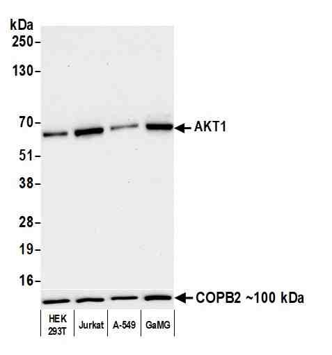 |
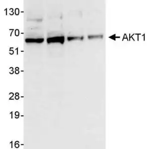
| Detection of human AKT1 by western blot. Samples: Whole cell lysate (10 µg) from HEK293T, Jurkat, A-549, and GaMG cells prepared using NETN lysis buffer. Antibody: Rabbit anti-AKT1 recombinant monoclonal antibody [BLR245L] (#1020) used at 1:1000. Secondary: HRP-conjugated goat anti-rabbit IgG. Detection: Chemiluminescence with an exposure time of 30 seconds. |
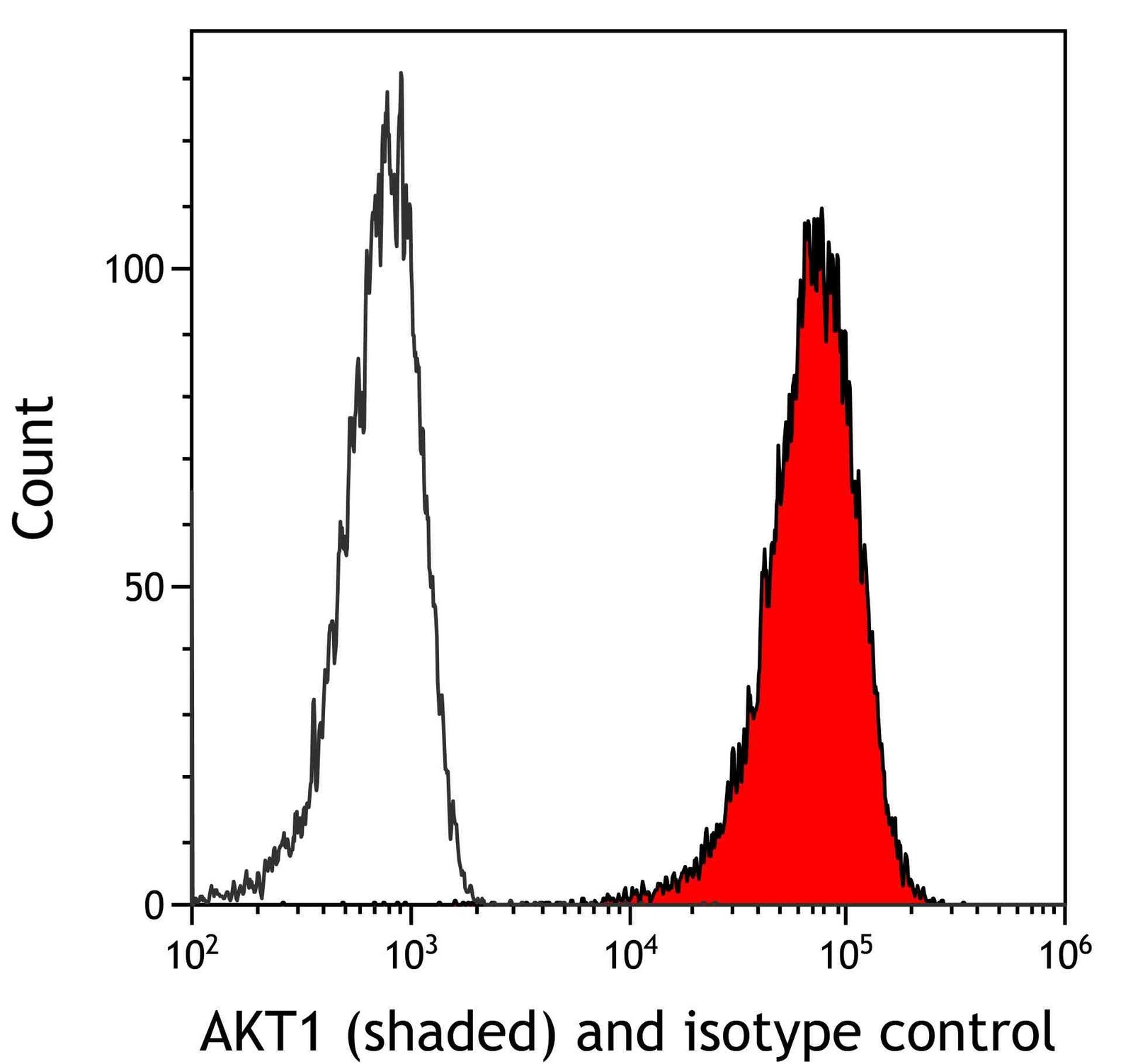 |
| Detection of human AKT1 (shaded) in FaDu cells by flow cytometry. Antibody: Rabbit anti-AKT1 recombinant monoclonal antibody [BLR245L] (#1020) or isotype control (unshaded). Secondary: DyLight 650-conjugated goat anti-rabbit IgG. |
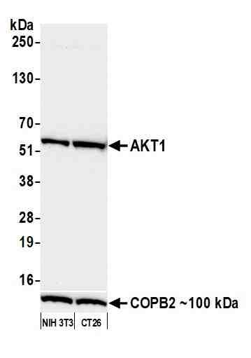 |
| Detection of mouse AKT1 by western blot. Samples: Whole cell lysate (25 µg) from NIH 3T3 and CT26 cells prepared using NETN lysis buffer. Antibody: Rabbit anti-AKT1 recombinant monoclonal antibody [BLR245L] (#1020) used at 1:1000. Secondary: HRP-conjugated goat anti-rabbit IgG. Detection: Chemiluminescence with an exposure time of 30 seconds. Lower Panel: Rabbit anti-COPB2 antibody. |
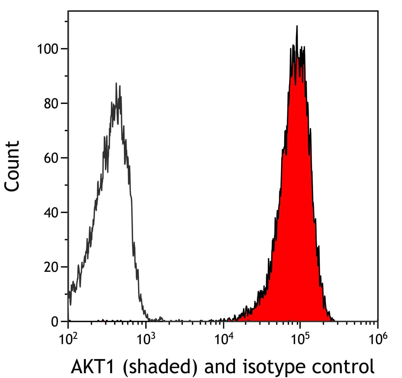 |
| Detection of mouse AKT1 (shaded) in CTLL-2 cells by flow cytometry. Antibody: Rabbit anti-AKT1 recombinant monoclonal antibody [BLR245L] (#1020) or isotype control (unshaded). Secondary: DyLight 650-conjugated goat anti-rabbit IgG. |
FAQ & Publications
Frequently Asked Questions
What species does the rabbit anti-AKT1 polyclonal antibody BLR245L 1020 react with, and in which applications has it been validated?
This antibody reacts with human and mouse species and has been validated for use in Western Blot (WB) and Immunocytochemistry (ICC) applications.
How should the rabbit anti-AKT1 polyclonal antibody BLR245L 1020 be stored to maintain its stability, and what is its concentration?
The antibody should be stored at 2-8°C and is stable for one year from the date of receipt. It is supplied as a liquid with a concentration of 0.4 mg/mL.
Publications
| pmid | title | authors | citation |
|---|---|---|---|
| We haven't added any publications to our database yet. | |||
Published literature highly relevant to the biological target of this product and referencing this antibody or clone are retrieved from the PubMed database provided by the United States National Library of Medicine at the National Institutes of Health.
Protocols
| relevant to this product |
|---|
| Western blot |
Documents
| Batch Number | QC File | SDS |
|---|---|---|
| To view batch-specific Safety Datasheets and Quality Certificates associated with your account, please Log In. | ||
Only logged in customers who have purchased this product may leave a review.


Reviews
There are no reviews yet.