| Weight | 1 lbs |
|---|---|
| Dimensions | 9 × 5 × 2 in |
| host | mouse |
| isotype | IgG2C |
| clonality | monoclonal |
| concentration | concentrate, predilute |
| applications | IHC |
| reactivity | human |
| available size | 0.1 mL, 0.5 mL, 1 mL concentrated, 7 mL prediluted |
mouse anti-Perforin monoclonal antibody (ZM159) 6329
Price range: $160.00 through $528.00
Antibody summary
- Mouse monoclonal to Perforin
- Suitable for: Immunohistochemistry (formalin-fixed, paraffin-embedded tissues)
- Reacts with: Human
- Isotype:IgG2C
- Control: Spleen
- Visualization: Granular cytoplasmic
- 0.1, 0.5, 1.0 mL concentrated, 7 mL prediluted
mouse anti-Perforin monoclonal antibody ZM159 6329
| target relevance |
|---|
| Protein names Perforin-1 (P1) (Cytolysin) (Lymphocyte pore-forming protein) (PFP) |
| Gene names PRF1,PRF1 PFP |
| Protein family Complement C6/C7/C8/C9 family |
| Mass 61377Da |
| Function FUNCTION: Pore-forming protein that plays a key role in granzyme-mediated programmed cell death, and in defense against virus-infected or neoplastic cells (PubMed:20889983, PubMed:21037563, PubMed:24558045, PubMed:9058810, PubMed:9164947). Plays an important role in killing other cells that are recognized as non-self by the immune system, e.g. in transplant rejection or some forms of autoimmune disease (PubMed:9058810). Can insert into the membrane of target cells in its calcium-bound form, oligomerize and form large pores (PubMed:20889983, PubMed:21037563). Promotes cytolysis and apoptosis of target cells by mediating the passage and uptake of cytotoxic granzymes (PubMed:20038786, PubMed:20225066, PubMed:24558045, PubMed:32299851). Facilitates the delivery of cationic cargo protein, while anionic or neural proteins are not delivered efficiently (PubMed:24558045). Perforin pores allow the release of mature caspase-7 (CASP7) into the extracellular milieu (By similarity). {ECO:0000250|UniProtKB:P10820, ECO:0000269|PubMed:20038786, ECO:0000269|PubMed:20225066, ECO:0000269|PubMed:20889983, ECO:0000269|PubMed:21037563, ECO:0000269|PubMed:24558045, ECO:0000269|PubMed:32299851, ECO:0000269|PubMed:9058810, ECO:0000269|PubMed:9164947}. |
| Subellular location SUBCELLULAR LOCATION: Cytolytic granule {ECO:0000269|PubMed:20038786, ECO:0000269|PubMed:24088571}. Secreted. Cell membrane {ECO:0000269|PubMed:20889983, ECO:0000269|PubMed:21037563}; Multi-pass membrane protein {ECO:0000269|PubMed:20889983, ECO:0000269|PubMed:21037563}. Endosome lumen {ECO:0000269|PubMed:20038786}. Note=Stored in cytolytic granules of cytolytic T-lymphocytes and secreted into the cleft between T-lymphocyte and target cell (PubMed:20038786). Inserts into the cell membrane of target cells and forms pores (PubMed:20889983). Membrane insertion and pore formation requires a major conformation change (PubMed:20889983). May be taken up via endocytosis involving clathrin-coated vesicles and accumulate in a first time in large early endosomes (PubMed:20038786). {ECO:0000269|PubMed:20038786, ECO:0000269|PubMed:20889983}. |
| Structure SUBUNIT: Monomer, as soluble protein (PubMed:20889983, PubMed:21037563). Homooligomer; homooligomerizes to form a pore-forming ring (PubMed:20889983, PubMed:21037563). {ECO:0000269|PubMed:20889983, ECO:0000269|PubMed:21037563}. |
| Post-translational modification PTM: N-glycosylated. {ECO:0000250|UniProtKB:P10820}. |
| Domain DOMAIN: Perforin consists of three domains: (1) the MACPF domain, which includes the central machinery of pore formation, (2) the EGF-like domain, which forms a 'shelf-like' assembly connecting the MACPF and C2 domains, and (3) the C2 domain, which mediates calcium-dependent binding to lipid membranes. The C2 domain is critical for initial calcium-dependent interaction with lipid membranes of the target cell: calcium-binding causes a significant structural rearrangement, leading to oligomerization and deployment of the two transmembrane beta-strands (named CH1/TMH1 and CH2/TMH2) that enter the membrane as amphipathic beta-hairpins. The third calcium-binding site (Ca(2+) 3), which constitutes the weakest affinity site, triggers structural rearrangements in the C2 domain that facilitate its interaction with lipid membranes. {ECO:0000250|UniProtKB:P10820}. |
| Involvement in disease DISEASE: Hemophagocytic lymphohistiocytosis, familial, 2 (FHL2) [MIM:603553]: A rare disorder characterized by immune dysregulation with hypercytokinemia, defective function of natural killer cell, and massive infiltration of several organs by activated lymphocytes and macrophages. The clinical features of the disease include fever, hepatosplenomegaly, cytopenia, and less frequently neurological abnormalities ranging from irritability and hypotonia to seizures, cranial nerve deficits and ataxia. {ECO:0000269|PubMed:10583959, ECO:0000269|PubMed:11179007}. Note=The disease is caused by variants affecting the gene represented in this entry. |
| Target Relevance information above includes information from UniProt accession: P14222 |
| The UniProt Consortium |
Data
|
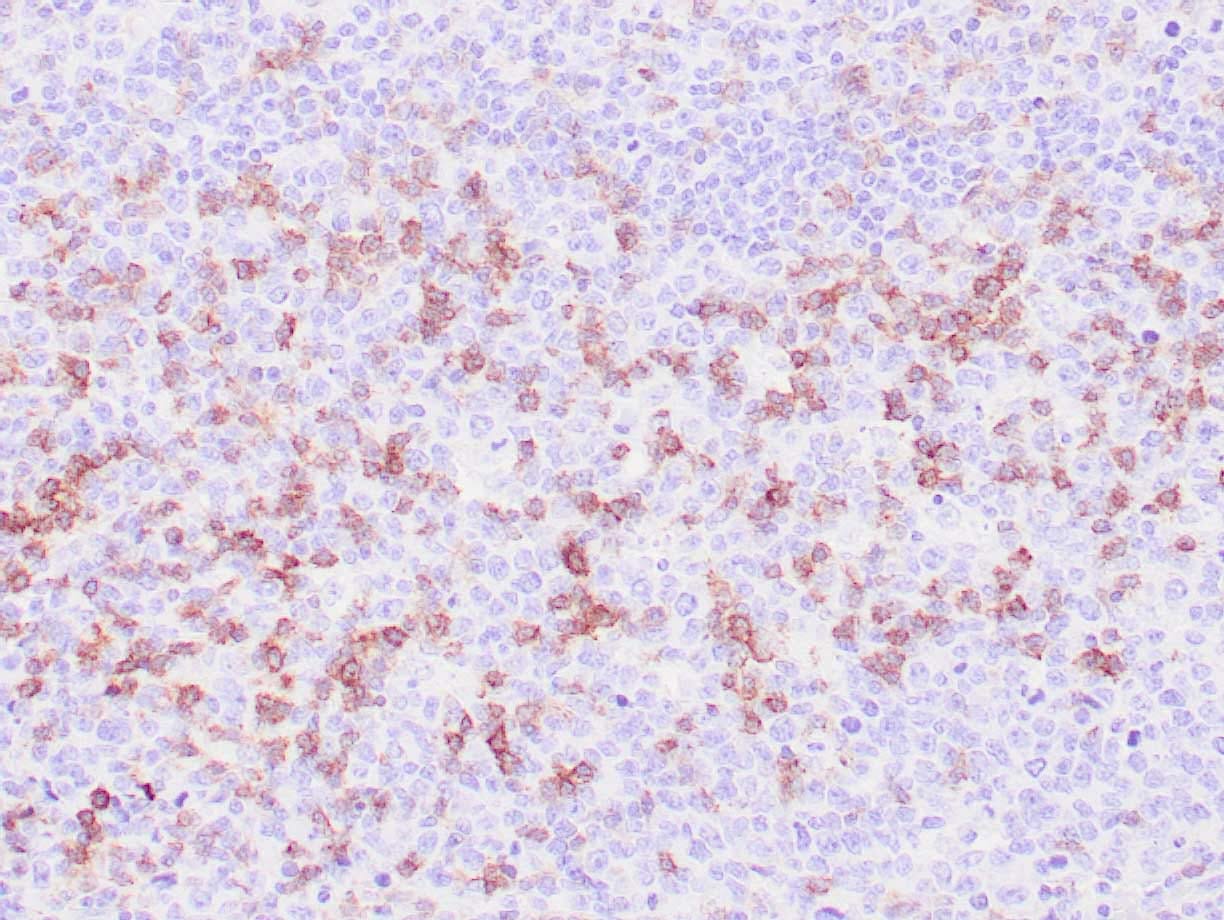
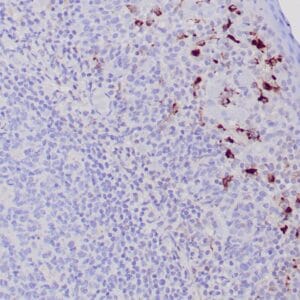
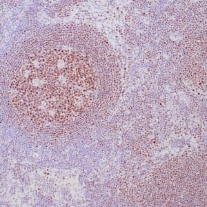
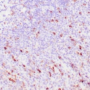
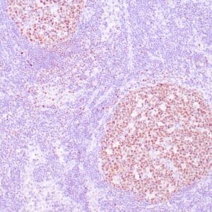
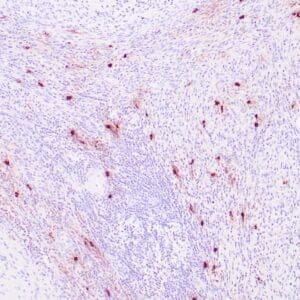
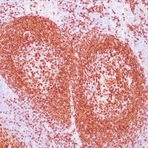
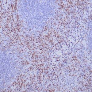
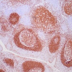
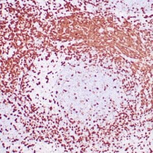
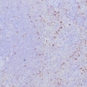
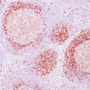
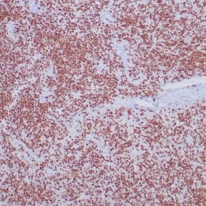
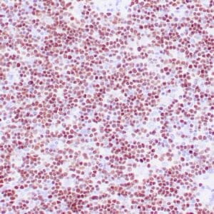
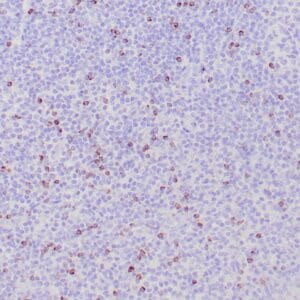
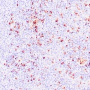
| Human spleen stained with anti-Perforin antibody using peroxidase-conjugate and DAB chromogen. Note the cytoplasmic granular staining of cytotoxic T cells. |
Publications
| pmid | title | authors | citation |
|---|---|---|---|
| We haven't added any publications to our database yet. | |||
Protocols
| relevant to this product |
|---|
| IHC |
Documents
| # | SDS | Certificate | |
|---|---|---|---|
| Please enter your product and batch number here to retrieve product datasheet, SDS, and QC information. | |||
Only logged in customers who have purchased this product may leave a review.
Reviews
There are no reviews yet.