| Weight | 1 lbs |
|---|---|
| Dimensions | 9 × 5 × 2 in |
| host | mouse |
| isotype | IgG1 |
| clonality | monoclonal |
| concentration | concentrate, predilute |
| applications | IHC |
| reactivity | human |
| available size | 0.1 mL, 0.5 mL, 1 mL concentrated, 7 mL prediluted |

mouse anti-PD-1 (PDCD1) monoclonal antibody (ZM357) 6326
Price range: $160.00 through $528.00
Antibody summary
- Mouse monoclonal to PD-1 (PDCD1)
- Suitable for: Immunohistochemistry (formalin-fixed, paraffin-embedded tissues)
- Reacts with: Human
- Isotype:IgG1
- Control: Lymph node
- Visualization: Cytoplasm & Membrane
- 0.1, 0.5, 1.0 mL concentrated, 7 mL prediluted
mouse anti-PD-1 (PDCD1) monoclonal antibody ZM357 6326
| target relevance |
|---|
| Homo sapiens PDCD1 Programmed cell death protein 1 |
| Protein names Programmed cell death protein 1 |
| Gene names PDCD1 |
| Function Inhibitory receptor on antigen activated T-cells that plays a critical role in induction and maintenance of immune tolerance to self (PubMed:21276005, PubMed:31754127, PubMed:32184441, PubMed:37208329). Delivers inhibitory signals upon binding to ligands CD274/PDCD1L1 and CD273/PDCD1LG2 (PubMed:21276005, PubMed:26602187). Following T-cell receptor (TCR) engagement, PDCD1 associates with TCR-CD3 in the immunological synapse and directly inhibits T-cell activation (PubMed:32184441). Suppresses T-cell activation through the recruitment of PTPN11/SHP-2: following ligand-binding, PDCD1 is phosphorylated within the ITSM motif, leading to the recruitment of the protein tyrosine phosphatase PTPN11/SHP-2 that mediates dephosphorylation of key TCR proximal signaling molecules, such as ZAP70, PRKCQ/PKCtheta and CD247/CD3zeta (PubMed:32184441) |
| Subcellular location Cell membrane |
| Structure Monomer |
| Post-translational modification Ubiquitinated at Lys-233 by the SCF(FBXO38) complex, leading to its proteasomal degradation (PubMed:30487606). Ubiquitinated via 'Lys-48'-linked polyubiquitin chains (PubMed:30487606). Deubiquitinated and thus stabilized by USP5 (PubMed:37208329) Tyrosine phosphorylated at Tyr-223 (within ITIM motif) and Tyr-248 (ITSM motif) upon ligand binding (PubMed:31754127, PubMed:32184441). Phosphorylation at Tyr-248 by FYN promotes the recruitment of the protein tyrosine phosphatase PTPN11/SHP-2 that mediates dephosphorylation of key TCR proximal signaling molecules, such as ZAP70, PRKCQ/PKCtheta and CD247/CD3zeta (PubMed:32184441). Phosphorylation at Thr-234 promotes the recruitment of the deubiquitinase USP5 (PubMed:37208329) N-glycosylation at Asn-58 contains at least two N-acetylglucosamine units and one fucose (PubMed:28165004). N-glycosylation does not affect binding to nivolumab drug (PubMed:28165004) |
| Involvement in disease Autoimmune disease, multisystem, infantile-onset, 4 An autosomal recessive immunologic disorder characterized by lymphoproliferative autoimmunity and onset of various autoimmune diseases in early childhood. Death from autoimmune pneumonitis may occur. |
| Keywords 3D-structure, Adaptive immunity, Apoptosis, Cell membrane, Disulfide bond, Glycoprotein, Immunity, Immunoglobulin domain, Isopeptide bond, Membrane, Phosphoprotein, Proteomics identification, Reference proteome, Signal, Transmembrane, Transmembrane helix, Ubl conjugation |
| Sequence MQIPQAPWPVVWAVLQLGWRPGWFLDSPDRPWNPPTFSPALLVVTEGDNATFTCSFSNTS ESFVLNWYRMSPSNQTDKLAAFPEDRSQPGQDCRFRVTQLPNGRDFHMSVVRARRNDSGT YLCGAISLAPKAQIKESLRAELRVTERRAEVPTAHPSPSPRPAGQFQTLVVGVVGGLLGS LVLLVWVLAVICSRAARGTIGARRTGQPLKEDPSAVPVFSVDYGELDFQWREKTPEPPVP CVPEQTEYATIVFPSGMGTSSPARRGSADGPRSAQPLRPEDGHCSWPL |
| UniProt accession: Q15116 |
Data
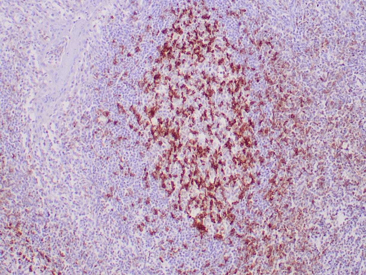 |
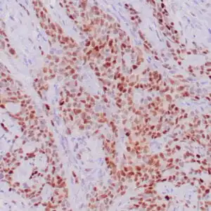
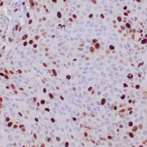
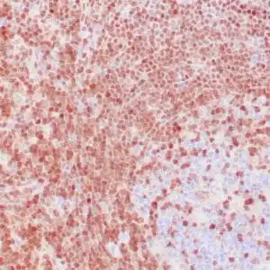
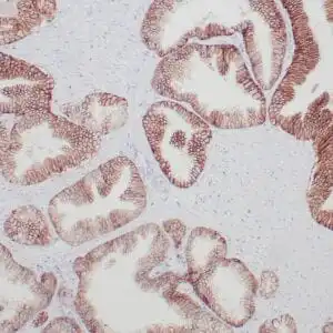
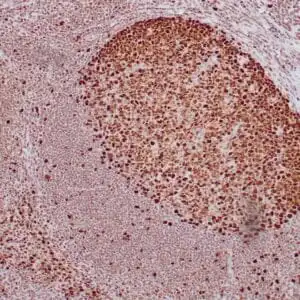
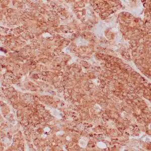
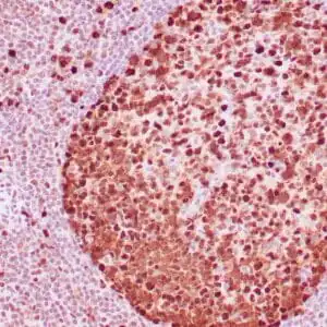
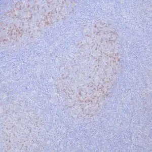
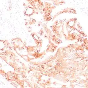
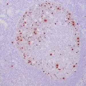
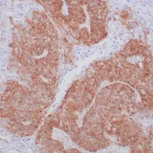
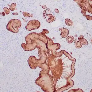
| Formalin-fixed, paraffin-embedded human tonsil stained with anti-PD-1 antibody using peroxidase-conjugate and DAB chromogen. Note cytoplasmic staining of T-cells |
FAQ & Publications
Frequently Asked Questions
What species reactivity and applications is the mouse anti-PD-1 (PDCD1) monoclonal antibody (ZM357) validated for?
This antibody is reactive with human PD-1 and is suitable for immunohistochemistry applications, specifically on formalin-fixed, paraffin-embedded tissues.
How should the mouse anti-PD-1 (PDCD1) monoclonal antibody (ZM357) be stored to maintain its stability?
For short-term storage, keep the antibody at 2-8°C. For longer-term preservation, store it at -20°C and avoid repeated freeze-thaw cycles to maintain antibody integrity.
Publications
| pmid | title | authors | citation |
|---|---|---|---|
| We haven't added any publications to our database yet. | |||
Published literature highly relevant to the biological target of this product and referencing this antibody or clone are retrieved from the PubMed database provided by the United States National Library of Medicine at the National Institutes of Health.
Protocols
| relevant to this product |
|---|
| IHC |
Documents
| Batch Number | QC File | SDS |
|---|---|---|
| To view batch-specific Safety Datasheets and Quality Certificates associated with your account, please Log In. | ||
Only logged in customers who have purchased this product may leave a review.
Reviews
There are no reviews yet.