| Weight | 1 lbs |
|---|---|
| Dimensions | 9 × 5 × 2 in |
| host | mouse |
| isotype | IgG1 |
| clonality | monoclonal |
| concentration | concentrate, predilute |
| applications | IHC |
| reactivity | human |
| available size | 0.1 mL, 0.5 mL, 1 mL concentrated, 7 mL prediluted |

mouse anti-Cytokeratin 5/6 monoclonal antibody (D5&16B4) 6147
Price range: $160.00 through $528.00
Antibody summary
- Mouse monoclonal to Cytokeratin 5/6
- Suitable for: Immunohistochemistry (formalin-fixed, paraffin-embedded tissues)
- Reacts with: Human
- Isotype:IgG1
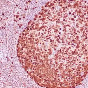
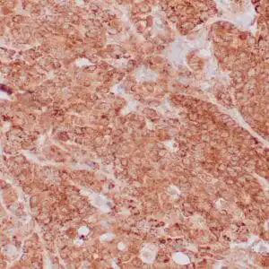
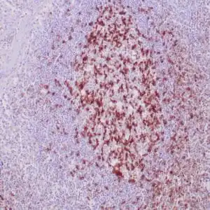
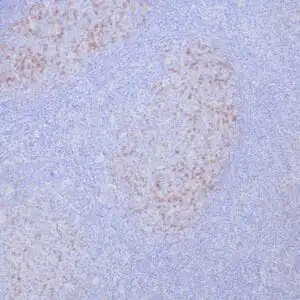
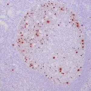
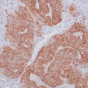
- Control: Mesothelioma, prostate
- Visualization: Cytoplasmic
- 0.1, 0.5, 1.0 mL concentrated, 7 mL prediluted
mouse anti-Cytokeratin 5/6 monoclonal antibody D5&16B4 6147
| target relevance |
|---|
| Homo sapiens KRT5 Keratin, type II cytoskeletal 5 |
| Protein names Keratin, type II cytoskeletal 5 |
| Alternative names 58 kDa cytokeratin, Cytokeratin-5, Keratin-5, Type-II keratin Kb5 |
| Gene names KRT5 |
| Protein family Belongs to the intermediate filament family |
| Function Required for the formation of keratin intermediate filaments in the basal epidermis and maintenance of the skin barrier in response to mechanical stress (By similarity). Regulates the recruitment of Langerhans cells to the epidermis, potentially by modulation of the abundance of macrophage chemotactic cytokines, macrophage inflammatory cytokines and CTNND1 localization in keratinocytes (By similarity) |
| Subcellular location Cytoplasm |
| Structure Heterodimer of a type I and a type II keratin (PubMed:22705788, PubMed:31995743). Heterodimer with type I keratin KRT25 leading to the formation of keratin intermediate filament (KIF) network (PubMed:28899683). Forms a heterodimer (via 2B domains) with KRT14 (via 2B domains) (PubMed:22705788, PubMed:24940650, PubMed:31995743). Interacts with PLEC isoform 1C, when in a heterodimer with KRT14 (PubMed:24940650). Interacts with TCHP (PubMed:15731013). Interacts with EPPK1 (By similarity). Interacts with AMELX (By similarity). Interacts with PKP1 (via N-terminus) and PKP2 (PubMed:10852826) |
| Post-translational modification Phosphorylated by CDK1, AURKB and Rho-kinase, phosphorylation is regulated by the cell cycle (By similarity). Thr-24 phosphorylation, mediated by CDK1, peaks during prometaphase or metaphase cells with phosphorylated filamentous structures evident throughout the cytoplasm during early mitosis (By similarity). CDK1 phosphorylates Thr-24 in mitotic cells at the site of injury (By similarity) O-glycosylated |
| Involvement in disease Epidermolysis bullosa simplex 2A, generalized severe A form of epidermolysis bullosa simplex, a group of skin fragility disorders characterized by skin blistering due to cleavage within the basal layer of keratinocytes, and erosions caused by minor mechanical trauma. There is a broad spectrum of clinical severity ranging from minor blistering on the feet, to subtypes with extracutaneous involvement and a lethal outcome. EBS2A is an autosomal dominant, severe form characterized by extensive intraepidermal blistering from the time of birth with herpetiform marginal spreading and central healing. Oral mucosal involvement, nail dystrophy, onychogryposis, formation of milia, and palmoplantar hyperkeratosis are common features. Epidermolysis bullosa simplex 2B, generalized intermediate A form of epidermolysis bullosa simplex, a group of skin fragility disorders characterized by skin blistering due to cleavage within the basal layer of keratinocytes, and erosions caused by minor mechanical trauma. There is a broad spectrum of clinical severity ranging from minor blistering on the feet, to subtypes with extracutaneous involvement and a lethal outcome. EBS2B is an autosomal dominant form characterized by generalized blistering manifesting at birth. The tendency to blistering diminishes in adolescence, when it may become localized to hands and feet. Epidermolysis bullosa simplex 2C, localized A form of epidermolysis bullosa simplex, a group of skin fragility disorders characterized by skin blistering due to cleavage within the basal layer of keratinocytes, and erosions caused by minor mechanical trauma. There is a broad spectrum of clinical severity ranging from minor blistering on the feet, to subtypes with extracutaneous involvement and a lethal outcome. EBS2C is an autosomal dominant form with intraepidermal blistering mainly restricted to hands and feet beginning in infancy. Nails may be thick and dystrophic. Epidermolysis bullosa simplex 2D, generalized, intermediate or severe, autosomal recessive A form of epidermolysis bullosa simplex, a group of skin fragility disorders characterized by skin blistering due to cleavage within the basal layer of keratinocytes, and erosions caused by minor mechanical trauma. There is a broad spectrum of clinical severity ranging from minor blistering on the feet, to subtypes with extracutaneous involvement and a lethal outcome. EBS2D is an autosomal recessive form characterized by widespread intraepidermal skin blistering and erosions from birth. Death may occur in the neonatal period. Epidermolysis bullosa simplex 2E, with migratory circinate erythema A form of epidermolysis bullosa simplex, a group of skin fragility disorders characterized by skin blistering due to cleavage within the basal layer of keratinocytes, and erosions caused by minor mechanical trauma. There is a broad spectrum of clinical severity ranging from minor blistering on the feet, to subtypes with extracutaneous involvement and a lethal outcome. EBS2E is an autosomal dominant form in which multiple vesicles are present from birth onward and acquire over time a typical migratory circinate pattern on an erythematous background. Postinflammatory hyperpigmentation develops gradually and may have a mottled pattern. Epidermolysis bullosa simplex 2F, with mottled pigmentation A form of epidermolysis bullosa simplex, a group of skin fragility disorders characterized by skin blistering due to cleavage within the basal layer of keratinocytes, and erosions caused by minor mechanical trauma. There is a broad spectrum of clinical severity ranging from minor blistering on the feet, to subtypes with extracutaneous involvement and a lethal outcome. EBS2F is an autosomal dominant form characterized by generalized skin blistering of intermediate severity beginning at birth, with mottled or reticulate pigmentation developing gradually. Focal keratoses of palms and soles and dystrophic, thickened nails develop over time. Dowling-Degos disease 1 An autosomal dominant genodermatosis. Affected individuals develop a postpubertal reticulate hyperpigmentation that is progressive and disfiguring, and small hyperkeratotic dark brown papules that affect mainly the flexures and great skin folds. Patients usually show no abnormalities of the hair or nails. |
| Keywords 3D-structure, Coiled coil, Cytoplasm, Disease variant, Epidermolysis bullosa, Intermediate filament, Keratin, Phosphoprotein, Proteomics identification, Reference proteome |
| Sequence MSRQSSVSFRSGGSRSFSTASAITPSVSRTSFTSVSRSGGGGGGGFGRVSLAGACGVGGY GSRSLYNLGGSKRISISTSGGSFRNRFGAGAGGGYGFGGGAGSGFGFGGGAGGGFGLGGG AGFGGGFGGPGFPVCPPGGIQEVTVNQSLLTPLNLQIDPSIQRVRTEEREQIKTLNNKFA SFIDKVRFLEQQNKVLDTKWTLLQEQGTKTVRQNLEPLFEQYINNLRRQLDSIVGERGRL DSELRNMQDLVEDFKNKYEDEINKRTTAENEFVMLKKDVDAAYMNKVELEAKVDALMDEI NFMKMFFDAELSQMQTHVSDTSVVLSMDNNRNLDLDSIIAEVKAQYEEIANRSRTEAESW YQTKYEELQQTAGRHGDDLRNTKHEISEMNRMIQRLRAEIDNVKKQCANLQNAIADAEQR GELALKDARNKLAELEEALQKAKQDMARLLREYQELMNTKLALDVEIATYRKLLEGEECR LSGEGVGPVNISVVTSSVSSGYGSGSGYGGGLGGGLGGGLGGGLAGGSSGSYYSSSSGGV GLGGGLSVGGSGFSASSGRGLGVGFGSGGGSSSSVKFVSTTSSSRKSFKS |
| UniProt accession: P13647 |
Data
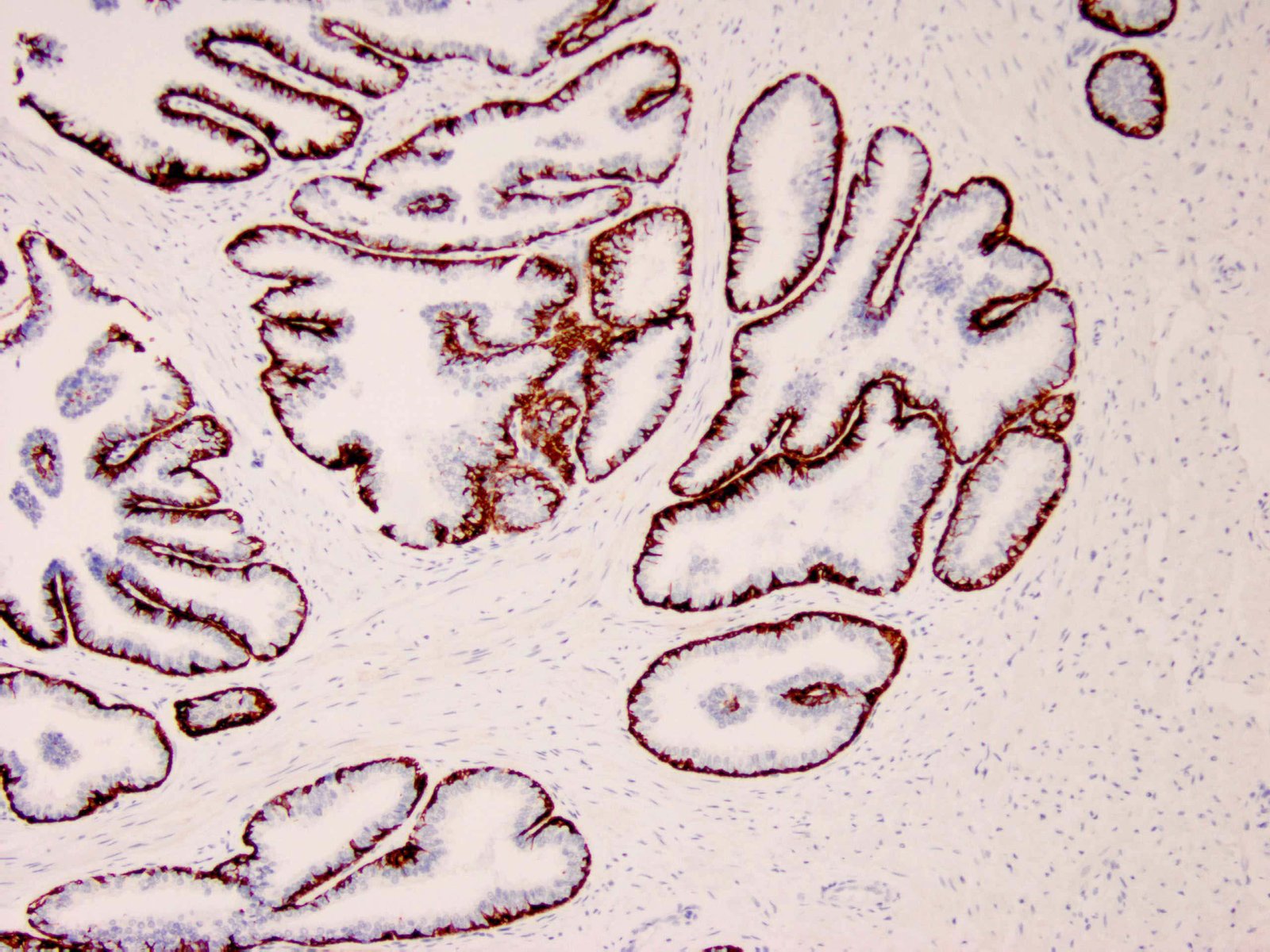 |
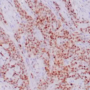
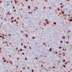
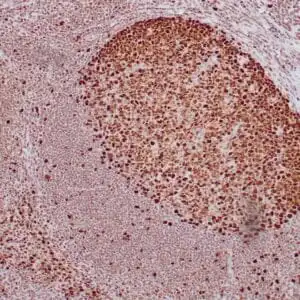
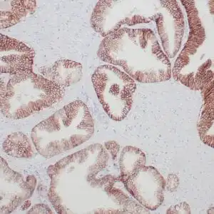
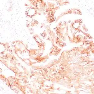
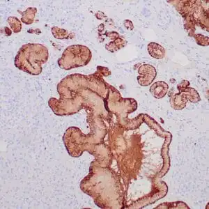
| Human prostate tissue stained with anti-Keratin 5/6 antibody using peroxidase-conjugate and DAB chromogen. Note the cytoplasmic staining of basal cells. |
FAQ & Publications
Frequently Asked Questions
What is the recommended application for the mouse anti-Cytokeratin 5/6 monoclonal antibody (D5&16B4)?
This antibody is suitable for immunohistochemistry (IHC) on formalin-fixed, paraffin-embedded human tissue samples.
What species does the Cytokeratin 5/6 antibody react with?
The antibody specifically reacts with human tissues.
How should the mouse anti-Cytokeratin 5/6 antibody be stored to maintain stability?
For short-term storage, keep the antibody at 2-8°C. For long-term storage, it should be kept at -20°C while avoiding repeated freeze/thaw cycles.
What is the host and isotype of this Cytokeratin 5/6 monoclonal antibody?
The antibody is a mouse monoclonal of the IgG1 isotype.
Are there recommended positive controls for validating the staining with this antibody?
Yes, mesothelioma and prostate tissues are recommended as positive controls for this antibody in IHC applications.
Publications
| pmid | title | authors | citation |
|---|---|---|---|
| We haven't added any publications to our database yet. | |||
Published literature highly relevant to the biological target of this product and referencing this antibody or clone are retrieved from the PubMed database provided by the United States National Library of Medicine at the National Institutes of Health.
Protocols
| relevant to this product |
|---|
| IHC |
Documents
| Batch Number | QC File | SDS |
|---|---|---|
| To view batch-specific Safety Datasheets and Quality Certificates associated with your account, please Log In. | ||
Only logged in customers who have purchased this product may leave a review.
Reviews
There are no reviews yet.