| Weight | 1 lbs |
|---|---|
| Dimensions | 9 × 5 × 2 in |
| host | mouse |
| isotype | IgG2b |
| clonality | monoclonal |
| concentration | concentrate, predilute |
| applications | IHC |
| reactivity | human |
| available size | 0.1 mL, 0.5 mL, 1 mL concentrated, 7 mL prediluted |

mouse anti-Beta-Catenin monoclonal antibody (ZM13) 6033
Price range: $160.00 through $528.00
Antibody summary
- Mouse monoclonal to Beta-Catenin
- Suitable for: Immunohistochemistry (formalin-fixed, paraffin-embedded tissues)
- Reacts with: Human
- Isotype:IgG2b
- Control: Fibromatosis or transitional cell carcinoma
- Visualization: Membrane, cytoplasmic or nuclear
- 0.1, 0.5, 1.0 mL concentrated, 7 mL prediluted
mouse anti-Beta-Catenin monoclonal antibody ZM13 6033
| target relevance |
|---|
| Homo sapiens CTNNB1 Catenin beta-1 |
| Protein names Catenin beta-1 |
| Alternative names Beta-catenin |
| Gene names CTNNB1 |
| Protein family Belongs to the beta-catenin family |
| Function Key downstream component of the canonical Wnt signaling pathway (PubMed:17524503, PubMed:18077326, PubMed:18086858, PubMed:18957423, PubMed:21262353, PubMed:22155184, PubMed:22647378, PubMed:22699938). In the absence of Wnt, forms a complex with AXIN1, AXIN2, APC, CSNK1A1 and GSK3B that promotes phosphorylation on N-terminal Ser and Thr residues and ubiquitination of CTNNB1 via BTRC and its subsequent degradation by the proteasome (PubMed:17524503, PubMed:18077326, PubMed:18086858, PubMed:18957423, PubMed:21262353, PubMed:22155184, PubMed:22647378, PubMed:22699938). In the presence of Wnt ligand, CTNNB1 is not ubiquitinated and accumulates in the nucleus, where it acts as a coactivator for transcription factors of the TCF/LEF family, leading to activate Wnt responsive genes (PubMed:17524503, PubMed:18077326, PubMed:18086858, PubMed:18957423, PubMed:21262353, PubMed:22155184, PubMed:22647378, PubMed:22699938). Also acts as a coactivator for other transcription factors, such as NR5A2 (PubMed:22187462). Promotes epithelial to mesenchymal transition/mesenchymal to epithelial transition (EMT/MET) via driving transcription of CTNNB1/TCF-target genes (PubMed:29910125). Involved in the regulation of cell adhesion, as component of an E-cadherin:catenin adhesion complex (By similarity). Acts as a negative regulator of centrosome cohesion (PubMed:18086858). Involved in the CDK2/PTPN6/CTNNB1/CEACAM1 pathway of insulin internalization (PubMed:21262353). Blocks anoikis of malignant kidney and intestinal epithelial cells and promotes their anchorage-independent growth by down-regulating DAPK2 (PubMed:18957423). Disrupts PML function and PML-NB formation by inhibiting RANBP2-mediated sumoylation of PML (PubMed:22155184). Promotes neurogenesis by maintaining sympathetic neuroblasts within the cell cycle (By similarity). Involved in chondrocyte differentiation via interaction with SOX9: SOX9-binding competes with the binding sites of TCF/LEF within CTNNB1, thereby inhibiting the Wnt signaling (By similarity). Acts as a positive regulator of odontoblast differentiation during mesenchymal tooth germ formation, via promoting the transcription of differentiation factors such as LEF1, BMP2 and BMP4 (By similarity). Activity is repressed in a MSX1-mediated manner at the bell stage of mesenchymal tooth germ formation which prevents premature differentiation of odontoblasts (By similarity) |
| Subcellular location Cytoplasm, Nucleus, Cytoplasm, cytoskeleton, Cell junction, adherens junction, Cell junction, Cell membrane, Cytoplasm, cytoskeleton, microtubule organizing center, centrosome, Cytoplasm, cytoskeleton, spindle pole, Synapse, Cytoplasm, cytoskeleton, cilium basal body |
| Structure (Microbial infection) Interacts with herpes virus 8 protein vPK; this interaction inhibits the Wnt signaling pathway |
| Post-translational modification Phosphorylation at Ser-552 by AMPK promotes stabilization of the protein, enhancing TCF/LEF-mediated transcription (By similarity). Phosphorylation by GSK3B requires prior phosphorylation of Ser-45 by another kinase (PubMed:10966653, PubMed:12027456, PubMed:12051714). Phosphorylation proceeds then from Thr-41 to Ser-37 and Ser-33 (PubMed:12077367, PubMed:25169422). Phosphorylated by NEK2 (PubMed:18086858). EGF stimulates tyrosine phosphorylation (PubMed:10187801). Phosphorylated on Ser-33 and Ser-37 by HIPK2 and GSK3B, this phosphorylation triggers proteasomal degradation (PubMed:20307497). Phosphorylation on Ser-191 and Ser-246 by CDK5 (PubMed:17009320). Phosphorylation by CDK2 regulates insulin internalization (PubMed:21262353). Phosphorylation by PTK6 at Tyr-64, Tyr-142, Tyr-331 and/or Tyr-333 with the predominant site at Tyr-64 is not essential for inhibition of transcriptional activity (PubMed:20026641). Phosphorylation by SRC at Tyr-333 promotes interaction with isoform M2 of PKM (PKM2); promoting transcription activation (PubMed:22056988) Ubiquitinated by the SCF(BTRC) E3 ligase complex when phosphorylated by GSK3B, leading to its degradation (PubMed:12077367). Ubiquitinated by a E3 ubiquitin ligase complex containing UBE2D1, SIAH1, CACYBP/SIP, SKP1, APC and TBL1X, leading to its subsequent proteasomal degradation (PubMed:11389839, PubMed:11389840, PubMed:20307497). Ubiquitinated and degraded following interaction with SOX9 (By similarity). Ubiquitinated via 'Lys-11'- and 'Lys-29'-linked ubiquitin chains by UBR5, leading to its stabilization (PubMed:21118991). Ubiquitinated by the SCF-FBXO16 E3 ubiquitin ligase, leading to proteasomal degradation (PubMed:30714168) S-nitrosylation at Cys-619 within adherens junctions promotes VEGF-induced, NO-dependent endothelial cell permeability by disrupting interaction with E-cadherin, thus mediating disassembly adherens junctions O-glycosylation at Ser-23 decreases nuclear localization and transcriptional activity, and increases localization to the plasma membrane and interaction with E-cadherin CDH1 Deacetylated at Lys-49 by SIRT1 Phosphorylated at Thr-556 by herpes virus 1/HHV-1 leading to CTNNB1 inhibition |
| Involvement in disease Colorectal cancer A complex disease characterized by malignant lesions arising from the inner wall of the large intestine (the colon) and the rectum. Genetic alterations are often associated with progression from premalignant lesion (adenoma) to invasive adenocarcinoma. Risk factors for cancer of the colon and rectum include colon polyps, long-standing ulcerative colitis, and genetic family history. Pilomatrixoma Common benign skin tumor. Medulloblastoma Malignant, invasive embryonal tumor of the cerebellum with a preferential manifestation in children. Ovarian cancer The term ovarian cancer defines malignancies originating from ovarian tissue. Although many histologic types of ovarian tumors have been described, epithelial ovarian carcinoma is the most common form. Ovarian cancers are often asymptomatic and the recognized signs and symptoms, even of late-stage disease, are vague. Consequently, most patients are diagnosed with advanced disease. Mesothelioma, malignant An aggressive neoplasm of the serosal lining of the chest. It appears as broad sheets of cells, with some regions containing spindle-shaped, sarcoma-like cells and other regions showing adenomatous patterns. Pleural mesotheliomas have been linked to exposure to asbestos. Neurodevelopmental disorder with spastic diplegia and visual defects An autosomal dominant disorder characterized by global developmental delay, severe intellectual disability with absent or very limited speech, microcephaly, spasticity, and visual abnormalities. Vitreoretinopathy, exudative 7 An autosomal dominant form of exudative vitreoretinopathy, a disorder of the retinal vasculature characterized by an abrupt cessation of growth of peripheral capillaries, leading to an avascular peripheral retina. This may lead to compensatory retinal neovascularization, which is thought to be induced by hypoxia from the initial avascular insult. New vessels are prone to leakage and rupture causing exudates and bleeding, followed by scarring, retinal detachment and blindness. Clinical features can be highly variable, even within the same family. Patients with mild forms of the disease are asymptomatic, and their only disease related abnormality is an arc of avascular retina in the extreme temporal periphery. |
| Keywords 3D-structure, Acetylation, Activator, Cell adhesion, Cell junction, Cell membrane, Cell projection, Chromosomal rearrangement, Cytoplasm, Cytoskeleton, Disease variant, Glycoprotein, Host-virus interaction, Intellectual disability, Membrane, Neurogenesis, Nucleus, Phosphoprotein, Proteomics identification, Reference proteome, Repeat, S-nitrosylation, Synapse, Transcription, Transcription regulation, Ubl conjugation, Wnt signaling pathway |
| Sequence MATQADLMELDMAMEPDRKAAVSHWQQQSYLDSGIHSGATTTAPSLSGKGNPEEEDVDTS QVLYEWEQGFSQSFTQEQVADIDGQYAMTRAQRVRAAMFPETLDEGMQIPSTQFDAAHPT NVQRLAEPSQMLKHAVVNLINYQDDAELATRAIPELTKLLNDEDQVVVNKAAVMVHQLSK KEASRHAIMRSPQMVSAIVRTMQNTNDVETARCTAGTLHNLSHHREGLLAIFKSGGIPAL VKMLGSPVDSVLFYAITTLHNLLLHQEGAKMAVRLAGGLQKMVALLNKTNVKFLAITTDC LQILAYGNQESKLIILASGGPQALVNIMRTYTYEKLLWTTSRVLKVLSVCSSNKPAIVEA GGMQALGLHLTDPSQRLVQNCLWTLRNLSDAATKQEGMEGLLGTLVQLLGSDDINVVTCA AGILSNLTCNNYKNKMMVCQVGGIEALVRTVLRAGDREDITEPAICALRHLTSRHQEAEM AQNAVRLHYGLPVVVKLLHPPSHWPLIKATVGLIRNLALCPANHAPLREQGAIPRLVQLL VRAHQDTQRRTSMGGTQQQFVEGVRMEEIVEGCTGALHILARDVHNRIVIRGLNTIPLFV QLLYSPIENIQRVAAGVLCELAQDKEAAEAIEAEGATAPLTELLHSRNEGVATYAAAVLF RMSEDKPQDYKKRLSVELTSSLFRTEPMAWNETADLGLDIGAQGEPLGYRQDDPSYRSFH SGGYGQDALGMDPMMEHEMGGHHPGADYPVDGLPDLGHAQDLMDGLPPGDSNQLAWFDTD L |
| UniProt accession: P35222 |
Data
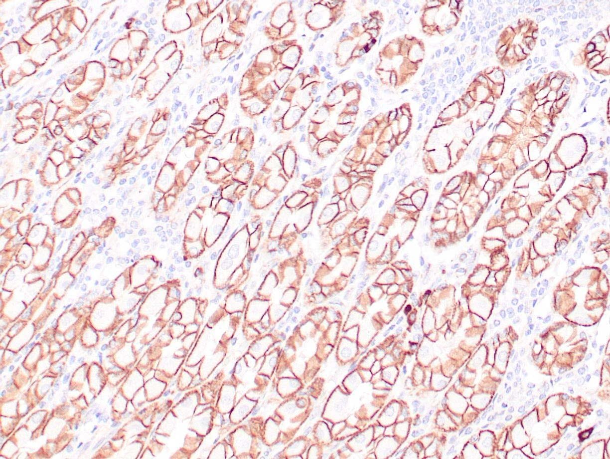 |
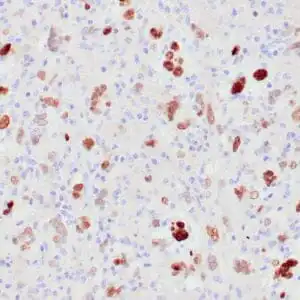
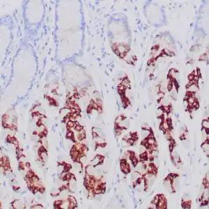
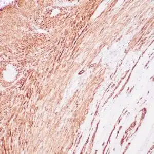
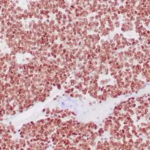
| Human colon adenocarcinoma stained with anti-beta-catenin antibody using peroxidase-conjugate and DAB chromogen. Note membranous staining of tumor cells. |
FAQ & Publications
Frequently Asked Questions
What are the recommended applications and dilutions for the mouse anti-Beta-Catenin monoclonal antibody (ZM13) 6033?
This antibody is suitable for immunohistochemistry (IHC) on formalin-fixed, paraffin-embedded tissues. The recommended dilution for the concentrated antibody is between 1:100 and 1:200.
How should the mouse anti-Beta-Catenin monoclonal antibody (ZM13) 6033 be stored to maintain stability?
For short-term storage, keep the antibody at 2-8°C. For long-term storage, it should be kept at -20°C. Avoid freeze and thaw cycles to preserve antibody integrity.
Publications
| pmid | title | authors | citation |
|---|---|---|---|
| We haven't added any publications to our database yet. | |||
Published literature highly relevant to the biological target of this product and referencing this antibody or clone are retrieved from the PubMed database provided by the United States National Library of Medicine at the National Institutes of Health.
Protocols
| relevant to this product |
|---|
| IHC |
Documents
| Batch Number | QC File | SDS |
|---|---|---|
| To view batch-specific Safety Datasheets and Quality Certificates associated with your account, please Log In. | ||
Only logged in customers who have purchased this product may leave a review.


Reviews
There are no reviews yet.