| Weight | 1 lbs |
|---|---|
| Dimensions | 9 × 5 × 2 in |
| host | rabbit |
| isotype | IgG |
| clonality | polyclonal |
| concentration | 1 mg/mL |
| applications | IP, WB |
| reactivity | human, mouse |
| available sizes | 10 µL, 100 µL |


rabbit anti-FOXO3a polyclonal antibody 1039
Price range: $100.00 through $2,600.00
Antibody summary
- Rabbit polyclonal to FOXO (Forkhead box O) Transcription Factor 3a
- Suitable for: IP,WB
- Reacts with: Hu, Ms
- Isotype: IgG
- 100 µL, 10 µL
rabbit anti- foxo3a polyclonal antibody 1039
| antibody |
|---|
| Database link: human O43524 mouse Q9WVH4 |
| Tested applications IP,WB |
| Recommended dilutions Immunoprecipitation (IP) 6 µl/mg lysate, Western Blot (WB) 1:1000 - 1:5000, IHC 1:200 - 1:1000. Epitope retrieval with Tris-EDTA pH 9.0 is recommended for FFPE tissue sections. |
| Immunogen Between 275 and 325 |
| Size and concentration 100µL and 1 mg/mL |
| Form liquid |
| Storage Instructions Store at 2-8°C. Expires 1 year from date of receipt. |
| Storage buffer Tris-citrate/phosphate buffer, pH 7 to 8 containing 0.09% Sodium Azide |
| Purity affinity purified |
| Clonality polyclonal |
| Isotype IgG |
| Compatible secondaries goat anti-rabbit IgG, H&L chain specific, peroxidase conjugated, conjugated polyclonal antibody 9512 goat anti-rabbit IgG, H&L chain specific, biotin conjugated polyclonal antibody 2079 goat anti-rabbit IgG, H&L chain specific, FITC conjugated polyclonal antibody 7863 goat anti-rabbit IgG, H&L chain specific, Cross Absorbed polyclonal antibody 2371 goat anti-rabbit IgG, H&L chain specific, biotin conjugated polyclonal antibody, crossabsorbed 1715 goat anti-rabbit IgG, H&L chain specific, FITC conjugated polyclonal antibody, crossabsorbed 1720 |
| Isotype control Rabbit monolconal - Isotype Control |
| target relevance |
|---|
| Homo sapiens FOXO3 Forkhead box protein O3 |
| Protein names Forkhead box protein O3 |
| Alternative names AF6q21 protein, Forkhead in rhabdomyosarcoma-like 1 |
| Gene names FOXO3 |
| Function Transcriptional activator that recognizes and binds to the DNA sequence 5'-[AG]TAAA[TC]A-3' and regulates different processes, such as apoptosis and autophagy (PubMed:10102273, PubMed:16751106, PubMed:21329882, PubMed:30513302). Acts as a positive regulator of autophagy in skeletal muscle: in starved cells, enters the nucleus following dephosphorylation and binds the promoters of autophagy genes, such as GABARAP1L, MAP1LC3B and ATG12, thereby activating their expression, resulting in proteolysis of skeletal muscle proteins (By similarity). Triggers apoptosis in the absence of survival factors, including neuronal cell death upon oxidative stress (PubMed:10102273, PubMed:16751106). Participates in post-transcriptional regulation of MYC: following phosphorylation by MAPKAPK5, promotes induction of miR-34b and miR-34c expression, 2 post-transcriptional regulators of MYC that bind to the 3'UTR of MYC transcript and prevent its translation (PubMed:21329882). In response to metabolic stress, translocates into the mitochondria where it promotes mtDNA transcription (PubMed:23283301). In response to metabolic stress, translocates into the mitochondria where it promotes mtDNA transcription. Also acts as a key regulator of chondrogenic commitment of skeletal progenitor cells in response to lipid availability: when lipids levels are low, translocates to the nucleus and promotes expression of SOX9, which induces chondrogenic commitment and suppresses fatty acid oxidation (By similarity). Also acts as a key regulator of regulatory T-cells (Treg) differentiation by activating expression of FOXP3 (PubMed:30513302) |
| Subcellular location Cytoplasm, cytosol, Nucleus, Mitochondrion matrix, Mitochondrion outer membrane |
| Structure Upon metabolic stress, forms a complex composed of FOXO3, SIRT3 and mitochondrial RNA polymerase POLRMT; the complex is recruited to mtDNA in a SIRT3-dependent manner (PubMed:23283301). Also forms a complex composed of FOXO3, SIRT3, TFAM and POLRMT (PubMed:29445193). Interacts with SIRT2; the interaction occurs independently of SIRT2 deacetylase activity (By similarity). Interacts with YWHAB/14-3-3-beta and YWHAZ/14-3-3-zeta, which are required for cytosolic sequestration (PubMed:16751106). Upon oxidative stress, interacts with STK4/MST1, which disrupts interaction with YWHAB/14-3-3-beta and leads to nuclear translocation (PubMed:16751106). Interacts with PIM1 (PubMed:18593906). Interacts with DDIT3/CHOP (PubMed:22761832). Interacts (deacetylated form) with SKP2 (PubMed:21841822). Interacts with CHUK and IKBKB (PubMed:15084260, PubMed:22313691). Interacts with CAMK2A, CAMK2B and calcineurin A (By similarity). Interacts with NUPR1; this interaction represses FOXO3 transactivation (PubMed:20181828). Interacts with CTDSPL2 (PubMed:28851713) |
| Post-translational modification In the presence of survival factors such as IGF1, phosphorylated on Thr-32 and Ser-253 by AKT1/PKB (PubMed:10102273). This phosphorylated form then interacts with 14-3-3 proteins and is retained in the cytoplasm (PubMed:10102273). Survival factor withdrawal induces dephosphorylation and promotes translocation to the nucleus where the dephosphorylated protein induces transcription of target genes and triggers apoptosis (PubMed:10102273). Although AKT1/PKB doesn't appear to phosphorylate Ser-315 directly, it may activate other kinases that trigger phosphorylation at this residue (PubMed:10102273, PubMed:11154281). Phosphorylated by STK4/MST1 on Ser-209 upon oxidative stress, which leads to dissociation from YWHAB/14-3-3-beta and nuclear translocation (PubMed:16751106). Phosphorylated by PIM1 (PubMed:18593906). Phosphorylation by AMPK leads to the activation of transcriptional activity without affecting subcellular localization (PubMed:17711846). In response to metabolic stress, phosphorylated by AMPK on Ser-30 which mediates FOXO3 mitochondrial translocation (PubMed:29445193). Phosphorylation by MAPKAPK5 promotes nuclear localization and DNA-binding, leading to induction of miR-34b and miR-34c expression, 2 post-transcriptional regulators of MYC that bind to the 3'UTR of MYC transcript and prevent its translation (PubMed:21329882). Phosphorylated by CHUK/IKKA and IKBKB/IKKB (PubMed:15084260). TNF-induced inactivation of FOXO3 requires its phosphorylation at Ser-644 by IKBKB/IKKB which promotes FOXO3 retention in the cytoplasm, polyubiquitination and ubiquitin-mediated proteasomal degradation (PubMed:15084260). May be dephosphorylated by calcineurin A on Ser-299 which abolishes FOXO3 transcriptional activity (By similarity). In cancer cells, ERK mediated-phosphorylation of Ser-12 is required for mitochondrial translocation of FOXO3 in response to metabolic stress or chemotherapeutic agents (PubMed:29445193). Phosphorylation at Ser-253 promotes its degradation by the proteasome (PubMed:30513302). Dephosphorylation at Ser-253 by protein phosphatase 2A (PPP2CA) promotes its stabilization; interaction with PPP2CA is enhanced by AMBRA1 (PubMed:30513302). Dephosphorylated at Ser-253 by CTDSPL2 (PubMed:28851713) Deacetylation by SIRT1 or SIRT2 stimulates interaction of FOXO3 with SKP2 and facilitates SCF(SKP2)-mediated FOXO3 ubiquitination and proteasomal degradation (PubMed:21841822). Deacetylation by SIRT2 stimulates FOXO3-mediated transcriptional activity in response to oxidative stress (By similarity). Deacetylated by SIRT3 (PubMed:23283301). Deacetylation by SIRT3 stimulates FOXO3-mediated mtDNA transcriptional activity in response to metabolic stress (PubMed:23283301) Heavily methylated by SET9 which decreases stability, while moderately increasing transcriptional activity. The main methylation site is Lys-271. Methylation doesn't affect subcellular location Polyubiquitinated. Ubiquitinated by a SCF complex containing SKP2, leading to proteasomal degradation The N-terminus is cleaved following import into the mitochondrion |
| Keywords 3D-structure, Acetylation, Activator, Alternative splicing, Apoptosis, Chromosomal rearrangement, Cytoplasm, DNA-binding, Membrane, Methylation, Mitochondrion, Mitochondrion outer membrane, Nucleus, Phosphoprotein, Proteomics identification, Proto-oncogene, Reference proteome, Transcription, Transcription regulation, Ubl conjugation |
| Sequence MAEAPASPAPLSPLEVELDPEFEPQSRPRSCTWPLQRPELQASPAKPSGETAADSMIPEE EDDEDDEDGGGRAGSAMAIGGGGGSGTLGSGLLLEDSARVLAPGGQDPGSGPATAAGGLS GGTQALLQPQQPLPPPQPGAAGGSGQPRKCSSRRNAWGNLSYADLITRAIESSPDKRLTL SQIYEWMVRCVPYFKDKGDSNSSAGWKNSIRHNLSLHSRFMRVQNEGTGKSSWWIINPDG GKSGKAPRRRAVSMDNSNKYTKSRGRAAKKKAALQTAPESADDSPSQLSKWPGSPTSRSS DELDAWTDFRSRTNSNASTVSGRLSPIMASTELDEVQDDDAPLSPMLYSSSASLSPSVSK PCTVELPRLTDMAGTMNLNDGLTENLMDDLLDNITLPPSQPSPTGGLMQRSSSFPYTTKG SGLGSPTSSFNSTVFGPSSLNSLRQSPMQTIQENKPATFSSMSHYGNQTLQDLLTSDSLS HSDVMMTQSDPLMSQASTAVSAQNSRRNVMLRNDPMMSFAAQPNQGSLVNQNLLHHQHQT QGALGGSRALSNSVSNMGLSESSSLGSAKHQQQSPVSQSMQTLSDSLSGSSLYSTSANLP VMGHEKFPSDLDLDMFNGSLECDMESIIRSELMDADGLDFNFDSLISTQNVVGLNVGNFT GAKQASSQSWVPG |
| UniProt accession: O43524 |
Data
 |
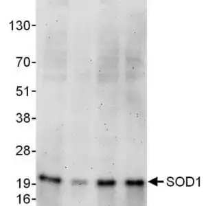
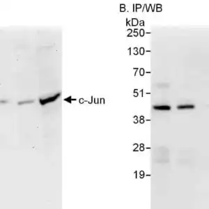
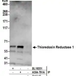
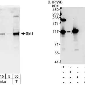
| Detection of human FOXO3a by western blot of immunoprecipitates. Samples: Whole cell lysate (1 mg for IP; 20% of IP loaded) from MCF-7 cells. Antibodies: Affinity purified rabbit anti-FOXO3a antibody (#1039) used for IP at 6 µg/mg lysate. FOXO3a was also immunoprecipitated by rabbit anti-FOXO3a antibody. For blotting immunoprecipitated FOXO3a was used at 1 µg/ml. Detection: Chemiluminescence with an exposure time of 3 minutes. |
 |
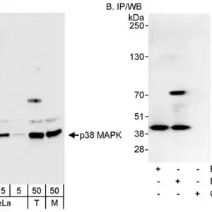
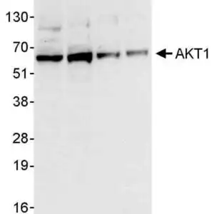
| Detection of human and mouse FOXO3a by western blot. Samples: Whole cell lysate (50 µg) from HEK293T, Jurkat, and mouse TCMK-1 cells. Antibodies: Affinity purified rabbit anti-FOXO3a antibody (#1039) used for WB at 0.4 µg/ml. Detection: Chemiluminescence with an exposure time of 3 minutes. |
 |
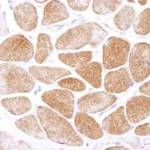
| Detection of human and mouse FOXO3a by immunohistochemistry. Sample: FFPE sections of human ovarian carcinoma (left) and mouse renal cell carcinoma (right). Antibody: Affinity purified rabbit anti-FOXO3a (#1039) used at a dilution of 1:500 (2µg/ml). Detection: DAB |
FAQ & Publications
Frequently Asked Questions
What species does the rabbit anti-FOXO3a polyclonal antibody 1039 react with?
The antibody is reactive with human and mouse FOXO3a proteins.
What are the recommended applications and dilutions for using this antibody in immunoprecipitation and western blot?
For immunoprecipitation (IP), the recommended dilution is 6 µL of antibody per mg of lysate. For western blot (WB), use a dilution range of 1:1000 to 1:5000. Additionally, for immunohistochemistry (IHC), a dilution of 1:200 to 1:1000 is suggested, with epitope retrieval using Tris-EDTA pH 9.0 recommended for FFPE tissue sections.
Publications
| pmid | title | authors | citation |
|---|---|---|---|
| We haven't added any publications to our database yet. | |||
Published literature highly relevant to the biological target of this product and referencing this antibody or clone are retrieved from the PubMed database provided by the United States National Library of Medicine at the National Institutes of Health.
Protocols
| relevant to this product |
|---|
| Western blot |
Documents
| Batch Number | QC File | SDS |
|---|---|---|
| To view batch-specific Safety Datasheets and Quality Certificates associated with your account, please Log In. | ||
Only logged in customers who have purchased this product may leave a review.


Reviews
There are no reviews yet.