| Weight | 1 lbs |
|---|---|
| Dimensions | 9 × 5 × 2 in |
| host | rabbit |
| isotype | IgG |
| clonality | polyclonal |
| concentration | 1 mg/mL |
| applications | ICC/IF, WB |
| reactivity | RIP3 |
| available sizes | 100 µg |


rabbit anti-RIP3 polyclonal antibody 3801
$445.00
Antibody summary
- Rabbit polyclonal to RIP3
- Suitable for: ELISA,IF,IHC-P,WB,IP
- Isotype: IgG
- 100 µg
rabbit anti-RIP3 polyclonal antibody 3801
| antibody |
|---|
| Tested applications IHC,IHC,ICC/IF,ELISA |
| Recommended dilutions Immunoblotting: use at 1ug/mL. Positive control: NIH/3T3 cell lysate. Immunohistochemistry: use at 5ug/mL. These are recommended concentrations. Enduser should determine optimal concentrations for their applications. |
| Immunogen Synthetic peptide corresponding to aa 473-486 of mouse RIP3 (accession no. AAF03133). |
| Size and concentration 100µg and lot specific |
| Form liquid |
| Storage Instructions This antibody is stable for at least one (1) year at -20°C. Avoid multiple freeze-thaw cycles. |
| Storage buffer PBS, pH 7.4. |
| Purity peptide affinity purification |
| Clonality polyclonal |
| Isotype IgG |
| Compatible secondaries goat anti-rabbit IgG, H&L chain specific, peroxidase conjugated, conjugated polyclonal antibody 9512 goat anti-rabbit IgG, H&L chain specific, biotin conjugated polyclonal antibody 2079 goat anti-rabbit IgG, H&L chain specific, FITC conjugated polyclonal antibody 7863 goat anti-rabbit IgG, H&L chain specific, Cross Absorbed polyclonal antibody 2371 goat anti-rabbit IgG, H&L chain specific, biotin conjugated polyclonal antibody, crossabsorbed 1715 goat anti-rabbit IgG, H&L chain specific, FITC conjugated polyclonal antibody, crossabsorbed 1720 |
| Isotype control Rabbit polyclonal - Isotype Control |
| target relevance |
|---|
| Mus musculus RIPK3 Receptor-interacting serine/threonine-protein kinase 3 |
| Protein names Receptor-interacting serine/threonine-protein kinase 3 |
| Alternative names RIP-like protein kinase 3, Receptor-interacting protein 3 |
| Gene names Ripk3 |
| Protein family Belongs to the protein kinase superfamily. TKL Ser/Thr protein kinase family |
| Function Serine/threonine-protein kinase that activates necroptosis and apoptosis, two parallel forms of cell death (PubMed:27321907, PubMed:27746097, PubMed:27917412, PubMed:28607035, PubMed:32200799, PubMed:32296175). Necroptosis, a programmed cell death process in response to death-inducing TNF family members, is triggered by RIPK3 following activation by ZBP1 (PubMed:19590578, PubMed:22423968, PubMed:24012422, PubMed:24019532, PubMed:24095729, PubMed:24557836, PubMed:27321907, PubMed:27746097, PubMed:27819681, PubMed:27819682, PubMed:32200799, PubMed:32296175). Activated RIPK3 forms a necrosis-inducing complex and mediates phosphorylation of MLKL, promoting MLKL localization to the plasma membrane and execution of programmed necrosis characterized by calcium influx and plasma membrane damage (PubMed:24813849, PubMed:24813850, PubMed:27321907). In addition to TNF-induced necroptosis, necroptosis can also take place in the nucleus in response to orthomyxoviruses infection: following ZBP1 activation, which senses double-stranded Z-RNA structures, nuclear RIPK3 catalyzes phosphorylation and activation of MLKL, promoting disruption of the nuclear envelope and leakage of cellular DNA into the cytosol (PubMed:32200799, PubMed:32296175). Also regulates apoptosis: apoptosis depends on RIPK1, FADD and CASP8, and is independent of MLKL and RIPK3 kinase activity (PubMed:27321907). Phosphorylates RIPK1: RIPK1 and RIPK3 undergo reciprocal auto- and trans-phosphorylation (By similarity). In some cell types, also able to restrict viral replication by promoting cell death-independent responses (PubMed:30635240). In response to flavivirus infection in neurons, promotes a cell death-independent pathway that restricts viral replication: together with ZBP1, promotes a death-independent transcriptional program that modifies the cellular metabolism via up-regulation expression of the enzyme ACOD1/IRG1 and production of the metabolite itaconate (PubMed:30635240). Itaconate inhibits the activity of succinate dehydrogenase, generating a metabolic state in neurons that suppresses replication of viral genomes (PubMed:30635240). RIPK3 binds to and enhances the activity of three metabolic enzymes: GLUL, GLUD1, and PYGL (By similarity). These metabolic enzymes may eventually stimulate the tricarboxylic acid cycle and oxidative phosphorylation, which could result in enhanced ROS production (By similarity) |
| Catalytic activity L-seryl-[protein] + ATP = O-phospho-L-seryl-[protein] + ADP + H(+) L-threonyl-[protein] + ATP = O-phospho-L-threonyl-[protein] + ADP + H(+) |
| Subcellular location Cytoplasm, cytosol, Nucleus |
| Structure (Microbial infection) Interacts (via RIP homotypic interaction motif) with murid herpesvirus protein RIR1; this interaction disrupts RIP3-RIP1 interactions characteristic of TNF induced necroptosis, thereby suppressing this death pathway |
| Post-translational modification RIPK1 and RIPK3 undergo reciprocal auto- and trans-phosphorylation (By similarity). Autophosphorylated following interaction with ZBP1 (PubMed:27819681). Phosphorylation of Ser-204 plays a role in the necroptotic function of RIPK3 (By similarity). Autophosphorylates at Thr-231 and Ser-232 following activation by ZBP1: phosphorylation at these sites is a hallmark of necroptosis and is required for binding MLKL (PubMed:23612963, PubMed:27819682). Phosphorylation at Thr-187 is important for its kinase activity, interaction with PELI1 and for its ability to mediate TNF-induced necroptosis (By similarity) Polyubiquitinated with 'Lys-48' and 'Lys-63'-linked chains by BIRC2/c-IAP1 and BIRC3/c-IAP2, leading to activation of NF-kappa-B. Ubiquitinated by STUB1 leading to its subsequent proteasome-dependent degradation |
| Keywords 3D-structure, Apoptosis, ATP-binding, Cytoplasm, Host-virus interaction, Kinase, Methylation, Necrosis, Nucleotide-binding, Nucleus, Phosphoprotein, Reference proteome, Serine/threonine-protein kinase, Transferase, Ubl conjugation |
| Sequence MSSVKLWPTGASAVPLVSREELKKLEFVGKGGFGVVFRAHHRTWNHDVAVKIVNSKKISW EVKAMVNLRNENVLLLLGVTEDLQWDFVSGQALVTRFMENGSLAGLLQPECPRPWPLLCR LLQEVVLGMCYLHSLNPPLLHRDLKPSNILLDPELHAKLADFGLSTFQGGSQSGSGSGSG SRDSGGTLAYLDPELLFDVNLKASKASDVYSFGILVWAVLAGREAELVDKTSLIRETVCD RQSRPPLTELPPGSPETPGLEKLKELMIHCWGSQSENRPSFQDCEPKTNEVYNLVKDKVD AAVSEVKHYLSQHRSSGRNLSAREPSQRGTEMDCPRETMVSKMLDRLHLEEPSGPVPGKC PERQAQDTSVGPATPARTSSDPVAGTPQIPHTLPFRGTTPGPVFTETPGPHPQRNQGDGR HGTPWYPWTPPNPMTGPPALVFNNCSEVQIGNYNSLVAPPRTTASSSAKYDQAQFGRGRG WQPFHK |
| UniProt accession: Q9QZL0 |
Data
 |
| Western Blot Validation in Human Cell Lines Loading: 15 µg of lysates per lane. Antibodies: RIP3 3801, (0.5 µg/mL), 1h incubation at RT in 5% NFDM/TBST.Secondary: Goat anti-rabbit IgG HRP conjugate at 1:10000 dilution. |
 |
| Western Blot Validation in C2C12 Cells Mouse Loading: 15 µg of lysates per lane. Antibodies: RIP3 3801, 1h incubation at RT in 5% NFDM/TBST.Secondary: Goat anti-rabbit IgG HRP conjugate at 1:10000 dilution.Lane 1: 3801, 0.1 µg/mL in the presence of peptide blockingLane 2: 3801, 0.1 µg/mLLane 3: 3801, 0.2 µg/mLLane 4: 3801, 0.5 µg/mL |
 |
| Western Blot Validation in Mouse Cell lines Loading: 15 µg of lysates per lane. Antibodies: RIP3 3801, (0.5 µg/mL), 1h incubation at RT in 5% NFDM/TBST.Secondary: Goat anti-rabbit IgG HRP conjugate at 1:10000 dilution. |
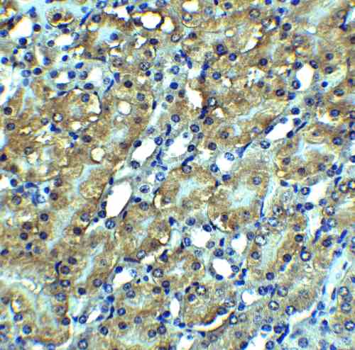 |
| Immunohistochemistry Validation of RIP3 in Mouse Kidney Tissue Immunohistochemical analysis of paraffin-embedded mouse kidney tissue using anti-RIP3 antibody (3801) at 2.5 µg/mL. Tissue was fixed with formaldehyde and blocked with 10% serum for 1 h at RT; antigen retrieval was by heat mediation with a citrate buffer (pH6). Samples were incubated with primary antibody overnight at 4C. A goat anti-rabbit IgG H&L (HRP) at 1/250 was used as secondary. Counter stained with Hematoxylin. |
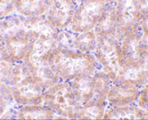 |
| Immunohistochemistry Validation of RIP3 in Rat Kidney Tissue Immunohistochemical analysis of paraffin-embedded rat kidney tissue using anti-RIP3 antibody (3801) at 5 µg/mL. Tissue was fixed with formaldehyde and blocked with 10% serum for 1 h at RT; antigen retrieval was by heat mediation with a citrate buffer (pH6). Samples were incubated with primary antibody overnight at 4C. A goat anti-rabbit IgG H&L (HRP) at 1/250 was used as secondary. Counter stained with Hematoxylin. |
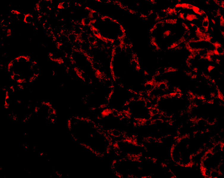 |
| Immunofluorescence Validation of RIP3 in Rat Kidney Tissue Immunofluorescent analysis of 4% paraformaldehyde-fixed Rat Kidney tissue labeling RIP3 with 3801 at 20 µg/mL, followed by goat anti-rabbit IgG secondary antibody at 1/500 dilution (red). |
 |
| KO Validation of RIP3 in RIP3 KO Mice (He et al., 2009) Western blot analysis of RIP3 with anti-RIP3 antibodies shows disrupted RIP3 expression in pancreas, liver spleen thymus and lung of RIP3 KO mice. Cerulein treatment upregulated RIP3 expression in the pancreas of WT mice. |
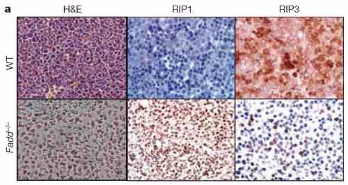 |
| Immunohistochemistry Validation of RIP3 in Fadd KO mice (Zhang et al., 2011) RIP3 expression detected by anti-RIP3 antibodies (3801) was decreased in Fadd KO mice as compared to WT mice. |
 |
| KO Validation of RIP3 in MEF Cells (He et al., 2012) Western blot analysis of RIP3 with anti-RIP3 antibodies shows disrupted RIP3 expression in RIP3 KO MEFs, but not in WT MEF cells. |
FAQ & Publications
Frequently Asked Questions
What applications is the rabbit anti-RIP3 polyclonal antibody 3801 validated for?
This antibody is suitable for ELISA, immunofluorescence (IF), immunohistochemistry on paraffin-embedded tissue (IHC-P), Western blotting (WB), and immunoprecipitation (IP). Recommended dilutions are 1 µg/mL for immunoblotting and 5 µg/mL for immunohistochemistry, though optimal concentrations should be determined by the end user.
How should the rabbit anti-RIP3 antibody 3801 be stored to maintain stability?
The antibody should be stored at -20°C and is stable for at least one year under these conditions. It is important to avoid multiple freeze-thaw cycles to preserve antibody integrity. The storage buffer is PBS at pH 7.4.
What is the immunogen used to generate the rabbit anti-RIP3 polyclonal antibody 3801?
The immunogen is a synthetic peptide corresponding to amino acids 473-486 of mouse RIP3, based on accession number AAF03133.
Publications
| pmid | title | authors | citation |
|---|---|---|---|
| We haven't added any publications to our database yet. | |||
Published literature highly relevant to the biological target of this product and referencing this antibody or clone are retrieved from the PubMed database provided by the United States National Library of Medicine at the National Institutes of Health.
Protocols
| relevant to this product |
|---|
| Western blot IHC ICC |
Documents
| Batch Number | QC File | SDS |
|---|---|---|
| To view batch-specific Safety Datasheets and Quality Certificates associated with your account, please Log In. | ||
Only logged in customers who have purchased this product may leave a review.


Reviews
There are no reviews yet.