| Weight | 1 lbs |
|---|---|
| Dimensions | 9 × 5 × 2 in |
| host | rabbit |
| isotype | IgG |
| clonality | polyclonal |
| concentration | 1 mg/mL |
| applications | ICC/IF, WB |
| reactivity | PUMA (NT) |
| available sizes | 100 µg |
rabbit anti-PUMA (NT) polyclonal antibody 9434
$445.00
Antibody summary
- Rabbit polyclonal to PUMA (NT)
- Suitable for: ELISA,WB,IHC-P,IF
- Isotype: IgG
- 100 µg
rabbit anti-PUMA (NT) polyclonal antibody 9434
| antibody |
|---|
| Tested applications WB,IHC,IHC,ICC/IF,ELISA |
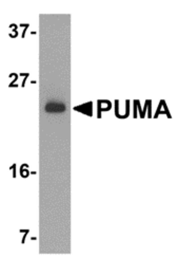
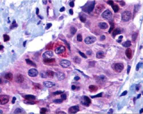
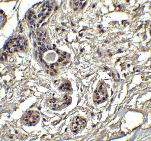
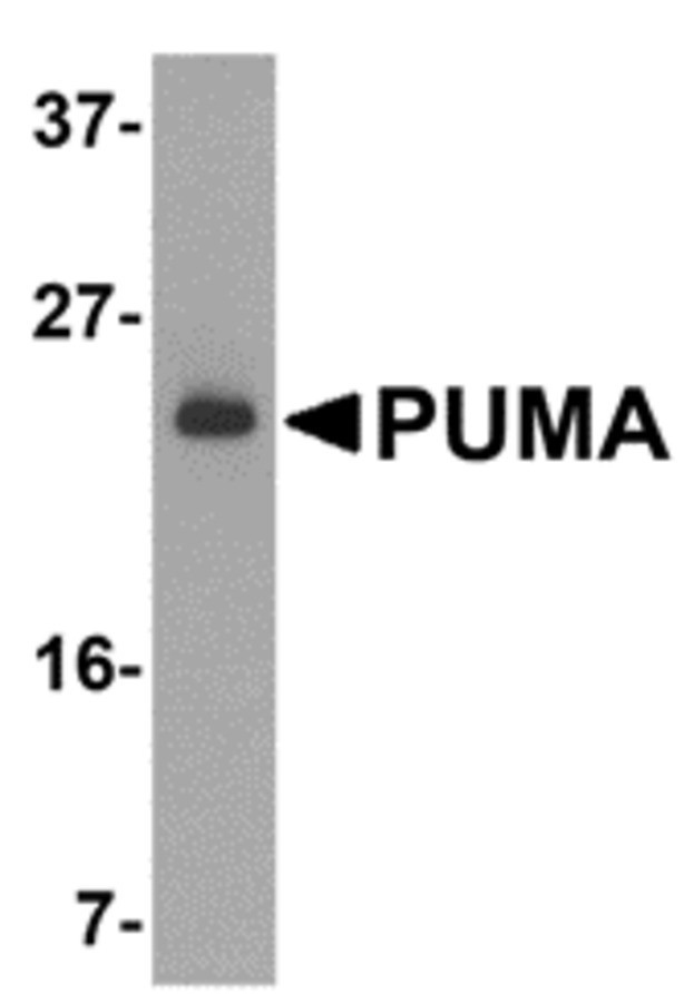
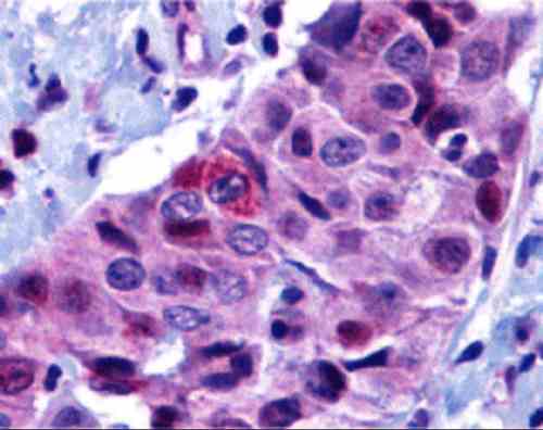
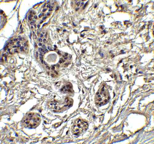
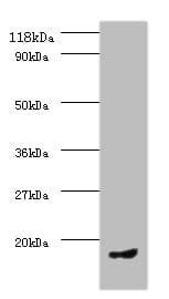
| Recommended dilutions Immunoblotting: use at 2ug/mL. Positive control: K562 cell lysate. Immunohistochemistry: use at 10ug/mL. These are recommended concentrations. Enduser should determine optimal concentrations for their applications. |
| Immunogen Synthetic peptide corresponding to aa 2-16 of human PUMA-a (accession no. NP_055232).. This sequence is identical in mouse PUMA-a. |
| Size and concentration 100µg and lot specific |
| Form liquid |
| Storage Instructions This antibody is stable for at least one (1) year at -20°C. Avoid multiple freeze-thaw cycles. |
| Storage buffer PBS, pH 7.4. |
| Purity peptide affinity purification |
| Clonality polyclonal |
| Isotype IgG |
| Compatible secondaries goat anti-rabbit IgG, H&L chain specific, peroxidase conjugated, conjugated polyclonal antibody 9512 goat anti-rabbit IgG, H&L chain specific, biotin conjugated polyclonal antibody 2079 goat anti-rabbit IgG, H&L chain specific, FITC conjugated polyclonal antibody 7863 goat anti-rabbit IgG, H&L chain specific, Cross Absorbed polyclonal antibody 2371 goat anti-rabbit IgG, H&L chain specific, biotin conjugated polyclonal antibody, crossabsorbed 1715 goat anti-rabbit IgG, H&L chain specific, FITC conjugated polyclonal antibody, crossabsorbed 1720 |
| Isotype control Rabbit polyclonal - Isotype Control |
| target relevance |
|---|
| Protein names Bcl-2-binding component 3, isoforms 1/2 (JFY-1) (p53 up-regulated modulator of apoptosis) |
| Gene names BBC3,BBC3 PUMA |
| Protein family Bcl-2 family |
| Mass 20532Da |
| Function FUNCTION: Essential mediator of p53/TP53-dependent and p53/TP53-independent apoptosis (PubMed:11463391, PubMed:23340338). Promotes partial unfolding of BCL2L1 and dissociation of BCL2L1 from p53/TP53, releasing the bound p53/TP53 to induce apoptosis (PubMed:23340338). Regulates ER stress-induced neuronal apoptosis (By similarity). {ECO:0000250|UniProtKB:Q99ML1, ECO:0000269|PubMed:11463391, ECO:0000269|PubMed:23340338}. |
| Subellular location SUBCELLULAR LOCATION: Mitochondrion {ECO:0000269|PubMed:11463391, ECO:0000269|PubMed:11463392, ECO:0000269|PubMed:11572983}. Note=Localized to the mitochondria in order to induce cytochrome c release. |
| Tissues TISSUE SPECIFICITY: Ubiquitously expressed. {ECO:0000269|PubMed:11463391}. |
| Structure SUBUNIT: Interacts with MCL1 and BCL2A1 (By similarity). Interacts (via BH3 domain) with BCL2 (PubMed:11463391). Interacts with BCL2L1/BCL-XL (PubMed:23340338). Interacts (via BH3 domain) with NOL3/ARC (via CARD domain); this interaction prevents BBC3 association with BCL2 and results in CASP8 activation (By similarity). {ECO:0000250|UniProtKB:Q80ZG6, ECO:0000250|UniProtKB:Q99ML1, ECO:0000269|PubMed:11463391, ECO:0000269|PubMed:23340338}. |
| Domain DOMAIN: The BH3 motif is intrinsically disordered in the absence of a binding partner but folds upon binding (PubMed:23340338). Folds when bound to BCL2L1 (PubMed:23340338). Also folds when bound to MCL1 (By similarity). {ECO:0000250|UniProtKB:Q99ML1, ECO:0000269|PubMed:23340338}. |
| Target Relevance information above includes information from UniProt accession: Q9BXH1 |
| The UniProt Consortium |
Data
Publications
| pmid | title | authors | citation |
|---|---|---|---|
| We haven't added any publications to our database yet. | |||
Protocols
| relevant to this product |
|---|
| Western blot IHC ICC |
Documents
| # | SDS | Certificate | |
|---|---|---|---|
| Please enter your product and batch number here to retrieve product datasheet, SDS, and QC information. | |||
Only logged in customers who have purchased this product may leave a review.






Reviews
There are no reviews yet.