| Weight | 1 lbs |
|---|---|
| Dimensions | 9 × 5 × 2 in |
| host | rabbit |
| isotype | IgG |
| clonality | polyclonal |
| concentration | 1 mg/mL |
| applications | ICC/IF, WB |
| reactivity | PUMA (CT) |
| available sizes | 100 µg |


rabbit anti-PUMA (CT) polyclonal antibody 7882
$445.00
Antibody summary
- Rabbit polyclonal to PUMA (CT)
- Suitable for: ELISA,WB,ICC,IF
- Isotype: IgG
- 100 µg
rabbit anti-PUMA (CT) polyclonal antibody 7882
| antibody |
|---|
| Tested applications WB,ICC/IF,ELISA |
| Recommended dilutions Immunoblotting: use at 2ug/mL. Positive control: K562 cell lysate. Immunocytochemistry: use at 1ug/mL. These are recommended concentrations. Enduser should determine optimal concentrations for their applications. |
| Immunogen Synthetic peptide corresponding to aa 180-193 of human PUMA-a (accession no NP_055232). This sequence is identical in human PUMA-a and PUMA-b. |
| Size and concentration 100µg and lot specific |
| Form liquid |
| Storage Instructions This antibody is stable for at least one (1) year at -20°C. Avoid multiple freeze-thaw cycles. |
| Storage buffer PBS, pH 7.4. |
| Purity peptide affinity purification |
| Clonality polyclonal |
| Isotype IgG |
| Compatible secondaries goat anti-rabbit IgG, H&L chain specific, peroxidase conjugated, conjugated polyclonal antibody 9512 goat anti-rabbit IgG, H&L chain specific, biotin conjugated polyclonal antibody 2079 goat anti-rabbit IgG, H&L chain specific, FITC conjugated polyclonal antibody 7863 goat anti-rabbit IgG, H&L chain specific, Cross Absorbed polyclonal antibody 2371 goat anti-rabbit IgG, H&L chain specific, biotin conjugated polyclonal antibody, crossabsorbed 1715 goat anti-rabbit IgG, H&L chain specific, FITC conjugated polyclonal antibody, crossabsorbed 1720 |
| Isotype control Rabbit polyclonal - Isotype Control |
| target relevance |
|---|
| Homo sapiens BBC3 Bcl-2-binding component 3, isoforms 1/2 |
| Protein names Bcl-2-binding component 3, isoforms 1/2 |
| Alternative names JFY-1, p53 up-regulated modulator of apoptosis |
| Gene names BBC3 |
| Protein family Belongs to the Bcl-2 family |
| Function Essential mediator of p53/TP53-dependent and p53/TP53-independent apoptosis (PubMed:11463391, PubMed:23340338). Promotes partial unfolding of BCL2L1 and dissociation of BCL2L1 from p53/TP53, releasing the bound p53/TP53 to induce apoptosis (PubMed:23340338). Regulates ER stress-induced neuronal apoptosis (By similarity) |
| Subcellular location Mitochondrion |
| Structure Interacts with MCL1 and BCL2A1 (By similarity). Interacts (via BH3 domain) with BCL2 (PubMed:11463391). Interacts with BCL2L1/BCL-XL (PubMed:23340338). Interacts (via BH3 domain) with NOL3/ARC (via CARD domain); this interaction prevents BBC3 association with BCL2 and results in CASP8 activation (By similarity) |
| Keywords 3D-structure, Alternative splicing, Apoptosis, Mitochondrion, Phosphoprotein, Proteomics identification, Reference proteome |
| Sequence MARARQEGSSPEPVEGLARDGPRPFPLGRLVPSAVSCGLCEPGLAAAPAAPTLLPAAYLC APTAPPAVTAALGGSRWPGGPRSRPRGPRPDGPQPSLSLAEQHLESPVPSAPGALAGGPT QAAPGVRGEEEQWAREIGAQLRRMADDLNAQYERRRQEEQQRHRPSPWRVLYNLIMGLLP LPRGHRAPEMEPN |
| UniProt accession: Q9BXH1 |
Data
 |
| Western Blot Validation of PUMA in K562 and 3T3/NIH Cells Loading: 15 µg of lysates per lane. Antibodies: 7882 (2 µg/mL), 1 h incubation at RT in 5% NFDM/TBST.Secondary: Goat anti-rabbit IgG HRP conjugate at 1:10000 dilution. |
 |
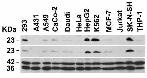
| Independent Antibody Validation (IAV) via Protein Expression Profile in Cell Lines Loading: 20 µg of lysates per lane. Antibodies: 7882 (3 µg/mL), 11008 (2 µg/mL), beta-actin (1 µg/mL) and GAPDH (0.02 µg/mL), 1 h incubation at RT in 5% NFDM/TBST.Secondary: Goat anti-rabbit IgG HRP conjugate at 1:10000 dilution. |
 |
| Validation with PUMA siRNA Knockdown in 293 Cells 293 cells were transfected with control siRNAs (lane 1) or PUMA siRNAs (lane 2) Loading: 15 µg of 293 whole cell lysates per lane. Antibodies: 7882 (2 µg/mL), beta-actin (1 µg/mL) and GAPDH (0.02 µg/mL), 1 h incubation at RT in 5% NFDM/TBST.Secondary: Goat anti-rabbit IgG HRP conjugate at 1:10000 dilution. |
 |
| Sensitivity Test for PUMA in 2983 Cells Loading: Lysates/proteins at 15 µg per lane. Antibodies: 7882 (lane 1-3: 1, 2 and 4 µg/mL). 1 h incubation at RT in 5% NFDM/TBST.Secondary: Goat anti-rabbit IgG HRP conjugate at 1:10000 dilution. |
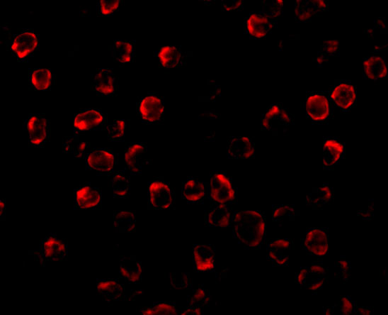 |
| Immunofluorescence Validation of PUMA in K562 Cells Immunofluorescent analysis of 4% paraformaldehyde-fixed K562 cells labeling PUMA with 7882 at 2 µg/mL, followed by goat anti-rabbit IgG secondary antibody at 1/500 dilution (red). Image showing cytosol staining on K562 cells. |
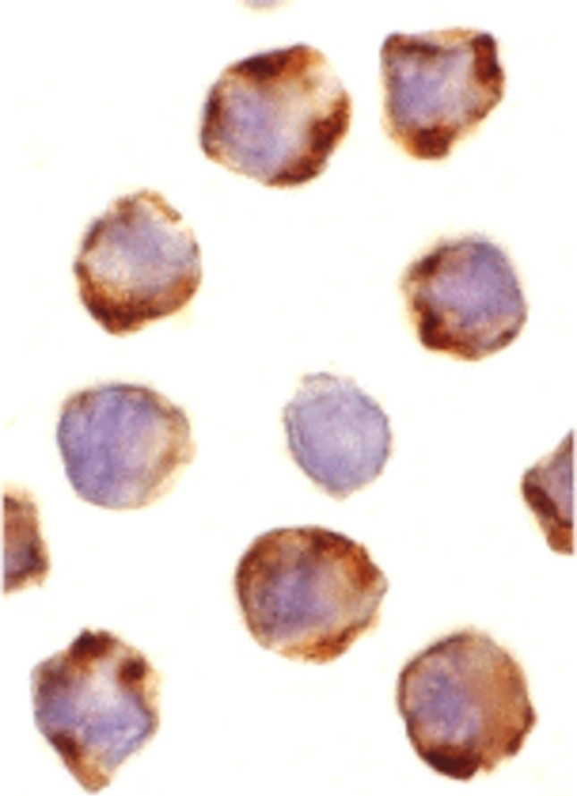 |
| Immunocytochemistry Validation of PUMA in K562 Cells Immunocytochemical analysis of K562 cells using anti-PUMA antibody (7882) at 1 µg/mL. Cells was fixed with formaldehyde and blocked with 10% serum for 1 h at RT; antigen retrieval was by heat mediation with a citrate buffer (pH6). Samples were incubated with primary antibody overnight at 4C. A goat anti-rabbit IgG H&L (HRP) at 1/250 was used as secondary. Counter stained with Hematoxylin. |
 |
| KD Validation of PUMA in HOSE-RasV12 Cells (Elgendy et al., 2011) HOSE-RasV12 cells were transfected with control shRNA plasmid or shRNA plasmids (KD) targeted against Noxa or Puma, as indicated. PUMA expression was not observed in PUMA KD cells detected by anti-PUMA antibodies (7882). |
 |
| Induction Validation of PUMA in Primary Cortical Neurons (Sabirzhanov et al., 2014) PUMA protein levels were increased in etoposide-treated primary cortical neurons detected by anti-PUMA antibodies (7882). |
 |
| KD Validation of PUMA PUMA in Tet Cells (Han et al., 2010) Immunoblot analyses of Tet-induced p53 cells treated with NOXA, Puma, Bim or non-targeting siRNAs that were utilized in this experiment. PUMA protein levels were markedly reduced in PUMA KD cells detected by anti-PUMA antibodies (7882). |
FAQ & Publications
Frequently Asked Questions
What applications has the rabbit anti-PUMA (CT) polyclonal antibody (SKU 7882) been validated for?
This antibody has been tested and validated for use in Western Blot (WB), Immunocytochemistry/Immunofluorescence (ICC/IF), and ELISA applications.
How should the rabbit anti-PUMA (CT) polyclonal antibody be stored to maintain stability?
The antibody is stable for at least one year when stored at -20°C. It is recommended to avoid multiple freeze-thaw cycles to preserve antibody integrity.
What is the immunogen used to generate the rabbit anti-PUMA (CT) polyclonal antibody (7882)?
The immunogen is a synthetic peptide corresponding to amino acids 180-193 of human PUMA-a (accession no NP_055232), which is identical in both human PUMA-a and PUMA-b isoforms.
Publications
| pmid | title | authors | citation |
|---|---|---|---|
| We haven't added any publications to our database yet. | |||
Published literature highly relevant to the biological target of this product and referencing this antibody or clone are retrieved from the PubMed database provided by the United States National Library of Medicine at the National Institutes of Health.
Protocols
| relevant to this product |
|---|
| Western blot IHC ICC |
Documents
| Batch Number | QC File | SDS |
|---|---|---|
| To view batch-specific Safety Datasheets and Quality Certificates associated with your account, please Log In. | ||
Only logged in customers who have purchased this product may leave a review.


Reviews
There are no reviews yet.