| Weight | 1 lbs |
|---|---|
| Dimensions | 9 × 5 × 2 in |
| host | rabbit |
| isotype | IgG |
| clonality | polyclonal |
| concentration | 1 mg/mL |
| applications | ICC/IF, WB |
| reactivity | PERP |
| available sizes | 100 µg |


rabbit anti-PERP polyclonal antibody 9812
$445.00
Antibody summary
- Rabbit polyclonal to PERP
- Suitable for: ELISA,WB,ICC
- Isotype: Whole IgG
- 100 µg
rabbit anti-PERP polyclonal antibody 9812
| antibody |
|---|
| Tested applications WB,ICC/IF,ELISA |
| Recommended dilutions Immunoblotting: use at 0.5-1 ug/mL. In immunoblots, a band of 21 kD is detected. Positive control: A431 cell lysate. |
| Immunogen Peptide corresponding to aa 175-193 of human PERP. This sequence differs from that of mouse by three amino acids. |
| Size and concentration 100µg and lot specific |
| Form liquid |
| Storage Instructions This antibody is stable for at least one (1) year at -20°C. Avoid multiple freeze- thaw cycles. |
| Storage buffer PBS, pH 7.4. |
| Purity peptide affinity purification |
| Clonality polyclonal |
| Isotype IgG |
| Compatible secondaries goat anti-rabbit IgG, H&L chain specific, peroxidase conjugated, conjugated polyclonal antibody 9512 goat anti-rabbit IgG, H&L chain specific, biotin conjugated polyclonal antibody 2079 goat anti-rabbit IgG, H&L chain specific, FITC conjugated polyclonal antibody 7863 goat anti-rabbit IgG, H&L chain specific, Cross Absorbed polyclonal antibody 2371 goat anti-rabbit IgG, H&L chain specific, biotin conjugated polyclonal antibody, crossabsorbed 1715 goat anti-rabbit IgG, H&L chain specific, FITC conjugated polyclonal antibody, crossabsorbed 1720 |
| Isotype control Rabbit polyclonal - Isotype Control |
| target relevance |
|---|
| Homo sapiens PERP p53 apoptosis effector related to PMP-22 |
| Protein names p53 apoptosis effector related to PMP-22 |
| Alternative names Keratinocyte-associated protein 1, P53-induced protein PIGPC1, Transmembrane protein THW |
| Gene names PERP |
| Protein family Belongs to the TMEM47 family |
| Function Component of intercellular desmosome junctions (By similarity). Plays a role in stratified epithelial integrity and cell-cell adhesion by promoting desmosome assembly (By similarity). Thereby plays a role in barrier function of the skin against infection (By similarity). Plays a role in mammary epithelial tissue homeostasis and remodeling during and after pregnancy, potentially via its involvement in desmosome cell-cell junctions (By similarity). Required for tooth enamel development via facilitating desmosome-mediated ameloblast adhesion to the stratum intermedium during the transitional stage of amelogenesis (By similarity). May also play a role in downstream transcriptional regulation of other genes involved in amelogenesis such as AMBN, ENAM, MMP20 and KLK4 (By similarity). Plays a role as an effector in the TP53-dependent apoptotic pathway (By similarity). Positively regulates apoptosis in T-helper 17 (Th17) cell populations via caspase-dependent signaling (By similarity). Promotes neutrophil transepithelial migration in response to chemoattractants such as hepoxilin A3 (HXA3), N-Formylmethionyl-leucyl-phenylalanine (fMLP) and CXCL8/IL-8 (PubMed:25486861). Required for neutrophil transepithelial migration in response to S.typhimurium infection (PubMed:25486861). May act as a positive regulator of endothelial cell apoptosis in response to blood flow-derived shear stress (By similarity) |
| Subcellular location Cell junction, desmosome, Cell membrane, Cytoplasm |
| Structure (Microbial infection) Interacts with S.typhimurium sipA and sctB1/sipC |
| Involvement in disease Erythrokeratodermia variabilis et progressiva 7 A form of erythrokeratodermia variabilis et progressiva, a genodermatosis characterized by the coexistence of two independent skin lesions: transient erythema and hyperkeratosis that is usually localized but occasionally occurs in its generalized form. Clinical presentation varies significantly within a family and from one family to another. Palmoplantar keratoderma is present in around 50% of cases. EKVP7 is an autosomal recessive form characterized by palmoplantar keratoderma that extends to the dorsal surface of the hands and feet, as well as erythematous annular skin lesions. Pruritus, woolly hair, and dystrophic nails may also be present. Olmsted syndrome 2 A form of Olmsted syndrome, a rare congenital disorder characterized by bilateral mutilating palmoplantar keratoderma and periorificial keratotic plaques with severe itching at all lesions. Diffuse alopecia, constriction of digits, and onychodystrophy have also been reported. Infections and squamous cell carcinomas can arise on the keratotic areas. The digital constriction may progress to autoamputation of fingers and toes. OLMS2 is an autosomal dominant form with onset in the first months of life or in early childhood. |
| Keywords Apoptosis, Cell adhesion, Cell junction, Cell membrane, Cytoplasm, Disease variant, Membrane, Palmoplantar keratoderma, Proteomics identification, Reference proteome, Transmembrane, Transmembrane helix |
| Sequence MIRCGLACERCRWILPLLLLSAIAFDIIALAGRGWLQSSDHGQTSSLWWKCSQEGGGSGS YEEGCQSLMEYAWGRAAAAMLFCGFIILVICFILSFFALCGPQMLVFLRVIGGLLALAAV FQIISLVIYPVKYTQTFTLHANPAVTYIYNWAYGFGWAATIILIGCAFFFCCLPNYEDDL LGNAKPRYFYTSA |
| UniProt accession: Q96FX8 |
Data
 |
| Independent Antibody Validation (IAV) via Protein Expression Profile in Human Cell Lines Loading: 15 µg of lysates per lane. Antibodies: PERP 9812 (1 µg/mL), PERP, 57-777 (2 µg/mL), beta-actin 3779 (1 µg/mL) and GAPDH (0.02 µg/mL), 1h incubation at RT in 5% NFDM/TBST.Secondary: Goat anti-rabbit IgG HRP conjugate at 1:10000 dilution. |
 |
| Western Blot Validation in Human Cell Lines Loading: 15 µg of lysates per lane. Antibodies: PERP 9812 (1 µg/mL), 1h incubation at RT in 5% NFDM/TBST.Secondary: Goat anti-rabbit IgG HRP conjugate at 1:10000 dilution. |
 |
| Western Blot Validation in Human A431 whole cell lysates in the Absence (A) and Presence (B) of Blocking Peptide Loading: 15 µg of lysates per lane. Antibodies: PERP 9812 (1 µg/mL), 1h incubation at RT in 5% NFDM/TBST.Secondary: Goat anti-rabbit IgG HRP conjugate at 1:10000 dilution. |
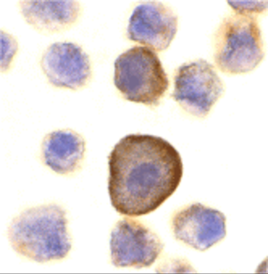 |
| Immunocytochemistry Validation of PERP in A431 Cells Immunocytochemical analysis of A431 cells using anti-PEPR antibody (9812) at 10 µg/mL. Cells was fixed with formaldehyde and blocked with 10% serum for 1 h at RT; antigen retrieval was by heat mediation with a citrate buffer (pH6). Samples were incubated with primary antibody overnight at 4C. A goat anti-rabbit IgG H&L (HRP) at 1/250 was used as secondary. Counter stained with Hematoxylin. |
FAQ & Publications
Frequently Asked Questions
What applications is the rabbit anti-PERP polyclonal antibody 9812 validated for, and what are the recommended dilutions?
The rabbit anti-PERP polyclonal antibody 9812 is validated for use in Western blot (WB), immunocytochemistry/immunofluorescence (ICC/IF), and ELISA. For immunoblotting, the recommended dilution is 0.5-1 µg/mL, where it detects a band at approximately 21 kD using A431 cell lysate as a positive control.
How should the rabbit anti-PERP polyclonal antibody 9812 be stored to maintain stability?
This antibody should be stored at -20°C and is stable for at least one year under these conditions. It is supplied in PBS buffer at pH 7.4 and users should avoid multiple freeze-thaw cycles to preserve antibody integrity.
Publications
| pmid | title | authors | citation |
|---|---|---|---|
| We haven't added any publications to our database yet. | |||
Published literature highly relevant to the biological target of this product and referencing this antibody or clone are retrieved from the PubMed database provided by the United States National Library of Medicine at the National Institutes of Health.
Protocols
| relevant to this product |
|---|
| Western blot IHC ICC |
Documents
| Batch Number | QC File | SDS |
|---|---|---|
| To view batch-specific Safety Datasheets and Quality Certificates associated with your account, please Log In. | ||
Only logged in customers who have purchased this product may leave a review.


Reviews
There are no reviews yet.