| Weight | 1 lbs |
|---|---|
| Dimensions | 9 × 5 × 2 in |
| host | rabbit |
| isotype | IgG |
| clonality | polyclonal |
| concentration | 1 mg/mL |
| applications | ICC/IF, WB |
| reactivity | p53R2 (NT) |
| available sizes | 100 µg |


rabbit anti-p53R2 (NT) polyclonal antibody 7406
$445.00
Antibody summary
- Rabbit polyclonal to p53R2 (NT)
- Suitable for: ELISA,WB,IHC-P,IF
- Isotype: Whole IgG
- 100 µg
rabbit anti-p53R2 (NT) polyclonal antibody 7406
| antibody |
|---|
| Tested applications WB,IHC,IHC,ICC/IF,ELISA |
| Recommended dilutions Immunoblotting: use at 1ug/mL. A band of ~39kDa is detected IHC: use at 1ug/mL. Immunofluorescence: use at 20ugml. |
| Immunogen Peptide corresponding to aa 2-17 of human p53R2. |
| Size and concentration 100µg and 1 mg/mL |
| Form liquid |
| Storage Instructions This antibody is stable for at least one (1) year at -20°C. Avoid multiple freeze-thaw cycles. |
| Storage buffer PBS, pH 7.4, 0.02% NaN3. |
| Purity peptide affinity purification |
| Clonality polyclonal |
| Isotype IgG |
| Compatible secondaries goat anti-rabbit IgG, H&L chain specific, peroxidase conjugated, conjugated polyclonal antibody 9512 goat anti-rabbit IgG, H&L chain specific, biotin conjugated polyclonal antibody 2079 goat anti-rabbit IgG, H&L chain specific, FITC conjugated polyclonal antibody 7863 goat anti-rabbit IgG, H&L chain specific, Cross Absorbed polyclonal antibody 2371 goat anti-rabbit IgG, H&L chain specific, biotin conjugated polyclonal antibody, crossabsorbed 1715 goat anti-rabbit IgG, H&L chain specific, FITC conjugated polyclonal antibody, crossabsorbed 1720 |
| Isotype control Rabbit polyclonal - Isotype Control |
| target relevance |
|---|
| Homo sapiens RRM2B Ribonucleoside-diphosphate reductase subunit M2 B |
| Protein names Ribonucleoside-diphosphate reductase subunit M2 B |
| Alternative names TP53-inducible ribonucleotide reductase M2 B, p53-inducible ribonucleotide reductase small subunit 2-like protein |
| Gene names RRM2B |
| Protein family Belongs to the ribonucleoside diphosphate reductase small chain family |
| Function Plays a pivotal role in cell survival by repairing damaged DNA in a p53/TP53-dependent manner. Supplies deoxyribonucleotides for DNA repair in cells arrested at G1 or G2. Contains an iron-tyrosyl free radical center required for catalysis. Forms an active ribonucleotide reductase (RNR) complex with RRM1 which is expressed both in resting and proliferating cells in response to DNA damage |
| Catalytic activity a 2'-deoxyribonucleoside 5'-diphosphate + [thioredoxin]-disulfide + H2O = a ribonucleoside 5'-diphosphate + [thioredoxin]-dithiol |
| Subcellular location Cytoplasm, Nucleus |
| Structure Heterotetramer with large (RRM1) subunit. Interacts with p53/TP53. Interacts with RRM1 in response to DNA damage |
| Involvement in disease Mitochondrial DNA depletion syndrome 8A A disorder due to mitochondrial dysfunction characterized by various combinations of neonatal hypotonia, neurological deterioration, respiratory distress, lactic acidosis, and renal tubulopathy. Mitochondrial DNA depletion syndrome 8B A disease due to mitochondrial dysfunction and characterized by ophthalmoplegia, ptosis, gastrointestinal dysmotility, cachexia, peripheral neuropathy. Progressive external ophthalmoplegia with mitochondrial DNA deletions, autosomal dominant, 5 A disorder characterized by progressive weakness of ocular muscles and levator muscle of the upper eyelid. In a minority of cases, it is associated with skeletal myopathy, which predominantly involves axial or proximal muscles and which causes abnormal fatigability and even permanent muscle weakness. Ragged-red fibers and atrophy are found on muscle biopsy. A large proportion of chronic ophthalmoplegias are associated with other symptoms, leading to a multisystemic pattern of this disease. Additional symptoms are variable, and may include cataracts, hearing loss, sensory axonal neuropathy, ataxia, depression, hypogonadism, and parkinsonism. Rod-cone dystrophy, sensorineural deafness, and Fanconi-type renal dysfunction An autosomal recessive disease characterized by visual impairment due to rod-cone dystrophy, sensorineural hearing loss, and Fanconi-type renal dysfunction resulting in rickets-like skeletal changes. Death may occur in childhood or young adulthood due to renal failure. Disease onset is before age 5 years. |
| Keywords 3D-structure, Alternative splicing, Cytoplasm, Deafness, Deoxyribonucleotide synthesis, Disease variant, DNA damage, DNA repair, Iron, Metal-binding, Neuropathy, Nucleus, Oxidoreductase, Primary mitochondrial disease, Progressive external ophthalmoplegia, Proteomics identification, Reference proteome |
| Sequence MGDPERPEAAGLDQDERSSSDTNESEIKSNEEPLLRKSSRRFVIFPIQYPDIWKMYKQAQ ASFWTAEEVDLSKDLPHWNKLKADEKYFISHILAFFAASDGIVNENLVERFSQEVQVPEA RCFYGFQILIENVHSEMYSLLIDTYIRDPKKREFLFNAIETMPYVKKKADWALRWIADRK STFGERVVAFAAVEGVFFSGSFAAIFWLKKRGLMPGLTFSNELISRDEGLHCDFACLMFQ YLVNKPSEERVREIIVDAVKIEQEFLTEALPVGLIGMNCILMKQYIEFVADRLLVELGFS KVFQAENPFDFMENISLEGKTNFFEKRVSEYQRFAVMAETTDNVFTLDADF |
| UniProt accession: Q7LG56 |
Data
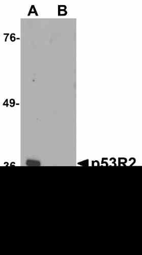 |
| Western blot analysis of p53R2 in 3T3 cell lysate with p53R2 antibody at 1 µg/mL in (A) the absence and (B) the presence of blocking peptide. |
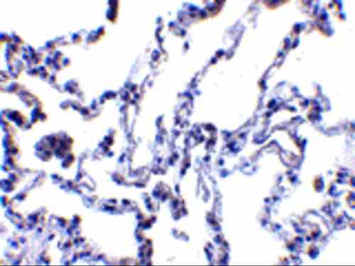 |
| Immunohistochemistry of p53R2 in human lung tissue with p53R2 antibody at 1 µg/mL. |
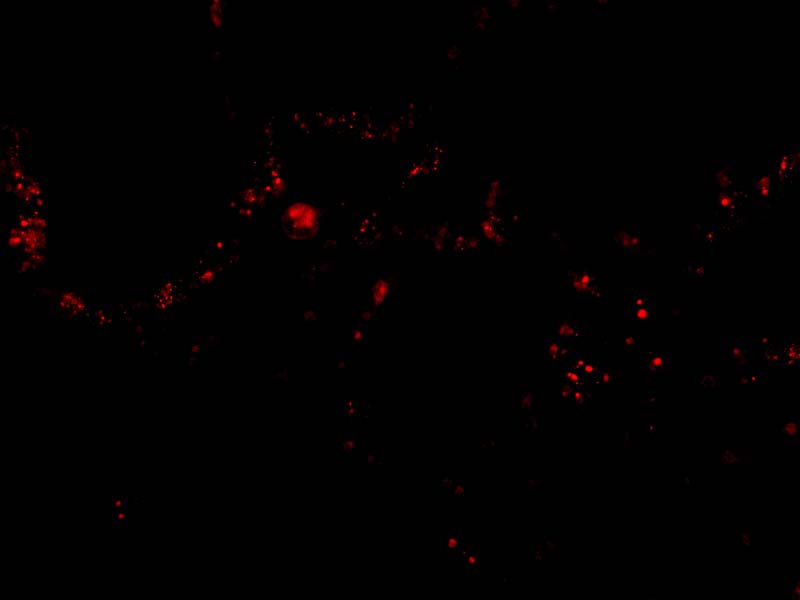 |
| Immunofluorescence of p53R2 in Human Lung tissue with p53R2 antibody at 20 µg/mL. |
FAQ & Publications
Frequently Asked Questions
What is the recommended dilution for using the rabbit anti-p53R2 (NT) polyclonal antibody in Western blot applications?
The recommended dilution for Western blot (immunoblotting) is 1 µg/mL, which detects a band of approximately 39 kDa.
Which secondary antibodies are compatible with the rabbit anti-p53R2 (NT) polyclonal antibody for detection?
Compatible secondary antibodies include goat anti-rabbit IgG, H&L chain specific, available in peroxidase conjugated, biotin conjugated, FITC conjugated, and cross-absorbed forms.
How should the rabbit anti-p53R2 (NT) polyclonal antibody be stored to maintain its stability?
The antibody should be stored at -20°C and is stable for at least one year. It is important to avoid multiple freeze-thaw cycles to preserve antibody integrity.
What is the immunogen used for generating the rabbit anti-p53R2 (NT) polyclonal antibody?
The immunogen is a peptide corresponding to amino acids 2 to 17 of the human p53R2 protein.
Is the rabbit anti-p53R2 (NT) polyclonal antibody suitable for immunofluorescence applications, and if so, what dilution is recommended?
Yes, the antibody is suitable for immunofluorescence (IF) and the recommended dilution is 20 µg/mL.
Publications
| pmid | title | authors | citation |
|---|---|---|---|
| We haven't added any publications to our database yet. | |||
Published literature highly relevant to the biological target of this product and referencing this antibody or clone are retrieved from the PubMed database provided by the United States National Library of Medicine at the National Institutes of Health.
Protocols
| relevant to this product |
|---|
| Western blot IHC ICC |
Documents
| Batch Number | QC File | SDS |
|---|---|---|
| To view batch-specific Safety Datasheets and Quality Certificates associated with your account, please Log In. | ||
Only logged in customers who have purchased this product may leave a review.


Reviews
There are no reviews yet.